Playlist
Show Playlist
Hide Playlist
Pityriasis Alba in Darker Skin

-
Slides Pityriasis Alba in Darker Skin.pdf
-
Download Lecture Overview
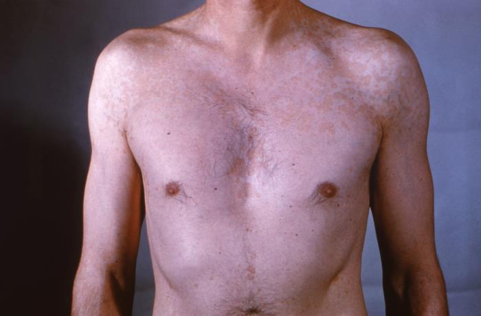
00:01 Welcome to our lecture on Pityriasis Alba. 00:05 Again, if you look at the classification of eczema there's endogenous and exogenous. 00:11 And Pityriasis alba falls under endogenous eczemas. 00:16 And that's what we're going to talk about now. 00:18 So it's a low-grade type of eczema or dermatitis. 00:23 And pityriasis refers to the characteristic fine scale and alba to its pale color or hypopigmentation. Pityriasis alba affects children and adolescents from the age of 3 to 16 years, with a male to female ratio of 1 :1. 00:42 It's actually more prominent and more common in dark skin because of the deep pigmentation. 00:49 The cause of Pityriasis alba is unknown, but it tends to coexist with dry skin and atopic dermatitis, and often presents following sun exposure. 00:59 So how does Pialba present clinically. 01:03 It presents with hypopigmented patches or plaques, and the usual sites are the face, the neck, shoulders and upper arms, and the lesions tend to be round or oval or sometimes irregular in shape, then range from 1 to 20 lesions and sizes varying from 0.5 to 5cm. 01:24 Patients present with dryness and scaling of the skin. 01:29 So the natural course of the disease. 01:31 It tends to be self-limiting and evolves through several stages, from hypopigmented patch plaque with fine surface scale to post-inflammatory hyperpigmented macule without scale and eventually resolution. 01:47 The diagnosis of Pityriasis alba. 01:50 It's important that you exclude other differentials, and one can do this by using a Wood's lamp, where one sees no enhancement of fluorescence. 02:03 The second way of confirming your diagnosis is to do scrapings for mycology, so that you can exclude fungal infection or things like tinea versicolor. 02:13 And here you will see negative microscopy and fungal culture will be negative. 02:19 Really one needs a skin biopsy to make a diagnosis. 02:24 The differential diagnosis includes Pityriasis versicolor Pityriasis alba Pityriasis versicolor. 02:31 They both scaly and on direct microscopy. 02:34 If you are dealing with Pityriasis versicolor, you're going to see hyphae and yeast cells in a pattern described as spaghetti and meatballs. 02:42 Spaghetti refers to the hyphae, and meatballs is equivalent to the yeast cells that you see on microscopy. 02:51 The second differential is scleroderma, where you may have salt and pepper pattern of hyperpigmentation, but usually followed by other cutaneous manifestations of the disease or autoimmune diseases. Nevus depigmentosus is another differential for Pityriasis alba. 03:13 It's not a common condition. 03:14 It's usually circumscribed with segmental area of depigmentation or hyperpigmentation. 03:20 The lesion is usually solitary and shows little change over time, unlike Pityriasis alba. So how do we treat these patients? It's usually a self-limiting disease with no treatment needed for asymptomatic cases. 03:34 However, using good moisturizers for the scaliness improves the look of the disease, and mild topical corticosteroids like hydrocortisone from 0.5 to 1% may reduce redness in each if present. Calcineurin inhibitors are also an option for treating Pityriasis alba.
About the Lecture
The lecture Pityriasis Alba in Darker Skin by Ncoza Dlova is from the course Hyper- and Hypopigmentation Skin Disorders.
Included Quiz Questions
What is the characteristic appearance of pityriasis alba lesions on physical examination?
- Hypopigmented patches with fine scaling
- Hyperpigmented macules with thick crusting
- Erythematous plaques with weeping
- Vesicular lesions with ulceration
- Deep brown patches with induration
Which finding would help distinguish pityriasis alba from pityriasis versicolor on microscopic examination?
- Negative microscopy for hyphae and yeast cells
- Presence of "spaghetti and meatballs" pattern
- Positive Wood's lamp fluorescence
- Presence of bacterial colonies
- Positive culture for dermatophytes
What is the first-line treatment approach for an asymptomatic case of pityriasis alba?
- No treatment is needed as it is self-limiting
- High-potency topical steroids
- Systemic antifungal therapy
- Oral antihistamines
- Phototherapy
Author of lecture Pityriasis Alba in Darker Skin

Ncoza Dlova
Customer reviews
5,0 of 5 stars
| 5 Stars |
|
5 |
| 4 Stars |
|
0 |
| 3 Stars |
|
0 |
| 2 Stars |
|
0 |
| 1 Star |
|
0 |
