Playlist
Show Playlist
Hide Playlist
Keloids in Darker Skin

-
Slides Keloids in Darker Skin.pdf
-
Download Lecture Overview
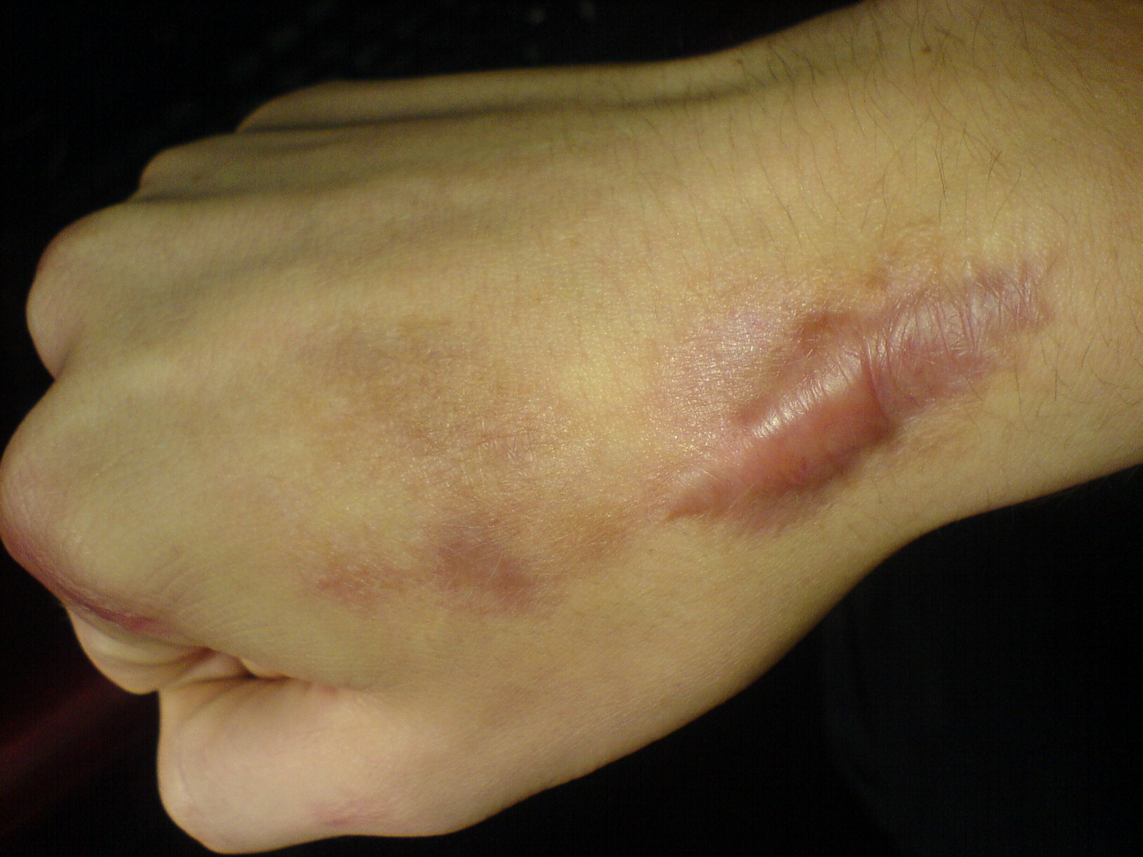
00:01 Welcome to our lecture. We will be discussing hypertrophic scars and keloids. 00:06 Fibro proliferative disorders that result from aberrant wound healing in predisposed individuals following trauma, inflammation, surgery or burns. 00:16 And that's how we define hypertrophic scars and keloids. 00:21 At times, spontaneous keloids develop with no underlying trauma or trigger, and it is important to note that there are also other conditions that can present as keloids, for example Kaposi's sarcoma, cutaneous sarcoidosis, and blastomycosis, etc. so it's not always a classical keloid, spontaneous or following injury. 00:43 We have to think beyond that and consider other causes of keloids. 00:50 Talking about hypertrophic scars, these tend not to extend beyond the margins of the original wound, and they're always confined to the area of the previous wound. 01:02 If one looks at keloids. Keloids- they extend beyond the original wound margin and can be spontaneous. So it's not always that keloids are due to secondary injury. 01:15 And this picture that you see here, this is a patient who had keloids after ear piercing. The keloids are more frequent in those with Fitzpatrick skin phototypes 3 to 6, and they're self reported in about 16% of black individuals. 01:37 People with lighter skin complexion and people with albinism appear to be less affected, and this sometimes is related to the melanocytes and of course the fibroblasts and the size of the fibroblasts. The exact pathogenesis is unknown, but it may develop after minor injuries, for example trauma, burns, insect bites, and surgery. 02:03 It's more common in wounds that have been allowed to heal by secondary intention. 02:08 So what are the clinical manifestations of keloids? They may present with purplish red, firm smooth, and they may be raised. 02:16 They may be associated with pain and pruritus, particularly when active. 02:21 And they may also occur long after the injury has healed. 02:26 What about hypertrophic scars? These tend to be pink to red. 02:30 They may be raised or flat, and sometimes may be associated with itchy lesions. They usually occur within weeks of the injury. 02:42 So how do we diagnose keloids and hypertrophic scars? The diagnosis is based on history and clinical features. 02:48 Sometimes the skin biopsy is indicated in some cases that have colloidal presentation, particularly if you want to exclude conditions like Kaposi sarcoma, keloidal, blastomycosis, and sarcoidosis. 03:01 These are conditions in dermatology that present with keloids. 03:05 And yet it's not actually the usual keloids that you see. 03:08 So we have to think about these at the back of our minds, but of course, in the right setting. 03:14 So some of the differential diagnosis of keloids or hypertrophic scars include skin tumors as listed over there, particularly dermatofibroma and dermatofibrosarcoma, cutaneous squamous cell carcinoma, morphea, which is a type of localized scleroderma. 03:34 So how do we treat keloids? Prevention of keloids is paramount, particularly in patients who have a predisposition of keloids. So we have to promote rapid wound healing because the risk is higher if the healing time takes more than three weeks. 03:51 We've got to keep the wound clean and moisturized, fix the wound and make sure that the edges come together and there's no tension in between the edges of the wound. That we can do by using paper tape. 04:06 Topical corticosteroids should be introduced in cases of a new scar showing signs of ongoing or relapse. Surgical incisions and repair techniques are crucial so that linear incisions that follows Langer's lines are important. 04:25 When you are doing your surgical incisions on the skin, this helps to make sure that there's no tension when we are following Langer's lines. 04:35 A version of wound edges during suturing is also crucial. 04:40 We also try and limit the number of sutures so that there's less sutures to remove after the healing. It's important that unnecessary surgery is avoided, particularly in patients who are keloid prone. 04:54 And sometimes it's easy to ask the patient that if you have trauma or injury, does it form a scar or keloid and the patient will inform you, then you know whether the patient is a keloid or scar former. 05:08 So how do we treat keloids? First, we look at conservative therapies as first line of treatment. 05:15 Corticosteroid tapes can be used if it's available or clusters. 05:19 And these contain fludrocxycorticoid. 05:23 And it's used as a tape with four or about 4mcg/cm2. 05:30 Intralesional corticosteroid injections are also used, and the strength ranges from 2.5 to 10mg per site. 05:37 Sometimes we go up to 40mg, depending on the size, the thickness, and the location of the keloid. 05:45 Compression therapy has also been used using pressure garments and bandages or special devices for certain locations, particularly the ears. 05:54 So for the ears, sometimes we use the ear clips which are attached on the ear and the pressure and the mechanical pressure helps to reduce the keloid. 06:04 Gel sheeting is another form of treating keloids, and this helps by stabilizing the keloids and moisturizes the scar, thus reducing the inflammation. 06:17 We do have other therapies, for example laser therapy, cryotherapy, intralesional agents and this we apply and use chemotherapy drugs like fluorouracil and bleomycin or botulinum toxin type A. 06:33 When you use these drugs, it's important that you use the right concentrations. Sometimes we mix five fluorouracil and bleomycin with steroids at different concentrations. And some of the side effects could actually include ulceration. 06:48 So you have to be careful when you use these drugs. 06:52 And these work by decreasing the fibroblast activity and scar tension. 06:57 Because we know that it's the fibroblasts that produce excessive collagen resulting in the scar or keloid. Surgical excision is another option that we use for treating keloids depending on the size of the keloid, the type of the keloid, the location of the keloid. 07:15 So it's usually used for larger multiple keloids. 07:19 And the goals really are to try and debulk most of the keloid if possible, and then reduce the thickness and the hard areas. 07:28 This makes it easy to use the intralesional therapy for keloids, and it helps to disrupt the tension on the scar with flaps. 07:42 Surgical excision must always almost be followed by intralesional steroids or intralesional chemotherapy or radiotherapy. 07:51 Otherwise the keloid will recur. 07:54 This is an important take home message for patients with keloids. 08:00 We need to know that firstly there are chronic skin conditions. 08:04 They require long term management. 08:07 And thirdly, follow up is crucial. 08:11 Otherwise the keloids will recur. 08:14 We cannot just treat them and discharge patients. 08:16 We need to follow them up constantly because colors can come back. 08:21 So I think this is an important take home message for patients with Keloids.
About the Lecture
The lecture Keloids in Darker Skin by Ncoza Dlova is from the course Hyper- and Hypopigmentation Skin Disorders.
Included Quiz Questions
What is the key distinguishing feature of keloids compared to hypertrophic scars?
- They extend beyond the original wound margins
- They develop within weeks of injury
- They remain confined to the wound area
- They are always triggered by trauma
- They only occur in light-skinned individuals
What is the critical time period for wound healing to prevent keloid formation?
- Less than 3 weeks
- Less than 1 week
- Less than 6 weeks
- Less than 2 months
- Less than 4 days
What is the recommended approach when performing surgical excision of keloids?
- Combine with post-surgical intralesional therapy
- Perform excision alone for complete cure
- Avoid any post-surgical interventions
- Use wide surgical margins only
- Treat with antibiotics post-surgery
In which Fitzpatrick skin types are keloids most commonly observed?
- Types III to VI
- Types I and II only
- Types I to III
- Types V and VI only
- All skin types equally
Author of lecture Keloids in Darker Skin

Ncoza Dlova
Customer reviews
5,0 of 5 stars
| 5 Stars |
|
5 |
| 4 Stars |
|
0 |
| 3 Stars |
|
0 |
| 2 Stars |
|
0 |
| 1 Star |
|
0 |
