Playlist
Show Playlist
Hide Playlist
Wound Healing: Primary Intention – Inflammation and Wound Repair
-
Slides Inflamation Cellular Pathology.pdf
-
Download Lecture Overview
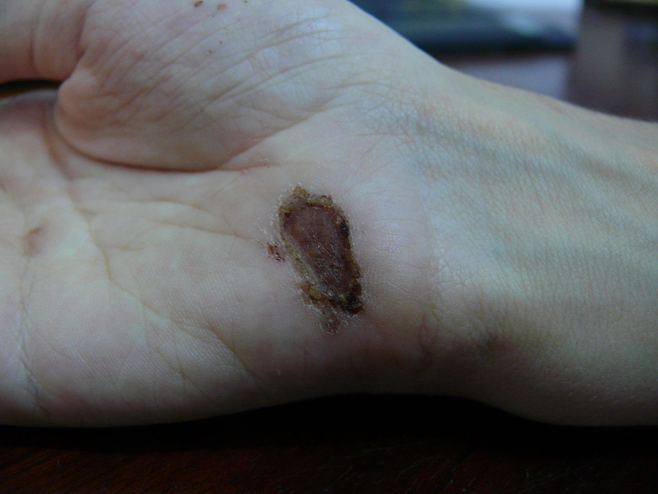
00:01 Our topic here quickly will be through what's known as your wound healing. Wound healing at first will be primary intention. 00:10 Now what that means is the fact that here with primary intention is the fact that the, let's say that you have taken a piece of paper and you've gotten yourself a paper cut. If you've gotten a paper cut you have created or introduced a wound, correct. 00:24 In your skin. Now, if you have introduced a wound the edges of the wound might be far apart or they might be close together. 00:34 If they are far apart, you will call that secondary intention. If there are close together it will be a primary intention. 00:42 So the primary intention, you don't have to lay down as much granulation tissue. Think of this as being a pothole in a road. 00:49 If it's a little pothole you can get away from you can get away with for the most part fibrosis, it bring the wound together, healing. 00:58 But if the edges of the wound are far apart and now it's a knife wound, far apart. You need to put in alot of asphalt into the pothole to clean it up, right. To fix it. The asphalt of a pothole in a road is equivalent to granulation tissue. We'll talk about granulation tissue in great detail which includes the fact that it has collagen and it has angiogenesis with VEGF. 01:24 All this is going to help you bring wounds back together. Once we get through the normal, we will go through some important in which you might lay down too much collagen. That will be a keloid. Or there might be deficiency of collagen such as Ehlers-Danlos. 01:42 Let's begin. Day 1 you have a fibrin clot, a hematoma. Also keep in mind if you cut yourself with a knife, then not only are you introducing a wound but you are also cutting through your blood vessels. Two things occuring at the same time. Wound and tension, repair also hemostasis. What's that mean to you? The platelets, we talked about hemodynamics, are going to then form a thrombus. 02:08 On day 2, the squamous cell will seal of the wound. The macrophages emigrate into the wound. Day 3, depending as to how much granulation tissue is required, it then begins to form. The initial type of collagen that you are going to deposit, and do not get your granulation tissue confused with granuloma, is going to be type III collagen. The type III collagen is then going to be replaced as you shall see soon enough. But here during day 3 for the most part almost always day 3, the neutrophils are going to start disappearing, apoptosis. Macrophages, monocytes will come in and then land into the tissue and we have our macrophages. About 4-6 we have granulation tissue which will peak and now fibronectin is a very key glycoprotein. 03:00 In the extracellular matrix, the fibronectin provides the infrastructure for proper intention of wound healing. 03:09 The fibronectin is also something that cancer cells will take advantage of when they metastasize. That is how important fibronectin is. Now we are going to skip over to week two. At this point we have approximately, a tensile strength of approximately 10%. 03:28 About a month later though we have remodelling. Which basically means that the type III collagen is being replaced by collagen type I. The major enzymes here would be things like collagenase and lysyl oxidase. Oxidase, not hydroxylase but oxidase you have to have copper. Tensile strength here may raise up to 80% in about 3 months. But we'll never get back to proper normal. 03:54 That's important. Now, the pictures are showing you excess keloid. Who is your patient here? Think of major contact sports. 04:04 Boxing, football. Anyhow, these sports in which you patients are getting hurt. When they get hurt, also think of ear piercings, piercings in general. Genetically speaking these patients and this individuals have a predisposition of during the granulation tissue phase, about day 3. Laying down collagen. You keep laying down collagen excessively. And when you do, you are going to form what's known as a keloid. Look for perhaps in a African-American male in which the earlobes might be affected. 04:46 Or as I told you earlier, you can have contact sports and during a cut may then have keloid formation. Versus you have a patient in which the skin is hyperextensible. That to you should be indicative of a diagnosis called Ehlers-Danlos in which this is the opposite of keloid. You might then be deficient of collagen type III or type V. Do not confuse with osteogenesis imperfecta in which you are only deficient of collagen type I.
About the Lecture
The lecture Wound Healing: Primary Intention – Inflammation and Wound Repair by Carlo Raj, MD is from the course Cellular Pathology: Basic Principles with Carlo Raj.
Included Quiz Questions
What is the method of wound healing when the edges of the severed tissues are well-approximated?
- Primary intention
- Secondary intention
- Scar
- Fibrin clot
- Keloid
When do macrophages start to appear during the healing process?
- Day 2
- Day 1
- Day 6
- Week 2
- Month 1
What is granulation tissue primarily made from?
- Type III collagen
- Fibrin
- Glycoproteins
- Fibronectin
- Squamous cells
Customer reviews
5,0 of 5 stars
| 5 Stars |
|
5 |
| 4 Stars |
|
0 |
| 3 Stars |
|
0 |
| 2 Stars |
|
0 |
| 1 Star |
|
0 |


