Playlist
Show Playlist
Hide Playlist
Vitiligo: Pathophysiology

-
Reference List Pathology.pdf
-
Slides Vitiligo Pathophysiology Dermatopathology.pdf
-
Download Lecture Overview
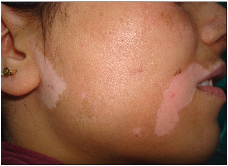
00:01 Welcome. Let's talk about a deep pigmenting disease called vitiligo. 00:08 What is vitiligo? Well, it's an autoimmune disorder. 00:12 And it is caused by destruction of melanocytes that results in the loss of skin pigmentation. It's a fairly common disorder. 00:23 It's seen in up to 2% of the population. 00:25 So invariably ultimately you've almost all seen examples of this. 00:30 And I'll show you some other examples of that, including on me personally, I have vitiligo. 00:37 It occurs in young children and adults. 00:40 There's an equal incidence across the genders, and there's no racial or ethnic predilection. 00:46 Having said that, it's very apparent in people of dark skin because there is no pigmentation. And it's quite obvious as white plaques on someone like me, who's kind of a very pale individual to begin with. 01:01 The vitiligo is not apparent. But having said that, I can expose my forearm and show you that I do have hyperpigmentation where I've lost melanocytes. The pathophysiology of this, it is an autoimmune disease that's acquired. 01:15 There is a genetic predisposition. 01:18 And interestingly, about half of affected individuals have a family history. 01:22 But they don't have to. Clearly 20% of affected individuals have another autoimmune disorder. And so Hashimoto's thyroiditis, graves disease, etc. are also very commonly associated with individuals who will have vitiligo. 01:38 What is happening. So the melanocytes, remember, live at the dermal epidermal junction at the basal layer. 01:44 And they are being attacked by either cytotoxic T cells macrophages and or antibodies and complement to drive their immune destruction. 01:55 If you don't have melanocytes, you don't make melanin. 01:58 Simple as that. What's the clinical presentation? Well, these are very sharply demarcated lesions which I've always found kind of kind of interesting, but don't understand because why did those attacking antibodies or attacking T cells or macrophages stop at that border? It's not known. But any event, sharply demarcated white plaques, their macules or patches where there's been no loss of pigmentation. 02:23 They're frequently even though it's an immune driven process. 02:26 They are not red. They are not painful. 02:29 They are not pruritic or itchy. 02:32 They just happen. And what you get is a chalky or milk white color. 02:36 All you're seeing now, the only pigmentation that is there is due to the underlying blood and or carotenes that are normally present within keratinocytes. 02:46 So vitiligo can occur on almost any part of the body, but there are some more common areas that are affected. 02:54 This list that we're going to go through is not from most common to least common, but in fact just represents the various areas that are affected. 03:01 So the face and especially around the mouth are areas where we'll see it. 03:06 That can be particularly distressing for individuals who are dark skinned, because that's obviously something that everybody sees elbows, hands, genitalia around the anus, knees. 03:19 Those are also sites that are commonly affected by the loss of pigmentation due to the destruction of the melanocytes. 03:27 The diagnosis, again, pretty much what you see is what you get in terms of the diagnosis. 03:32 You can use a Wood's lamp. 03:34 So this is a UV device. 03:37 That low powered UV device that you can use to scan over areas where there is hyperpigmentation. This is helpful not necessarily for individuals who are darkly colored or pigmented skin, but very helpful for lightly pigmented individuals where it may be more difficult to evaluate. 03:57 It accentuates the depigmented areas which appear white on the Wood's lamp exam. 04:04 So we can also do a biopsy to demonstrate this, although in most cases this isn't done. 04:09 What you're seeing now is a normally pigmented skin. 04:13 So there is a layer at the dermoepidermal junction in the basilar layer of pigmented epithelial cells in melanocytes. 04:21 If this same patient had vitiligo there would be no melanocytes at that junction. 04:28 We can also do special stains to highlight the absence or presence of melanocytes, because there are a number of basic markers on those cells. 04:36 So what do we do about vitiligo? How do we manage this? First of all, any patient who has vitiligo is not making melanin in those areas. 04:44 They are at very increased risk of having UV damage because of the absence. 04:49 The skin in that area is much like an albino skin without any melanocytes or any melanin production. You should rule out other sorts of autoimmune diseases. 05:01 So evaluate for thyroid function in particular, because Graves and Hashimoto's thyroiditis are associated with a high frequency with vitiligo. In terms of treatment, Topical steroids can be helpful by limiting the inflammatory damage of new of existing melanocytes, but once the damage has been done, steroids don't bring back the pigmentation. 05:26 For very localized disease, they can be helpful, but again, won't bring back the pigmentation. 05:31 When using them, the areas become somewhat sensitized because high dose corticosteroids tends to cause diminish the proliferation of the keratinocytes and also the production of the stratum corneum overlying it, so the skin is thinned and there may be increased sun sensitivity in those areas. 05:53 In the same way that topical corticosteroids are effective by limiting inflammation, you can also give topical calcineurin inhibitors such as tacrolimus cyclosporin, an alternative to topical steroids. 06:07 They still will not bring back the pigmentation once it's gone. 06:12 For areas such as the face where you don't want to have the same risk of skin atrophy. 06:17 They may be a better alternative than giving steroids. 06:22 You can go to bigger guns, that is to say high dose systemic immunosuppression, glucocorticoids or some chemotherapies that kill proliferating T cells. And those have variable benefit. 06:34 Again, will not bring back pigmentation in areas that have already been destroyed. 06:39 Puva therapy, which is used to treat widespread vitiligo. 06:44 So this is hopefully an area where you might be able to get restored melanization of the tissue. Use psoralen, which is either administered topically or orally, and then give UVA exposure and you can use a light booth. 07:05 Or you can use sunlight to do this. 07:07 And this helps to re pigment the skin by driving the local proliferation of melanocytes coming in from the edges of edges of the lesion. 07:16 It also causes increased T cell apoptosis. 07:20 So it kills off the proliferating T cells in those areas. 07:23 So you kind of get a two dose bang for your buck. 07:28 Alternatively, for individuals who have such severe depigmentation you go the other way. 07:34 So you then dip pigment the rest of the body to provide uniformity in terms of the overall pigmentation. That's when you have greater than 40% of the surface area already having lost melanocytes. You use it on all the unaffected skin and basically it's skin bleaching. Um, and in many cases will give improved kind of cosmetic satisfaction. 08:03 But there's a price to be paid with that. 08:06 We've covered an interesting topic. 08:08 So the loss of melanocytes rather than the proliferation of melanocytes at the autoimmune disease vitiligo.
About the Lecture
The lecture Vitiligo: Pathophysiology by Richard Mitchell, MD, PhD is from the course Disorders of Maturation and Pigmentation of the Skin.
Included Quiz Questions
Which population shows the highest incidence of vitiligo?
- There is no racial or ethnic predilection
- People with dark skin tones
- People with light skin tones
- Asian populations
- African populations
What percentage of vitiligo patients have another autoimmune disorder?
- 20%
- 10%
- 35%
- 50%
- 75%
What is the primary purpose of using Wood's lamp in vitiligo diagnosis?
- To accentuate depigmented areas
- To measure skin thickness
- To detect inflammation
- To identify melanomas
- To evaluate blood flow
Which treatment is most appropriate for widespread vitiligo affecting over 40% of body surface?
- Depigmentation therapy
- Topical steroids
- Calcineurin inhibitors
- Local UV therapy
- Skin grafting
Author of lecture Vitiligo: Pathophysiology

Richard Mitchell, MD, PhD
Customer reviews
5,0 of 5 stars
| 5 Stars |
|
5 |
| 4 Stars |
|
0 |
| 3 Stars |
|
0 |
| 2 Stars |
|
0 |
| 1 Star |
|
0 |
