Playlist
Show Playlist
Hide Playlist
Triangles – Topographic Back Anatomy
-
Slides 04 Abdominal Wall Canby.pdf
-
Download Lecture Overview
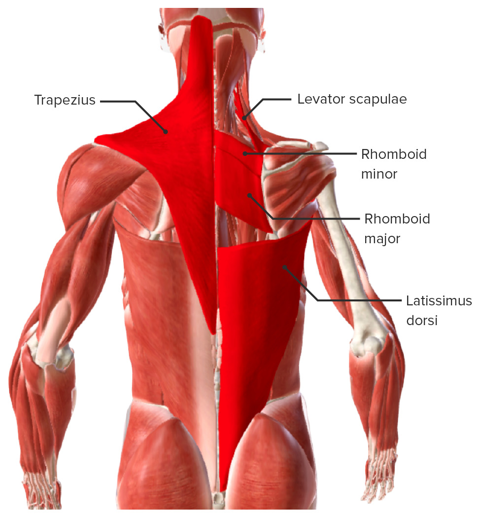
00:01 This slide demonstrates the borders of the triangle of auscultation. The three structures that define this triangle would be the scapula, the latissimus dorsi muscle and then lastly, we have another muscular contribution, the trapezius muscle. So now, let’s look at these three borders. 00:24 This slide represents the superior most of these three triangles and this is going to be the triangle of auscultation. It’s going to be defined by three anatomic structures - the inferior aspect or angle of the scapula and then two muscular contributions, one from the latissimus dorsi muscle as well as from the trapezius muscle. So if we take a look, we have all three structures defined. Here is the inferior angle of the scapula. Here is the margin of the trapezius; we’re looking at the lateral margin in through here. And then here is the inferior margin of the latissimus that’s coursing over here more laterally. 01:11 So, our triangle of auscultation is right in through this area. 01:17 The next triangle of interest is going to be the superior lumbar triangle. And there are three structures that define the borders of this triangle - the 12th rib, quadratus lumborum muscle and the internal abdominal oblique muscle. And two of these three structures are discernible in this particular illustration. The third component is obstructed by another more superficial component. But, if we take a look in this image, our superior triangle, lumbar triangle is down in this area. Your 12th rib is right in through here. And the internal abdominal oblique is coursing right in through here. And what we have here are more superficial structures that are obstructing our view of the third and final border of the triangle and that border is defined by the quadratus lumborum. And these muscular slips that we see here belong to the serratus posterior inferior. So, if we were to peel away, remove these muscular slips, we would be able to see the quadratus lumborum running from the 12th rib, which is right in through here, down to the iliac crest down through here. So, those are the three borders of our superior lumbar triangle. 02:48 The last triangle of interest is going to be the inferior lumbar triangle. It is going to be formed by three structures that define its borders: the iliac crest, the latissimus dorsi muscle and lastly by the external abdominal oblique muscle. And if we take a look here, we can see all three contributions defining the inferior lumbar triangle at this location. 03:16 Here is the latissimus dorsi, its lateral most border. Here is the external abdominal oblique and then here is your iliac crest. Some clinical relevance to the lumbar triangles, not very common, but the superior lumbar triangle and the inferior lumbar triangle are potential sites of weakness. So, in some cases, you can have herniations through either one of these triangles. We’ll also see here very, very shortly another clinical application as it relates to the inferior lumbar triangle. Here we want to understand the topography as it relates to the intercristal line. The intercristal line will be a line that spans from the superior aspect of one iliac crest, that we see here to the right, run a line from this superior point over to the opposite superior most point of the iliac crest. This line will have a vertebral relationship and that’s typically going to be defined as a vertebral relationship to the fourth lumbar vertebra or between L4 and L5, the intervertebral disc that’s located between L4 and L5. These vertebral relationships have been confirmed by imaging studies. However, it is interesting that palpation is less accurate in defining these vertebral relationships.
About the Lecture
The lecture Triangles – Topographic Back Anatomy by Craig Canby, PhD is from the course Abdominal Wall with Dr. Canby.
Included Quiz Questions
Which structures border the superior lumbar triangle? Select all that apply.
- 12th rib
- Psoas major muscle
- Quadratus lumborum muscle
- Internal abdominal oblique muscle
- External oblique muscle
What vertebral level does the intercristal line identify?
- LIV
- T12
- L2
- LV
- S1
Which structure makes the boundary of the triangle of auscultation?
- Inferior angle of the scapula
- Superior angle of the scapula
- Internal abdominal oblique
- External abdominal oblique
- Iliac crest
Customer reviews
5,0 of 5 stars
| 5 Stars |
|
5 |
| 4 Stars |
|
0 |
| 3 Stars |
|
0 |
| 2 Stars |
|
0 |
| 1 Star |
|
0 |


