Playlist
Show Playlist
Hide Playlist
Trachea – Pulmonary Structures and Esophagus
-
Slides 03 Thoracic Viscera Canby.pdf
-
Download Lecture Overview
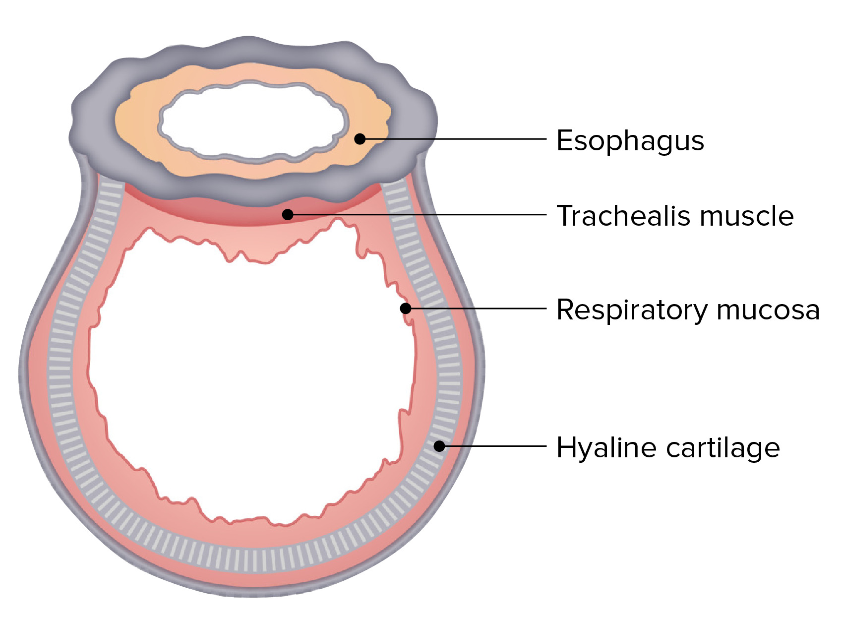
00:01 Now, let’s take a look at the trachea. And what you’ll want to remember here will be the vertebral levels where the trachea begins and where it bifurcates. 00:12 The trachea is beginning at this point right below the cricoid cartilage of the larynx, which is here. And so, this is the initial point of the trachea. It has a vertebral level of C6. It then will travel distally and then it’s bifurcating at this level. And this will have a vertebral relationship between T4 and T5. The distance travelled from C6 to T4 or T5 is only about 10 to 11 centimetres. We’re only looking at a respiratory passageway that is about 4 inches in overall length. Not as long as we might think. 00:59 Along its length, you’ll see various cartilaginous rings that are incomplete posteriorly and the number of these cartilaginous rings is anywhere from 16 upwards to 20. 01:22 Where the trachea bifurcates at its T4-T5 relationship, we can see a keel-like projection at that level bifurcation. And that is shown right in through here on the illustration. 01:35 That keel-like projection is called the carina. And then you can see the right bronchus here and you can see the left bronchus on the left side of the carina. 01:49 What we’ll want to understand here is which one of the bronchi is wider than the other and which bronchus is more vertical than the other. And in this case the answer to both questions is the right bronchus. 02:09 The right bronchus is wider in diameter than the left. The right bronchus is more vertical than the left. The left bronchus comes off at a more acute angle. We can utilize our understanding of the characteristics of the right bronchus when we think about aspiration of a foreign object and the likelihood of the bronchus then that will receive it. And if you did follow a foreign object that’s small enough, it has a greater likelihood of entering the right bronchus. 02:47 The wall of the trachea is made up of layers or strata. And those layers include the innermost layer of the mucosa. External to that is the submucosa. The third layer that’s even more external is the cartilaginous/fibromuscular layer. And then the outer layer or the coat of the trachea is going to be the adventitia. 03:13 All four of those layers are depicted in this slide. Here we have the mucosa and the portion of the mucosa in direct contact with the air moving though the lumen will have the epithelium that lines the mucosa. 03:32 The submucosa is going to be characterized by the presence of numerous glands that we see here. 03:40 The cartilaginous fibromuscular layer is demonstrated here where we have the hyaline cartilage. 03:46 And then you can see posteriorly that the rings of hyaline cartilage are incomplete and the ends here of the rings are bridged by fibromuscular components. So, here is the collagenous component and then in red we have the trachealis or muscular contribution to this region. 04:13 The outermost layer is your adventitia. That is simply a connective-tissue coat. 04:21 There are various types of bronchi. So, let’s describe the various divisions of the bronchi. 04:29 The first bronchi that form will be those bronchi that divide where the trachea ends. 04:36 And these are primary bronchi: a right one and a left one. 04:41 So, we have one primary bronchus for each lung. 04:45 Shortly after, each primary bronchus will divide into secondary bronchi. And we can see some secondary bronchi here and here and here, for example. There are three secondary bronchi to the right lung, two secondary bronchi to the left lung. And secondary bronchi are also known as lobar bronchi. The right lung, as we’ll see shortly, has three lobes hence it has to have three secondary or three lobar bronchi. The left lung, normally, only has two lobes and that is why we only have two secondary or two lobar bronchi associated with the left lung. 05:31 Each secondary bronchus will then divide into tertiary segments. And we see numerous tertiary segments in each one of these branching patterns. 05:42 Tertiary bronchi are also known as segmental bronchi because tertiary bronchi are going to supply each bronchopulmonary segment. Tertiary bronchi will then undergo further successive divisions. And there are many of these divisions before they’ll finally lead to our system of bronchioles. And the largest bronchiole is referred to as a primary bronchiole.
About the Lecture
The lecture Trachea – Pulmonary Structures and Esophagus by Craig Canby, PhD is from the course Thoracic Viscera with Dr. Canby.
Included Quiz Questions
Which vertebral level corresponds to the start of the trachea?
- C6
- C2
- T5
- T1
- C3
What is the average length of the trachea?
- 10–11 cm
- 4–5 cm
- 14–15 cm
- 17–18 cm
- 20–21 cm
Which side of the C-shaped rings of the trachea has the gap?
- Vertebral
- Mediastinal
- Pleural
- Anterior
- Superficial
Customer reviews
5,0 of 5 stars
| 5 Stars |
|
1 |
| 4 Stars |
|
0 |
| 3 Stars |
|
0 |
| 2 Stars |
|
0 |
| 1 Star |
|
0 |
I enjoy your lectures very much. Thank you for your great work


