Playlist
Show Playlist
Hide Playlist
Thoracic Cavity – Lungs, mediastinum and cardiac valves
-
Slides 01 Thoracic Viscera Canby.pdf
-
Download Lecture Overview
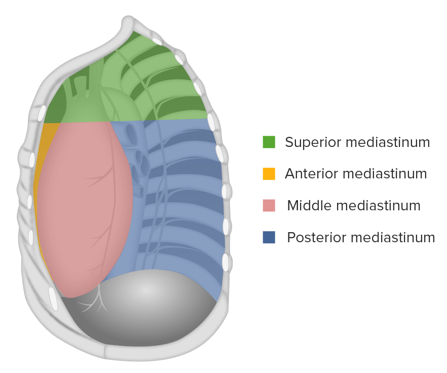
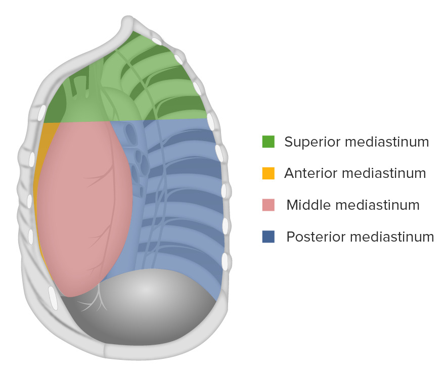
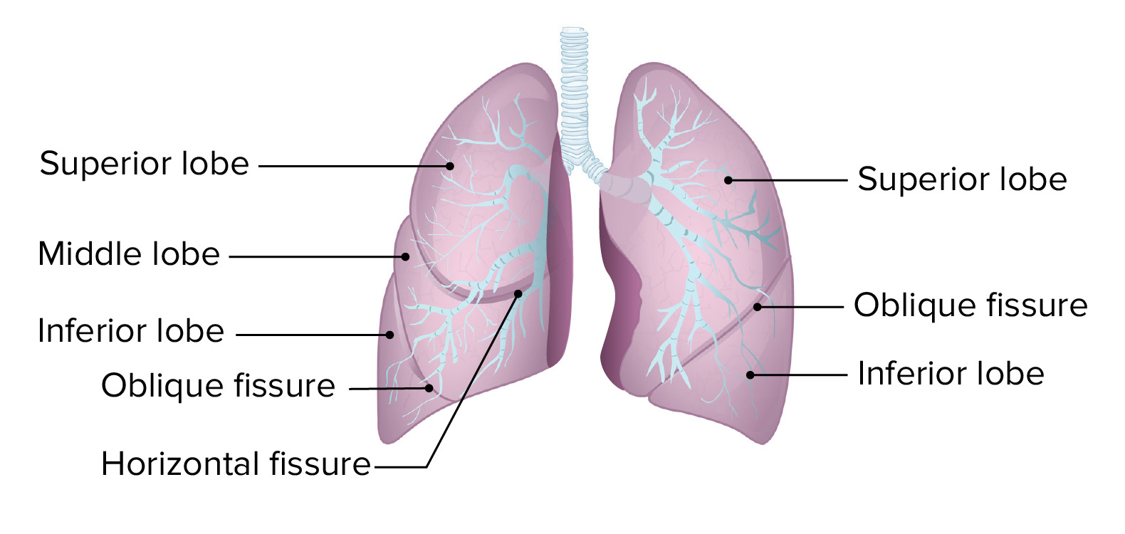
00:00 Welcome to this overview on the thoracic viscera. Our purpose here is to provide you major take-home messages and to avoid excessive detail within this particular lecture. 00:18 This slide lists the objectives that you should be able to answer at the conclusion of this presentation. First, list the three major compartments of the thoracic cavity. Describe the topography of the lungs. 00:33 Describe the boundaries and divisions of the mediastinum. 00:38 List the major viscera or organs located in each of the divisions of the mediastinum. 00:44 Describe the surface topography for the auscultation of cardiac valves. 00:49 Describe the general function of the thymus gland and its mediastinal location. 00:56 Then we’ll summarise the key take-home messages and then provide attribution for the images that were used throughout this lecture. Here is our body map and our focus will be anteriorly, approximately in this area of our body map. And then posteriorly, our focus will be generally deep to the area within this region. 01:32 This slide is an illustration that demonstrates the three potential spaces that exist within the thoracic cavity. Two of these compartments are pleural cavities. The right pleural cavity is shown here, surrounding the right lung. 01:57 Here, we see the left pleural cavity surrounding the left lung. 02:05 Between our two pleural cavities, we have this area standing in the middle of the thoracic cavity. This is the mediastinum. Also of note is the fact that we have a nice cardiac or heart window right in through here. And part of that window is provided by the fact that the left lung gets displaced toward the left side of the thoracic cavity, towards its own pleural cavity, and there’s a notch created by the impression of the heart. This allows access to the heart, if an intracardiac injection needs to be administered. It also provides access to the heart, if there’s fluid accumulating within its pericardial cavity and that fluid has to be withdrawn. Here, we’re looking at a radiograph of the thoracic cavity and we can see the two pleural cavities as well as the mediastinum standing in the middle. The pleural cavities are going to appear relatively black. The mediastinum is going to have a much lighter appearance in its contrast. So, here is our right pleural cavity and we can see how dark it is. We can also see here, on the left side, the left pleural cavity, and again, we can see how dark it is. And the darkness is due to the fact that the lungs hold a lot of air and the x-ray beam will readily pass through any air-filled structures, thereby darkening the film and providing for this nice, dark contrast. We can also visualise, though it’s obscured by the vertebrae, but we can also visualise the air-filled trachea. So, if you look here, between this vertebra and the vertebra below, you can see how darker it is. You can also see some blackness imparted here between those two vertebrae and even up here more superiorly, you can see some areas of darkness between the bony vertebrae. This would be the air that is filling the trachea. The mediastinum, then, would be represented by the lighter areas that we see in through here. The major occupant within this portion of the mediastinum is the heart and so, we can see a nice cardiac silhouette. Here’s the left ventricle over in through here and if we come over here, we can see the right atrium bulging toward the right side of the body. 05:06 Here is a cross-section or axial section through the thoracic cavity. The plane of section is right along here, where you see the red line and it’s very, very important that this axial section is oriented as such that you’re standing at the feet of the individual, looking upwards toward the individual’s head. That means that the left side of the image is, then, the right side of the individual and the right side of the image is the left side of the individual. So, if we go back here, here is your right lung surrounded by the right pleural cavity. Here is the left lung, surronded by your left pleural cavity. And then here, we can see the pericardium of the heart in green and we can see the heart muscle itself. The portion outlined here is your left ventricle. 06:14 So, the heart and its pericardium lie within the mediastinum. Let’s now apply the illustration that we saw here to radiologic imaging. All right. Let’s now take a look at the thoracic cavity as viewed with CT and we are looking at an axial section through the thoracic cavity. 06:37 And again, keep in mind what appears dark versus what appears bright and the most obvious dark areas are these huge regions right in through here. This would represent air in a lung and since this is the left side of the image, this is the patient’s right lung. 06:57 We also have a large area that’s dark on this side of the image, which would be the patient’s left lung. And then we see some areas that are bright. 07:09 This area represents blood flow. The blood is flowing through your ascending aorta. It is coming towards us as we view this image. The ascending aorta would go into the arch and then the arch will curve to the left posteriorly and will pick that descending portion of the aorta up along the left side of the vertebral column. And again, it appears bright because of the blood that’s flowing through it. We also have, in this area, a bright spot, again, representing blood flow. This is blood flowing through the superior vena cava and it’s going to empty into the right atrium. And if we look within the substances of the lung, we can see these bright areas that have a branching pattern. We see the same kind of bright branching pattern within the left lung and since these are bright areas that have branching, these branching patterns represent blood flow into the lungs. 08:13 And then, if we take a look here and here, we see two luminous profiles and the lumen of each one of these profiles is dark. That, by interpretation, means that these structures have air within them and these, then, will represent the bronchi. The substance of each lung has these bright networks within them. And they’re a little bit brighter here, perhaps, on the right side of the image within the left lung. And again, these bright streaks or networks represent blood.
About the Lecture
The lecture Thoracic Cavity – Lungs, mediastinum and cardiac valves by Craig Canby, PhD is from the course Thoracic Viscera with Dr. Canby.
Included Quiz Questions
What structure or structures appear(s) black on a CT scan of an axial section through the thorax?
- Bronchial lumen
- Heart
- Chest wall
- Decending aorta
- Pulmonary arteries
Which procedure is done on the site of the notch created by the displacement of the left lung by the heart?
- Drainage of pericardial effusion fluid
- Drainage of pleural abscess
- Coronary artery bypass graft
- Angioplasty
- Angiography
What is anterior to the esophagus?
- Trachea
- Thoracic duct
- Subclavian artery
- Posterior intercostal arteries
- Descending thoracic aorta
Customer reviews
5,0 of 5 stars
| 5 Stars |
|
4 |
| 4 Stars |
|
0 |
| 3 Stars |
|
0 |
| 2 Stars |
|
0 |
| 1 Star |
|
0 |
I rate it cause I found it good and helpful. I liked it very much because the x- ray explanation its very helpful for me .
Clear and succinct overview of thoracic cavity. Excellent foundation to build upon
I am studying for my Anatomy and physiology class, and your lecture is truly helping me.
1 customer review without text
1 user review without text


