Playlist
Show Playlist
Hide Playlist
The Skin is a Large Sensory Receptor Organ
-
Slides 05 Types of Tissues Meyer.pdf
-
Reference List Histology.pdf
-
Download Lecture Overview
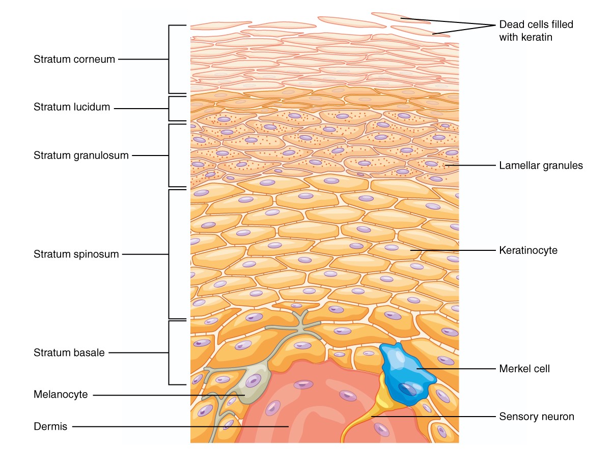
00:00 Well, skin is also a very large sensory receptor organ. 00:06 In fact, skin is the largest organ in the body that occupies between 15 and 20 percent of the volume of our body. So it is a huge area and it is very very important that the exterior coat of our body has a number of sensory components in it. Sensory receptors that detect certain sensations like pressure, like temperature, all sorts of sensations. 00:41 But I just want to introduce you to a couple of receptors, which you can actually see when you look at sections of skin. First of all, there are a number of receptors that have free nerve endings. These are simply nerve endings ending within the tissue that detects certain sensations and they are hard to see. You will never see these looking through sections of skin using normal routine stains. So I am not going to introduce those at this stage. 01:22 What I am going to emphasize are two encapsulated nerve endings. By encapsulating, I mean that they are enclosed by certain connective tissue components or capsules. And I am referring mainly to the Pacinian corpuscle and Meissner's corpuscle because Ruffini's corpuscles again are structures that are very hard to see using routine histological processing. Here are two images of Pacinian corpuscles. They stain differently, but they show the same histological features. They are mechanical receptors. They detect pressure and vibration. And if you look at any one of these two Pacinian corpuscles, you can see that they have a very tiny small dot in the center. That is the axon, the neuron. And then that neuron cell process is surrounded by numbers and numbers and numbers of sheaths called lamellae. You can see them clearer on the right-hand image. Those lamellae are really wrapped up encircling Schwann cells and on the outside is the thicker stained capsule. It looks a bit like a section through a cut onion. 03:02 Now, fluid lies between all those lamellae. And when pressure or vibration is applied to the surface of the skin, then that distorts the capsule, and then distorts these lamellae of Schwann cells and that distortion is then transmitted through the fluid to then initiate a stimulus on the axon. And therefore the information is conveyed back to the central nervous system for interpretation. Let us see now how we can identify a Meissner's corpuscle. These are touch receptors and you find them concentrated in places on the body like the lip or the fingertips or the toes, places where it is really important to have a very precise sensation of touch. They are bit harder to see, but they lie up in the dermal papillae. Those projections of the dermis up into the underlying areas of the epidermal pegs or up into the superficial layer of the actual junction between the epidermis and the dermis. On the left hand image, you can see these two projections of the dermal papillae into the epidermis. And look very very carefully particularly in the more central projection and you can make out a fine light paink stained structure with some bluish stained nuclei. That represents coils of Schwann cells and those coils of Schwann cells wrap around an axon. You cannot see the axon in that edge of each section, but have a look across to the right hand image stained differently. You can just see a very fine thin black line towards the base of the image. That is the axon, stained with the special stain, and that axon is going to project all the way up and be encapsulated by the Schwann cells. That is typical of a Meissner's corpuscle. Again as I said its a touch receptor. Well, Ruffini's corpuscles are mechanoreceptors, and as I said, they are very difficult to see. But they respond to stretch or torque of the collagen fibres within the dermis and therefore giving some information about the sorts of shear forces or stress forces on our skin.
About the Lecture
The lecture The Skin is a Large Sensory Receptor Organ by Geoffrey Meyer, PhD is from the course Epithelial Tissue.
Included Quiz Questions
Pacinian corpuscles are sensitive to which of the following?
- Pressure and vibration
- Touch and vibration
- Pain
- Temperature changes
- Shearing forces
Which of the following is NOT an encapsulated nerve ending?
- Merkel discs
- Pacinian corpuscles
- Meissner's corpuscles
- Ruffini endings
Ruffini endings respond to which of the following stimuli?
- Stretch and torque of collagen fibers
- Vibration and pain
- Touch and temperature changes
- Painful pressure
- Ultraviolet light
Customer reviews
3,5 of 5 stars
| 5 Stars |
|
1 |
| 4 Stars |
|
0 |
| 3 Stars |
|
0 |
| 2 Stars |
|
1 |
| 1 Star |
|
0 |
I agree that using a pointer might help, but the way he is describing the structures makes it very clear where to look to, so I don't agree at all with the 2-star rating. Also, the rest is very informative and nicely explained.
Needs to use pointer during lecture. He's telling us to look at something but he's not pointing it out.


