Playlist
Show Playlist
Hide Playlist
Surface Relief – Topographic Back Anatomy
-
Slides 04 Abdominal Wall Canby.pdf
-
Download Lecture Overview
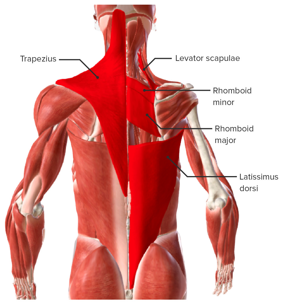
00:01 Thank you for joining me on this lecture on the “Topographic Anatomy of the Back.” This slide lists the objectives that you should be able to answer at the conclusion of this lecture. First, list the regions of the back. List the skeletal and muscular elements that contribute to the surface relief of the back. Define the borders of the following triangles - triangle of auscultation, the superior lumbar triangle and inferior lumbar triangle. Describe the clinical relevance of the superior and inferior lumbar triangles. Describe the clinical relevance of the intercristal line. Describe the applied anatomy in performing a thoracentesis. Describe the applied anatomy in a retroperitoneal approach to the kidneys. And then lastly, list other abdominal organs that project onto the back. Then we’ll take a look at the summary and then identify and provide attribution to the images that are contained within this lecture. 01:13 We will begin with a body map and the focus here would be on the image to our right. And so, we want to look at the relevant anatomy topographically in this particular view. 01:29 So, let’s start with defining the regions of the back and we’ll take a quick look at the vertebral region, sacral region, deltoid region, scapular region, infrascapular region as well as the lumbar region. So, let’s begin here by focusing in on this middle portion of the back. This is going to be the vertebral area. This area right in through here that extends down into the intergluteal cleft represents the sacral region. 02:09 Here, superior and lateral, we have the deltoid region as well as over here. Our scapular areas are defined within this territory as well as the territory to the right side of the body. The infrascapular region is defined by this area of [Inaudible 0:02:36] as well as over and through here. And then lastly, we have the lumbar regions of the back. So, here’s the one on the left side of the body and here is the one on the right side of the body. Surface relief of the back in this particular slide can be provided by skeletal contributions. There are also muscular contributions which we’ll see on the next slide. But, for our skeletal contributions, let’s take a look. 03:11 Here, we have the vertebra prominens, this is the spinous process of C7. We also have skeletal relief provided here and over here and this represents the acromion which is the lateral most projection of the scapula. And the acromion here laterally represents the point of the shoulder. We also have the spine of the scapula providing skeletal relief. 03:49 And we see that labelled right in through this area. And then if we take a look here more inferiorly, we will find the iliac crest. And then when we follow the iliac crest as it curves posteriorly and downward, you will find an area of skeletal relief provided by the posterior superior iliac spine. Same image, but this image is representing the contributions of muscles of the back to surface relief. And as you can see here, we will have a contribution from the deltoid, trapezius, teres major, infraspinatus, latissimus dorsi as well as the erector spinae. So, let’s take a look at the relief provided by these particular muscles. Here we see a nice contour to the shoulder. This is provided by the deltoid muscle. We also have muscular relief provided by the trapezius. And the trapezius here on the right side is coming down toward the spine of the scapula and then from the spine of the scapula is coursing inferiorly immediately back down here, more to an inferior midline attachment. Typically, the inferior most attachment of the trapezius would be to the spinous process up T12 and then we can go upwards along the relief provided by the opposite trapezius muscle. And you can see that the two muscles collectively define a four-sided structure called the trapezoid. 05:44 We also have the teres major that provides muscular relief. And so, it’s this rounded bulge that we see right in through here. And then we also have below the spine, right in through here the scapula and running a bit deep to the trapezius here more medially, we also have the infraspinatus region. And then we have an important contribution from the latissimus dorsi. And we can see here, more laterally, a more well-defined surface topography provided by the latissimus and this bulge of muscle is provided by the latissimus as it narrows to attach to the humerus. Lastly, we have the erector spinae and surface relief is shown in this area as well as to the right side of the body in through here.
About the Lecture
The lecture Surface Relief – Topographic Back Anatomy by Craig Canby, PhD is from the course Abdominal Wall with Dr. Canby.
Included Quiz Questions
Consider the posterior view of a man standing with both arms side-by-side in extension. Where is the deltoid region?
- Lateral to the scapula
- Inferior to the scapula
- Lateral to the neck
- Inferior to the iliac crest
- Inferior to the knee joint
What structure causes the vertebral prominence on the back of the neck?
- Spinous process of C7
- Spinous process of C6
- Spinous process of C5
- Spinous process of C3
- Spinous process of C4
What is the acromion?
- It is a projection of the scapula.
- It is a projection of the clavicle.
- It is a projection of a rib.
- It is an elbow joint.
- It is another name for the knee joint.
Where is the inferior-most attachment of the trapezius?
- T12
- T11
- T9
- T8
- T7
Customer reviews
5,0 of 5 stars
| 5 Stars |
|
5 |
| 4 Stars |
|
0 |
| 3 Stars |
|
0 |
| 2 Stars |
|
0 |
| 1 Star |
|
0 |


