Playlist
Show Playlist
Hide Playlist
Superficial Structures of the Neck
-
Slides Anatomy Superficial Structures of the Neck.pdf
-
Reference List Anatomy.pdf
-
Download Lecture Overview
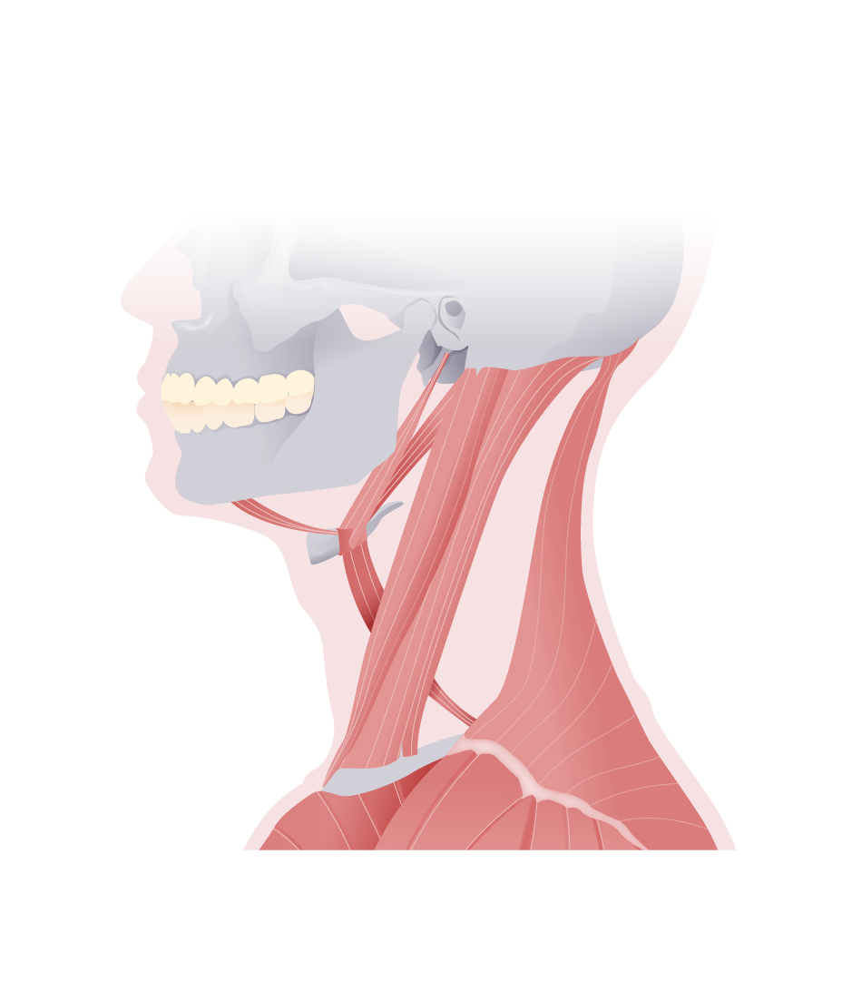
00:01 Now let's take a look at some of the superficial structures of the neck. 00:06 We'll start with a very, very thin muscle called the platysma. 00:11 We'll also look at some more substantial muscles such as the sternocleidomastoid and the trapezius. 00:17 On this anterior review of the platysma, we can see that it's a very wide flat and thin sheet of muscle. 00:25 And in fact, it inserts into the fascia that covers the areas around the pectoral muscle, clavicle, and acromion. 00:32 Rather than a bone like most muscles. 00:35 More approximately it does attach to the lower border of the mandible as well as a little bit of the orbicularis oris muscle. 00:43 And the platysma is a muscle of facial expression. 00:45 So it's innervated by the facial nerve or cranial nerve VII Because it's a very thin muscle that really only attaches into fascia distally its action is really just to tense the skin of the lower face and neck. 01:00 Here we see the sternocleidomastoid which has a sternal head and a clavicular head and cleido is another word for clavicular. 01:11 When we look at the origins insertions, we can see how it gets its name. 01:15 The sternal head is attaching to the manubrium of the sternum while the clavicular head is attaching to the medial third of the clavicle. 01:24 It also attaches superiorly to the mastoid process as well as a little bit of the superior nuchal line, which is how it gets its name sternocleidomastoid. 01:36 The sternocleidomastoid is innervated by the accessory nerve or cranial nerve XI. 01:41 And there are some contributions from the cervical plexus. 01:46 The sternocleidomastoid, if it's acting unilaterally, will act to rotate the head to the opposite side while tilting the head to the same side. 01:58 So it has the effect of looking up and away from the muscle that's contracting. 02:04 When both work together bilaterally, they serve to extend the head. 02:11 Now let's swing around to the back to find the trapezius which is a very long muscle. 02:17 It has a descending part, a transverse part, and an ascending part. 02:24 Here we see it begins all the way up at the superior nuchal line. 02:28 And that bump on the back of the school called the external occipital protuberance. 02:32 It runs down the nuchal ligament, and along the spinous processes of C7 to T12. 02:40 And then it attaches out laterally to the lateral third of the clavicle, as well as the spine of the scapula. 02:48 The trapezius like the sternocleidomastoid is innervated by the 11th cranial nerve or the accessory nerve. 02:55 It also has some contributions from cervical spinal nerves for proprioception or sensing where it is in space. 03:03 In terms of the function, it really depends on which fibers are contracting. 03:06 If it's the descending part, or the upper fibers, it's causing elevation of the scapula. 03:12 If it's the transverse part of the middle fibers, it's causing retraction of the scapula bringing it towards the midline. 03:18 And if it's the ascending part with these lower fibers, it will cause depression of the scapula. 03:25 But because of the odd shape of the scapula, when the upper and lower fibers work together, they also serve to rotate the scapula. 03:33 We would say, be rotated superiorly because it will bring the inferior angle a little bit upward. 03:38 And that's an important sort of accessory motion when we talk about upper limb movements.
About the Lecture
The lecture Superficial Structures of the Neck by Darren Salmi, MD, MS is from the course Neck Anatomy.
Included Quiz Questions
What is the origin of the platysma?
- Fascia covering the pectoral muscle
- Orbicularis oris muscle
- Lower border of the mandible
- Upper border of the mandible
- Fascia covering the temporalis
What innervates the sternocleidomastoid?
- CN XI
- CN XII
- CN IX
- CN III
- CN VI
What are the functions of the trapezius? Select all that apply.
- Elevation of the scapula
- Retraction of the scapula
- Depression of the scapula
- Tilting the head to the opposite side
- Tilting the head to the same side
Customer reviews
5,0 of 5 stars
| 5 Stars |
|
5 |
| 4 Stars |
|
0 |
| 3 Stars |
|
0 |
| 2 Stars |
|
0 |
| 1 Star |
|
0 |


