Playlist
Show Playlist
Hide Playlist
Superficial Back Muscles – Back Muscles
-
Slides 03 Abdominal Wall Canby.pdf
-
Download Lecture Overview
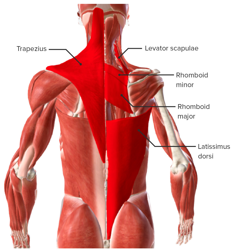
00:01 Welcome to this lecture on “Back Muscles”. This slide identifies the objectives that you should be able to answer at the conclusion of this presentation. First, list the structures of the back. 00:15 Categorize back muscles into groups. 00:18 List the suboccipital muscles and define the suboccipital triangle. 00:22 Describe the attachments, innervations and actions of the above muscles. 00:29 Identify the relevant neurovascular structures. 00:33 And then we will summarize the key take-home messages. 00:37 And lastly, provide attribution for the images that were used throughout this presentation. 00:45 This represents the body map in our focus of today, we will be on the region defined in through here, we will go pretty low onto the back and we will come very high outwards to the shoulder and even to the anterolateral clavicle and then we will work our way also to the back, the neck and up to the skull. 01:13 This list just provides you an idea of the structures that are associated with the back. 01:19 Moving from superficial to deep, we would have the skin, underlying the skin we would have superficial fascia and then deep to the superficial fascia, deep fascia, we would have our muscles. 01:32 Also, in this area, we’d have relevant neurovascular structures. In the midline of the back, we would have our vertebral column and its associated ligaments. We would also have within the vertebral canal, the spinal cord and its associated meninges. And we would also have the posterior aspects of the ribs in these particular regions here. 02:00 When we look at the muscles of the back they are going to be arranged in three layers. 02:07 We have a superficial layer which is visualized here. Here is one of the superficial muscles, here is a second superficial muscle. We will also have three more that fall within this layered category. 02:22 Deep to the superficial muscles, we have intermediate muscles. Both the superficial and the intermediate layers are known as extrinsic muscles and then the deepest layer is the deep layer and these are known as the intrinsic or true back muscles. 02:45 Here is a listing of the five muscles that make up the superficial layer. We have the trapezius muscle and the latissimus dorsi muscle, both of those are seen in this particular illustration. Here is our trapezius. It occurs on the opposite side as well and when we follow the contours of the trapezius, it does form a trapezoid shape. 03:10 Below is the extensive latissimus dorsi muscle on this side and we see the latissimus dorsi on the opposite side as well. The next three superficial muscles are the rhomboid major, the rhomboid minor and the levator scapulae. These are seen in this particular illustration and we will focus on the right side of the image. 03:42 Here is your rhomboid major, this slender strap like muscle represents your rhomboid minor. And then this muscle that we see running up into the neck represents the levator scapulae muscle. Now, let’s take a look at each one of these superficial muscles and we will wanna understand the attachments, innervation as well as associated actions. 04:12 We are looking at the trapezius, it has multiple points of origin. It’s going to attach to a prominent projection on the occipital bone called the external occipital protuberance. 04:25 It will then proceed laterally to that protuberance and attached to the superior nuchal line. 04:34 It will also originate along the ligamentum nuchae and then the spinous processes of C7 and then T1 all the way down normally to the level of the spinous process of T12. It has three points of insertion, one of those would be to the spine of the scapula, in this general vicinity. It then extends laterally and will insert onto the acromion of the scapula and then will wrap around to the anterior portion and also insert on the lateral one third of the clavicle. Motor innervation to your trapezius is going to be supplied by a cranial nerve and this is the accessory nerve. 05:25 The actions of the trapezius will depend on the orientation of the fibers that are contracting principally. For example, if you have these fibers of the trapezius contracting, these middle fibers, they are oriented horizontally, they will then pull the scapula towards the midline of the posterior back that is referred to as adduction or retrusion. If the superior fibers are primarily contracting, they will elevate or pull the scapula upwards. If the lower fibers are contracting and note their orientation down and in these lower fibers will depress, pull down the scapula. 06:13 And there is one more action that can be produced. If the superior and the inferior fibers are working in unison, they will rotate the glenoid cavity of the scapula or the humerus articulates, it will rotate that cavity superiorly and then that will allow for abduction of the humerus above the horizontal. This is done in association with the serratus anterior. 06:45 The latissimus dorsi has multiple points of origin. It’s going to attach or originate from the spinous processes of T7 through T12 and also, the spinous processes of all five lumbar vertebrae. It has a partial attachment to the iliac crest and the sacrum and will partially originate from the inferior three to even inferior four of ribs. 07:18 These points of origin are very wide or broad and you look as these fibers cross laterally and upwards towards the humerus, they all converge into a narrow band and the latissimus is going to insert into the floor of the inner tubercular groove or the sulcus of the humerus. 07:40 The nerve that innervates your latissimus dorsi is your thorcodorsal nerve and the actions produced by the latissimus would be several. One action would be to extend the humerus, pull it posteriorly. 07:57 It’s also a very powerful adductor of the humerus, so it pull it toward the midline and action shared with the pectoralis major, for example. It will also medially rotate your humerus turning it inwards toward your body and if you have your hands and arms extended above your body, the latissimus would help pull your trunk up towards your upper limbs and again, this function is shared with the pectoralis major. 08:32 Here we are looking at the rhomboids and levator scapulae. Our focus here, first, is with the rhomboid major which we see running right in through here, the rhomboid major has points of origin from spinous processes and we can see those points of origin along and through here. These particular points will be the spinous processes of T2 all the way down to T5, the insertion is on the medial border of the scapula at the level of the spine down toward the inferior angle. 09:14 The innervation of the rhomboid major is the dorsal scapular nerve and the action or actions of this muscle would be adduction. It would pull the scapula in toward the midline, also known as retrusion and because of the oblique course of these fibers, it can help to elevate or pull the scapula upwards. 09:45 The rhomboid minor is shown here, it is a slender band of muscle fibers. It’s going to attach to the inferior aspects of your ligamentum nuchae running right along in through here. It also has attachments for its points of origin, to the spinous processes of C7, the vertebra prominens as well as the first thoracic vertebra. 10:13 It will attach to the medial aspect of the scapula, add in slightly above the spine of the scapula. It’s innervated by the same nerve as the rhomboid major, dorsal scapular and also will share the same actions in producing adduction of the scapula as well as elevate the scapula. 10:39 Your levator scapulae is shown here as this band of muscle fibers. The levator scapulae is originating from the transverse processes of C1 through C4, that is better seen on the opposite side where you see those tendinous slips representing the points of origin from those transverse processes. 11:05 The insertion of your levator scapulae, as the name suggests, will be to the scapula and specifically it’s going to be along the superior border on the medial side of the scapula. The levator scapulae is innervated by the dorsal scapular nerve, it also receives contributions from anterior rami of the 3rd and 4th cervical nerves. For its action, the name tells you exactly what it does, it’s going to lift or elevate, pull the scapula superiorly.
About the Lecture
The lecture Superficial Back Muscles – Back Muscles by Craig Canby, PhD is from the course Abdominal Wall with Dr. Canby.
Included Quiz Questions
A patient has difficulty extending the right arm against resistance. Which back muscle is involved?
- Latissimus dorsi
- Trapezius
- Rhomboid major
- Rhomboid minor
- Levator scapulae
Which muscle layer of the back contains the true or intrinsic back muscles?
- Deep
- Superficial
- Intermediate
- Lateral
- Medial
How many layers of muscle are present in the back?
- 3
- 4
- 5
- 6
- 7
Which muscle is broad and has multiple points of origin?
- Latissimus dorsi
- Erector spinae
- Rhomboid major
- Rhomboid minor
- Levator scapulae
Which muscle is supplied by the thoracodorsal nerve?
- Latissimus dorsi
- Splenius cervicis
- Supraspinatus
- Levator scapulae
- Quadratus lumborum
What is the motor innervation of the trapezius?
- Accessory nerve
- Axillary nerve
- Spinal nerves
- Dorsal scapular nerve
- Glossopharyngeal nerve
Which function does the levator scapulae perform?
- Elevate the scapula.
- Elevate the arm.
- Elevate the head.
- Depress the scapula.
- Depress the arm.
What is the nerve supply of the rhomboid major and rhomboid minor?
- Dorsal scapular nerve
- Accessory nerve
- Axillary nerve
- Spinal nerves
- Sixth and seventh cranial nerves
Customer reviews
4,3 of 5 stars
| 5 Stars |
|
3 |
| 4 Stars |
|
0 |
| 3 Stars |
|
0 |
| 2 Stars |
|
1 |
| 1 Star |
|
0 |
Easy to understand and to take notes, even for a non native English talker
Excellent lectures from the best professional. Thanks a lot for your great help in learning so difficult topics
Please, stop speaking in such a monotone way. It gets people bored.
This lecture is easy to memorize and understand, also it has accurate and clear information.


