Playlist
Show Playlist
Hide Playlist
Skin Lesion Configuration

-
Slides Skin Lesion Configuration.pdf
-
Download Lecture Overview
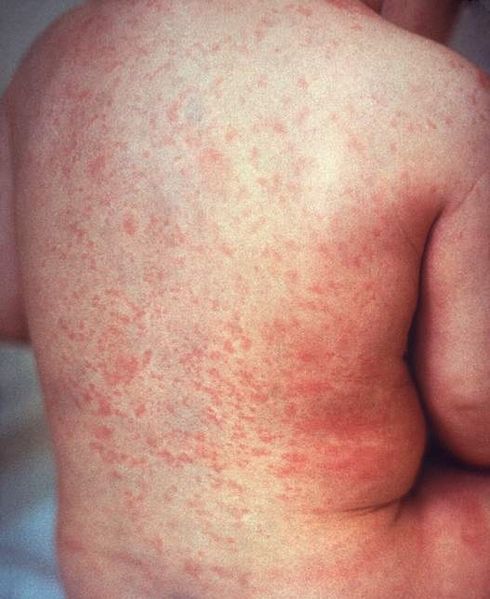
00:01 To move on now to the different configurations of lesions. 00:05 We can have linear lesions. 00:07 And these are lesions forming a straight line. 00:10 And an example of that is the linear epidermal nevus. 00:14 You can see the nice linear architecture of the skin. 00:18 There's another condition which is much more common than the previous one called lichen striatus, where you see a linear hyper or hypopigmented lesion on the arm or on the trunk. The other type of lesions could be annular. 00:35 And this means we have a ring lesion with central clearing. 00:39 And some examples of this are condition called granuloma annulare. 00:44 It's not so common. 00:45 But we do see it in dermatology. 00:46 And you can see the round configuration of the lesion on this picture. 00:51 Another typical which is much more common than the previous one is dermatophyte infection. Tinea corporis corpora means the skin, and the Latin word for that is ringworm. Typical coin shaped annular lesion. 01:10 A targetoid lesion is a lesion with a ring with central duskiness, and that central darkness is usually due to necrosis. 01:20 An example of that is erythema multiforme. 01:24 Typical targetoid lesion. 01:28 Serpiginous lesion. 01:29 Like a snake, you can see that it's linear, it's branched and with carving elements. 01:35 And an example of that is cutaneous larva migrans. 01:42 Of course scabies, which is much more common than cutaneous larva migrans, can also give you serpiginous lesions. 01:49 And when you look with your dermatoscope you can see that some lesions can be reticulated. 01:54 They look lacy or have a network pattern. 01:58 And an example of that is cutis marmorata, a condition that is seen in the pediatric population. The second one is Livedo reticularis. 02:09 This could be a benign presentation. 02:11 Actually, if you sit in front of the heater for a long time, you can get this on the lower legs. Or it could imply that the patient has got some connective tissue disease. So the history is very important. 02:23 The other type of lesion is herpetiform lesions. 02:26 These are grouped herpetiform means grouped lesions or vesicles. 02:31 An example of that again is your herpes labialis. 02:35 You can see those lesions on the left angle of the mouth. 02:38 They are collected and grouped together. 02:40 And this is your typical viva blister. 02:44 Due to herpes simplex virus type 1 or 2 zosteriform lesions. 02:49 They are clustered in a dermatomal distribution zosteriform and examples are herpes zoster, which we see in patients who either have older patients, or patients who are immunocompromised, or patients who have underlying malignancy, and of course, dermatomal vitiligo, which can also follow a zosteriform pattern.
About the Lecture
The lecture Skin Lesion Configuration by Ncoza Dlova is from the course Introduction to Dermatology.
Included Quiz Questions
Which of the following best describes an annular lesion?
- Ring lesion with central clearing
- Flat, discolored patch without any raised borders
- Raised, solid lesion with uniform coloration throughout
- Fluid-filled lesions that are scattered randomly across the skin
- Thickened, crusty area of skin with no clear shape
Which of the following best describes reticular lesions?
- Lacy or network pattern
- Circular with central clearing
- Flat, irregular patches with no distinct pattern
- Solid, raised lesions with sharp borders
- Clustered, fluid-filled blisters forming a ring pattern
Author of lecture Skin Lesion Configuration

Ncoza Dlova
Customer reviews
5,0 of 5 stars
| 5 Stars |
|
5 |
| 4 Stars |
|
0 |
| 3 Stars |
|
0 |
| 2 Stars |
|
0 |
| 1 Star |
|
0 |
