Playlist
Show Playlist
Hide Playlist
Skin: Epidermis

-
Reference List Pathology.pdf
-
Slides Skin Epidermis Dermatopathology.pdf
-
Download Lecture Overview
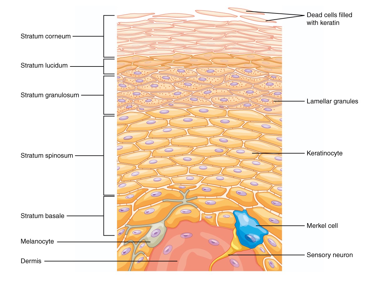
00:01 Okay. So let's look at some of that anatomy of the skin. 00:04 We've talked about what the skin does. What does it really look like. 00:07 And again this is important for understanding the pathology that we're going to talk about with all the other lectures in this series. 00:15 It's divided into the epidermis, that's where we have the epithelial cells. 00:20 That sits on top of the dermis. 00:22 So epidermis on top of the dermis. 00:26 And then underneath the dermis is the hypodermis. 00:29 And I will more commonly refer to that as the subcutaneous fat okay. 00:34 So those are the three major layers of the skin. 00:36 Let's kind of dissect that out even a little bit further. 00:40 The epidermis that's going to be the one that we see the most. 00:43 It is going to be the most important layer of the skin. 00:47 The epidermis is derived from the ectoderm germ layer in the trilaminar disk of the developing embryo. It is the outer layer of the skin and it is a stratified squamous epithelium, meaning it's more than more than one layer, so it's stratified. And it's squamous, meaning their flat plate-like cells kind of balance one on top of the other, as if we're building a brick wall. 01:10 An important point is that the epidermis, like every epithelial structure in the body, is avascular. There are no blood vessels in that layer of the skin. 01:19 There's blood vessels underneath, but none in the epidermis proper. 01:24 So it means it's dependent, the viability of the epidermis is dependent on the underlying dermal capillary cells for nutrient delivery. 01:33 Clearly there's a lot of oxygen coming from the outside world to the skin, but getting nutrition in and getting waste out is going to be really important, an important part of what the dermal capillary network does. 01:49 So let's take a closer look at the cells of the epidermis. 01:53 And the cells of the epidermis, it looks like we have a whole bunch of different kinds of cells. Well in fact, although there are different cells, the majority of them are one kind of cell, as we'll talk about. 02:04 But this is this is an example of what you would see if we took a sample of my skin, cut it, stained it, put it on a slide. 02:11 We would pretty much see something that looks like this. Okay. 02:14 The major, most important, most dominant cell within the skin are the keratinocytes. They're called keratinocytes because they are filled up with keratin or an intracellular protein that is an intermediate filament. 02:31 All those shapes that you're seeing on the right hand side, those are all keratinocytes. They all look remarkably different look like they're all different cells. In fact, they're the same cell at different levels of maturation. And the skin is constantly maturing from its basal layer, that sits on top of the dermis, all the way up to that stratum corneum at the very top. And the cells, as they mature, change in terms of what they're making and the way that they look. 03:01 But they're all keratinocytes. 03:03 So as I've already said, they're the majority of the epidermis. 03:06 They're rich with keratin, hence the name keratinocytes. 03:10 They're stacked in layers of increasingly mature states. 03:13 And we'll look in a moment at how that organization occurs. 03:17 Another important cell in the epidermis are the melanocytes. 03:22 The melanocytes actually have all these kind of weird looking dendrites associated with them. And then we'll see another cell type that does that as well, the Langerhans cell. 03:31 Melanocytes have a different origin from the ectodermal derived keratinocytes. 03:37 These are in fact going to be neural crest-derived cells as we'll see in just a second. They are responsible for making pigment. 03:44 That's what's going to be one of the major ways that we're protected against UV radiation. So they predict. 03:51 They protect. They product pigment. 03:54 They're present primarily in the basal layer, but they do have these interdigitation that allow them to extend up more into the major part of the of the keratin layer. 04:07 They are neural crest-derived, as I mentioned. 04:09 So they have migrated from the original notochord and are now sitting in the skin in the basal layer. Another important cell type is the Merkel cell. 04:19 No, this is not Angela Merkel. 04:20 This is a different German who described them a long time ago. 04:24 They're a relatively rare component in the skin, but nevertheless really important. 04:29 They're part of how we have touch sensation, especially light touch. 04:34 They have a cell body that sits up within the layer, the major layer of keratinocytes. 04:41 But then they have a process, a neural process that snakes all the way through the basal layer into the dermis and will connect with neurons that are in there to say, hey, we're touching something. 04:52 They're associated with dermal nerve fibers, as I said, and they are also ectoderm-derived. 04:57 So they come from the same original trilaminar component, the ectoderm, in the developing fetus. 05:06 I've already mentioned the cell, but now we'll talk more about it. This is the Langerhans cell. This is the one that's going to be important for immune surveillance. And you can see it's got its little arms. 05:14 Its processes of the cytoplasm kind of snaking throughout the major layer of the skin that's going to allow it to sample and say, hey, there seems to be a virus here. 05:27 There seems to be a bacterium here. There may be a fungus here. 05:29 And to be able to send that signal down into lymphatics and that will eventually get to the lymph nodes. So the Langerhans cells are very important for immune surveillance. 05:40 Clearly because of that role that they play in immune surveillance, they're derived from the bone marrow. They have a very important immunologic function. 05:48 Okay. So those are the kind of the players that are in that major layer of the epidermis. Let's talk about the different layers. 05:58 So the stratum basale, I just like saying that. 06:02 But it's just the basal layer, it's the easier way to think about it. 06:06 It is the germinative layer meaning that's where new keratinocytes are born. 06:11 They have a significant reproductive capacity so they will generate more and more cells. 06:17 It's a single layer that sits on top of a basement membrane at the dermal epidermal junction. So this is part of your skin. 06:26 This is part of the epidermis. 06:28 It's going to be what makes new epidermal cells. 06:33 The stratum spinosum, which is the thickest layer, sits on top of the basal layer. 06:38 And these are progressively maturing keratinocytes. 06:42 They start off kind of cuboidal just like the basal cells. 06:46 But now as they get bigger and bigger and flatter and flatter, they progress from bottom to top. 06:52 And the thickest layer over all of the skin is this. 06:56 It's called stratum spinosum because there are little spiny processes that connect. 07:01 When we look at this histologically, at very high power, there's a little, there appears to be a little gap between the cells and that the cells are held together by little spines. 07:11 Those spines are actually desmosomes. 07:13 So they're a structure there. 07:15 And that separation is kind of an artifact of when we do the pathologic processing. 07:19 But that's why this layer is called the stratum spinosum. 07:22 And it's the thickest layer. 07:24 On top of that is the stratum granulosum or the granular layer. 07:29 And it is characterized by the presence of cytoplasmic granules that are called keratohyalin granules. 07:35 They have an important protein that we'll see in some of the other talks, a protein called filaggrin, which is important for the maintenance of the layer on top, which is going to be the stratum corneum. 07:45 So the stratum granulosum we can see this in almost every good preparation of skin. 07:51 It looks bluer and dark purple. 07:52 We'll see some of the histology on other lectures. 07:56 Right on top of that is the stratum lucidum. 07:59 This is really kind of only apparent when we have super duper thick skin. 08:03 So if I was going to take a sample off your foot, off the bottom of your foot where you have a big old callus, then we could see a pretty good clear cell layer. 08:12 This is where we've lost those keratohyalin granules. 08:15 And it hasn't, we haven't had the same precipitation of the intercellular intermediate filaments of the keratin, so it doesn't look quite as blue or pink as the layers below or above it. 08:27 It's clear. But we usually only see this when we have really thick skin. 08:33 And then on top of that is the stratum corneum. 08:36 And it's the horny layer and not horny in the way some of you are thinking about. 08:41 But this is a layer where the, basically the nuclei have been popped out of the cells and it's pretty much just cross-linked intermediate filaments, cross-linked keratin. This is the most superficial layer. 08:56 It's dead. It's anucleate. 08:58 They're flattened keratinocytes. 09:00 They do have, there's some basement membrane there, but it's mostly cross-linked keratin. 09:04 And they are constantly being sloughed. 09:08 So the skin itself from the stratum basalis all the way up to the stratum corneum, is constantly being sloughed. 09:16 It's constantly turning over from bottom to top. 09:19 Cells are maturing getting older, losing their nuclei, getting those granules, dying entirely and then being on the top and then being sloughed. And if you ever wonder where all that goes, you wonder what you know when you're cleaning your room and you have all that dust layer, a lot of that is dead skin from you, and it's the dead stratum corneum that's been sloughed and floating around.
About the Lecture
The lecture Skin: Epidermis by Richard Mitchell, MD, PhD is from the course Review: Physiology and Structure of the Skin.
Included Quiz Questions
How do epidermal cells receive nutrients despite the absence of blood vessels in this layer?
- They depend on diffusion from dermal capillaries.
- They produce their own nutrients through metabolism.
- They receive direct arterial blood supply.
- They obtain nutrients from sweat glands.
- They absorb nutrients from the environment.
Which epidermal cell type is derived from neural crest cells and produces protective pigment?
- Keratinocytes
- Melanocytes
- Langerhans cells
- Merkel cells
- Fibroblasts
Which layer marks the beginning of keratinocyte maturation in the epidermis?
- The stratum basale acts as the germinative layer.
- The stratum corneum initiates cell division.
- The stratum spinosum starts cell production.
- The stratum granulosum begins cell formation.
- The stratum lucidum generates new cells.
What is the distinguishing characteristic of the stratum spinosum?
- It contains keratohyalin granules.
- It is the thickest epidermal layer with cells connected by desmosomes.
- It consists of dead, anucleate cells.
- It forms the waterproof barrier.
- It produces new keratinocytes.
What characterizes the cells in the stratum corneum?
- They are actively dividing cells.
- They contain melanin granules.
- They have prominent nuclei.
- They are dead, anucleated cells filled with cross-linked keratin.
- They produce keratohyalin granules.
Author of lecture Skin: Epidermis

Richard Mitchell, MD, PhD
Customer reviews
5,0 of 5 stars
| 5 Stars |
|
5 |
| 4 Stars |
|
0 |
| 3 Stars |
|
0 |
| 2 Stars |
|
0 |
| 1 Star |
|
0 |
