Playlist
Show Playlist
Hide Playlist
Seborrheic Keratosis: Pathophysiology

-
Reference List Pathology.pdf
-
Slides Seborrheic Keratosis Pathophysiology.pdf
-
Download Lecture Overview
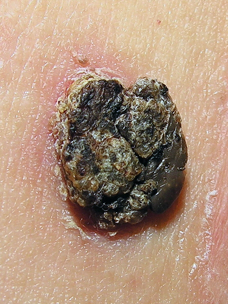
00:01 Welcome. With this talk, we're going to start a discussion concerning benign epithelial lesions and really common ones at that. So this is all about seborrheic keratosis. 00:14 And you in your general day to day practice or as the now erudite medical student that you are, you're going to say self-care. 00:21 Okay, we're going to refer to these as sectors seborrheic keratosis or subcarriers or benign skin condition, giving rise to kind of raised scaly patches that appear stuck on. 00:33 They don't appear to be arising within the skin so much as stuck on the skin. And that appearance will help you, but they truly are due to a proliferation of immature keratinocytes. 00:47 So these are benign tumors. 00:49 They are proliferating, but they almost never acquire a malignant potential. So the epidemiology of these incredibly common in middle aged and older adults, it's probably, as we'll see, driven by UV exposure like all good skin lesions . 01:07 If you are young in the age group probably watching this, you will in many cases have at least one self-care up to a quarter of the population. 01:18 As you get older into my age range, 90% will have at least one and many will have greater than ten. 01:27 It is more frequently seen in people with lighter skin tones, but there are no sex differences. 01:32 The lighter skin tones probably speak to increased UV sensitivity. 01:37 The pathophysiology as we'll see, it's a little bit understood. 01:42 But having said that, cumulative UV radiation exposure clearly drives this process. 01:48 So it's probably acquired mutations in a number of genes. 01:53 Um, it has been hypothesized that human papillomavirus may drive this. 01:58 The data for that is quite inconsistent. 02:01 And we can't always find HPV transcripts in these lesions. 02:05 So that makes them different than Verruca vulgaris, which is another talk elsewhere in the Lecturio portfolio. There are probably somatic. 02:17 So acquired activating variants in the fibroblast derived growth factor three receptor or the PI3 kinase oncogenes. 02:28 Those are found almost always in self-care. 02:30 So that's probably what's driving these UV damage is causing DNA breaks that cause mutations in these two genes in particular, but also others that give rise to a proliferative potential. 02:43 So we get a clonal expansion of somatically mutated cells. 02:47 This is not just an epidermal hyperplasia. 02:50 Epidermal hyperplasia is what is happening with verruca the common household wart. And this is different. 02:57 These are somatically mutated cells. 02:59 They have they have known acquired defects that are driving proliferation. 03:05 So the growth, the hyper proliferation is driven by those mutations, but they the tumors benign tumors are genetically stable. 03:18 They don't, unlike malignant tumors, they are not having acquiring additional mutations. 03:24 So these tumors are clonal expansions being driven specifically by things like the FGFR3 mutation or PI 3- kinase mutation that are going to cause proliferation. 03:38 All right. I've said that enough. 03:40 The lack of malignant potential is possibly due to we're not getting additional mutations in tumor suppressor genes like p53 or retinoblastoma. So we don't see that in these lesions. 03:53 And also interestingly enough, when you mutate or activate FGFR3 with a gain of function mutation that actually drives the differentiation of the keratinocytes, you induce the transcription factor FOXN1 which induces better differentiation of the keratinocytes. 04:15 And so long as they're maturing, they're eventually going to slough off. 04:19 So the ability to acquire additional nasty, potentially malignancy-driving mutations is very low. 04:26 So the clinical presentation yeah, these are those lesions that appear to be stuck on. Now they don't have to always look black. 04:35 They may be flesh-colored . 04:37 They may be slightly red tan. 04:39 They can be a variety of different colors. 04:41 They can be small 0.2cm up to greater than three centimeters. 04:45 So they can be fairly big things. 04:48 They tend to be domed or flat top. 04:50 And again they have that kind of like they were velcroed onto the surface rather than arising out of the skin. 04:57 They have round or irregular borders. 05:00 They're hyperkeratotic. 05:01 So they they seem horn like. 05:04 And I'll show you histology that will convince you and maybe and also make it understandable why they feel kind of firm and scaly or horn like. 05:15 As I say, the colors can be light tan to brown to black, just as you see here, and the surface tends to be dull. 05:22 The follicular orifices, the things where hairs and things would normally be coming through seem to be plugged. 05:28 And that's probably because of keratin overproduction. 05:31 So the location distribution, they are typically only in areas of hair bearing skin. 05:39 So you will not see a hair on your palm or on the sole of your foot. 05:44 The trunk is a common site for the. 05:46 The head is a good site. 05:48 Neck, face. Extremities. 05:51 See what else is left. 05:53 Oh, genitalia. Yeah. I should have given you a trigger warning. 05:58 Okay. So the symptoms associated with these, they're usually asymptomatic. 06:01 They are not usually pruritic unless you scratch at them because you think they look ugly. They can become irritated with excoriation, and you may have mild pain associated with those. 06:16 There is an an interesting syndrome called the sign of a Leser-Trélat and the sign of leser trélat is the sudden appearance of multiple subcarriers all over the body. 06:28 It may also be associated with acanthosis nigricans that we're going to talk about in a subsequent talk. But what this is is linked to various cancers, particularly gastrointestinal tumors, some lung tumors. And it's thought that those tumors are producing alpha transforming growth factor that gets systemic and driving the proliferation of kind of tiny little nascent subcarriers to get now this explosion of subcarriers. 06:58 So if you have a patient who suddenly presents with that, be very suspicious of an occult, typically GI or lung malignancy. 07:06 In making the diagnosis, it's really one of physical examination. 07:10 For little ones, you may want to use dermoscopy, but for the larger ones, it's really kind of that gross appearance. 07:18 It looks stuck on. It's pigmented. 07:20 It's scaly. Yeah. There you go. 07:23 This is the histology that I promised we were going to show. 07:26 So these lesions feel quite firm and kind of scaly or horn like. It is a very prominent proliferation of the epithelial cells. 07:36 But they're also making lots and lots and lots of keratin on the surface. 07:40 So the the stratum corneum on top is very thickened and it's exuberant. Um, there is no cytologic atypia. These are just proliferating and differentiating pretty much normally, but just an exaggeration of the normal architecture. 07:58 And again, depending on how much the the growth factor or other stimuli are driving melanocyte production of pigment, you may have variable pigmentation from none at all to very black. 08:13 This is just showing you kind of these accumulations of keratin within what are called horn cysts. 08:20 So kind of invaginations of the stratum corneum. 08:24 And you can actually in some of these lesions express just chunks, little chunks of keratin. 08:32 So the management generally you don't have to manage these. 08:36 You can assure the patient they're not going to die that these are not malignant. 08:41 But if they're symptomatic if they are a little bit irritated the patient doesn't like the location they look cosmetically concerning. 08:48 Then you can you have a variety of ways to get rid of them. 08:51 And they tend to be perfect in terms of getting rid of them completely and irreversibly. So cryotherapy liquid nitrogen that will work. Electrocautery you can do curettage kind of scoop the whole thing out. You can do a shave removal. 09:07 So there are lots of different options. 09:09 And as future dermatologists you will see a number of these and remove them because patients think they look ugly. 09:15 With that we've talked about a very important entity, not so much in terms of of seriousness but very common. 09:24 So a sub care. Thanks.
About the Lecture
The lecture Seborrheic Keratosis: Pathophysiology by Richard Mitchell, MD, PhD is from the course Benign Epithelial Tumors of the Skin.
Included Quiz Questions
What percentage of older adults are likely to have at least one seborrheic keratosis?
- 90%
- 25%
- 50%
- 75%
- 10%
Which description best characterizes the typical appearance of seborrheic keratosis?
- Stuck-on, hyperkeratotic lesions with plugged follicular orifices
- Smooth, dome-shaped, translucent papules
- Flat, scaly, erythematous patches
- Soft, pedunculated, flesh-colored growths
- Deep, indurated, purple nodules
What molecular mechanism helps prevent seborrheic keratoses from becoming malignant?
- FGFR3 mutations promote keratinocyte differentiation through FOXN1 activation
- Increased p53 expression blocks cell division
- Enhanced DNA repair mechanisms
- Decreased melanin production
- Activation of apoptotic pathways
Which finding should prompt investigation for internal malignancy?
- Sudden appearance of multiple seborrheic keratoses
- Gradual development of a single large lesion
- Change in color of existing lesions
- Development of pruritus in old lesions
- Regression of existing lesions
In which location would seborrheic keratoses NOT typically be found?
- Palms and soles
- Upper back
- Face and neck
- Chest
- Scalp
Author of lecture Seborrheic Keratosis: Pathophysiology

Richard Mitchell, MD, PhD
Customer reviews
5,0 of 5 stars
| 5 Stars |
|
5 |
| 4 Stars |
|
0 |
| 3 Stars |
|
0 |
| 2 Stars |
|
0 |
| 1 Star |
|
0 |
