Playlist
Show Playlist
Hide Playlist
Pyruvate Kinase Deficiency: Pathogenesis
-
Slides PyruvateKinaseDeficiency RedBloodCellPathology.pdf
-
Reference List Pathology.pdf
-
Download Lecture Overview
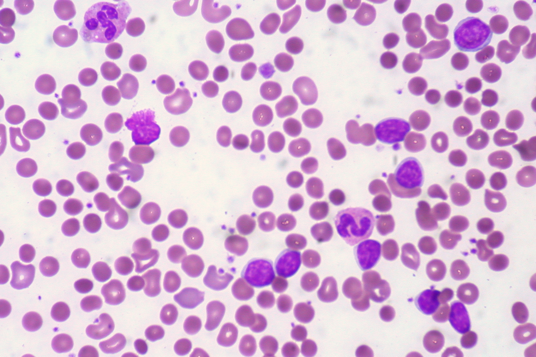
00:01 Next, your RBC only has glycolysis as you know for energy. 00:04 It has no mitochondria. 00:06 In pyruvate kinase deficiency, PK, very little ATP. 00:09 Stop there. 00:11 The block was before the pyruvate, wasn’t it? Can you picture it? If you can’t produce the pyruvate, can you then go into TCA cycle? No. 00:20 You don’t feel or excuse me – You don’t create the necessary FADH or NADH to then feed into your electron transport chain, right? Therefore, I cannot produce ATP properly or abundantly or adequately. 00:35 In fact, you have inadequate amounts of ATP, point number one. 00:40 Point number two, you’re stuck at pyruvate. 00:43 You can’t go into TCA cycle. 00:45 So what does that mean? Remember from the TCA cycle, what are you giving off? NADH. 00:52 Do not confuse that with NADPH. 00:54 What is then NADPH? It was pentose phosphate pathway. 00:57 HMP shunt. 00:59 This is NADH. 01:01 So we have too little ATP and you have too little NADH. 01:05 What kind of picture is this going to give you? We shall see. 01:09 Now, just keep this in mind. 01:11 This is actually now current day practice a separate diagnosis. 01:13 It really is. 01:15 It’s a particular sodium-potassium type of channel type of pathology. 01:20 However, to keep things really, really simple here is the following. 01:26 We know that we are not going to produce enough ATP. 01:27 There is going to be a problem with the membrane, meaning to say that if the sodium potassium pump was or did not have enough ATP, then it’s kind of like the ischemia that we’ve been talking about in basic pathology. 01:42 And so therefore, you will have increased amount of osmotic pressure within the cell. 01:46 The RBC is now – Well, it has increased osmotic pressure and then therefore accepts more fluid. 01:53 Now, based on all of that, we do know for a fact that pyruvate kinase deficiency, the RBC does take on a bizarre form. 02:01 And there is some kind of influence of the inadequate ATP with membrane problem or malformations. 02:11 Keep that in mind as you read through the third bullet point. 02:14 Now, as I said, current day practice in pathology, this is actually a separate diagnosis. 02:19 But to make your life simpler and much more, let’s say, relevant. 02:24 We do know that it’s going to influence the ATPase pump. 02:27 Let’s move on. 02:30 What about that NADH though? Now the NADH is important. 02:34 NADH in basic pathology, we talked about the conversion of FE3+, which is a methemoglobin into ferrous, your normal iron. 02:45 So remember in order for us to utilize the type of iron that gets incorporated into heme, you have to have it in the ferrous form, FE2+. 02:55 When we consume iron from the diet and such in the stomach, it will be in the form of FE3+, ferric. 03:03 So now, we need methods in which we convert the ferric into ferrous. 03:07 Now, remember, when we’re born or delivered, it’s rather difficult for us to immediately convert our ferric into ferrous. 03:17 We need to build up enough NADH and there is an enzyme called cytochrome-b5 reductase. 03:24 So the combination of cytochrome-b5 reductase and NADH, all of this helps us to convert ferric into ferrous. 03:35 In pyruvate kinase deficiency, what’s my NADH supply? Decreased. 03:41 Uh-oh. 03:42 What does this mean? That means that the patient is stuck in the state of methemoglobin. 03:47 What’s methemoglobin? Good. 03:51 Hemoglobin with FE3+. 03:54 Next, tell me about that 2,3-BPG. 03:56 If you have block at the pyruvate or at the enzyme because it’s not there You’re going to build up the proximal substrates including 2,3-BPG. 04:04 What kind of shift? Right shift. 04:07 And so therefore, it is then going to release the oxygen prematurely. 04:10 Now, just to be technical, would you tell me that methemoglobin, you would have what kind of effect in your oxygen dissociation curve? Good. 04:21 A left shift, okay? So from basic pathology and physiology, a couple of things here with shifting of your oxygen dissociation curve. 04:30 Fascinating, isn’t it? Pyruvate kinase deficiency. 04:35 If you take a look at the oxygen dissociation curve here, you’ll notice the solid line is the green one and that then represents a normal oxygen dissociation curve. 04:46 X-axis, PO2. 04:48 Y-axis, saturation of oxygen. 04:49 I need you to focus upon the right shift. 04:53 The right shift, the dash line, you’ll focus upon the increase in DPG. 04:58 You see that in the middle. 04:58 That DPG, same thing as your BPG, and in pyruvate kinase deficiency, that will be elevated causing the right shift.
About the Lecture
The lecture Pyruvate Kinase Deficiency: Pathogenesis by Carlo Raj, MD is from the course Hemolytic Anemia – Red Blood Cell Pathology (RBC).
Included Quiz Questions
Which of the following statements regarding RBCs in pyruvate kinase deficiency is true?
- RBCs undergo extravascular hemolysis.
- RBCs have decreased NADPH.
- RBCs have increased ATP.
- RBCs have excess NADH.
Which of the following statements is FALSE?
- The oxidation state of dietary iron that can be absorbed in the intestine is Fe3+.
- NADH is used to convert iron from Fe3+ to Fe2+.
- The oxidation status of iron in hemoglobin is Fe2+.
- Pyruvate kinase deficiency may result in methemoglobinemia.
- In pyruvate kinase deficiency, 2,3-BPG is increased.
Which of the following conditions is associated with a right shift of the hemoglobin–oxygen dissociation curve?
- Increased RBC levels of 2,3-BPG
- Methemoglobinemia
- Carboxyhemoglobinemia
- Increased RBC levels of pyruvate
- Fe3+ oxidation state
Customer reviews
5,0 of 5 stars
| 5 Stars |
|
5 |
| 4 Stars |
|
0 |
| 3 Stars |
|
0 |
| 2 Stars |
|
0 |
| 1 Star |
|
0 |


