Playlist
Show Playlist
Hide Playlist
Psoriasis: Pathophysiology

-
Reference List Pathology.pdf
-
Slides Psoriasis Pathophysiology Dermatopathology.pdf
-
Download Lecture Overview
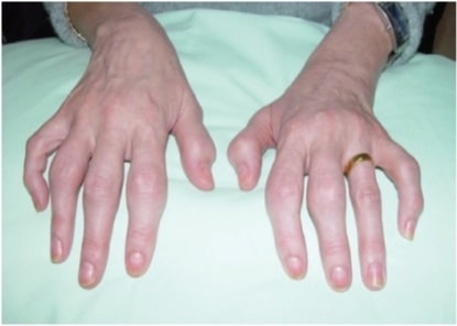
00:01 Welcome. For this topic, we're going to be covering psoriasis, which is an incredibly common T-cell-mediated disease. 00:11 Very important because now we also have a variety of drugs that can effectively treat it. Psoriasis, as I said, is a very common T-cell-mediated inflammatory disorder. 00:22 It can have systemic manifestations including significantly joint disease. The epidemiology is that it occurs in up to 3% of people worldwide and in some northern European countries, probably because of the predilection for certain HLA haplotypes, it's more common up to 11% in some of the northern European countries. It can be seen at any age, but there is a bimodal distribution, typically early adulthood and then later adulthood, 20s to 30s and 50s and 60s. 00:57 And there's definitely a genetic component to this, as we'll discuss shortly. 01:02 One third of patients will have a first-degree relative who's also affected. 01:06 There is seasonal variation. 01:07 It's worse in the winter than in the summer. Probably because of changes in, uh, emollients or kind of the lubrication of the skin. It is slightly more prevalent in women, as most autoimmune diseases or most inflammatory diseases are more common in women, and may be due to the effect of estrogens driving improved inflammatory responses. 01:31 Men are more likely, on the other hand, to have ocular disease, as we'll discuss later. 01:36 And overall, it's less common in people of color. 01:39 Again, that is probably not related to skin pigmentation, but rather due to the difference in HLA that is expressed histocompatibility complexes that are expressed in different populations. 01:53 The pathophysiology then. 01:55 So as I've already intimated, MHC major histocompatibility complex genes are really, really important, and there are a number of loci that are seen to provide a particular degree of susceptibility. HLA-B27 is a haplotype that is specifically and relatively frequently associated with psoriasis, that also has arthritis or psoriatic arthritis. 02:20 Environmental triggers are important, so you have to have the appropriate immune substrate. But then on top of that you add infections and strep infections of the skin, human immunodeficiency virus, systemically cold weather, trauma, a variety of medications can all drive the initial kind of breakthrough of autoregulation of the inflammatory response. So here's a schematic of what happens during the kind of initiation and then subsequent develop of the psoriatic lesions. 02:53 You have a trigger, as we've already discussed infection, trauma, various pharmacologic agents, that stimulate the Langerhans cells. 03:04 Remember those immune surveillance cells that live in the stratum spinosum? Those cells are stimulated, and they will produce a variety of cytokines, in particular TNF, tumor necrosis factor alpha and interleukin one, IL -1. 03:19 And those will subsequently then have antigen presented to dendritic cells indicated here as DCS. 03:28 The dendritic cells now are going to go off and running to the rest of the immune system to draining lymph nodes, etc., and in that setting are going to present whatever antigen came from the infection or the trauma or the drug. 03:41 They're going to present that to a variety of inflammatory cells present within the node in the context of appropriate cytokine stimulation. 03:49 And you see all those cytokines interleukin 12 tumor necrosis factor, interleukin 23. Et cetera. 03:56 Et cetera. Driving the various immune cells present within the lymph node. Those, in turn, may be recruited into the area where the initial stimulus occurred, and in that setting, then they're going to make their own witch's brew of various inflammatory mediators, the cytokines that you see there. 04:16 Do you need to memorize all of this? No, you absolutely don't. 04:19 You need to understand, though, that it's triggered. 04:22 It's a T-cell response. 04:24 T-cells get recruited. 04:26 They make their evil humors in the tissue thinking that they're doing a good thing. But as a consequence of those cytokines, we then get, we drive the proliferation of the keratinocytes in the stratum spinosum. And we also get disordered maturation once we get up to the granular layer and into the stratum corneum. 04:47 So we get a variety of effects driven as a consequence of the cytokines as a consequence of the inflammatory cells, as a consequence of the dendritic cells being stimulated by the initial trigger acting on the Langerhans cells. 05:02 Whoa! That was a lot. Okay, so now we get in the skin the manifestations that we really care about. 05:08 We're going to get epidermal hyperplasia for the dermatologists out there or the dermatopathologist we call that acanthosis. 05:16 So they're going to get a thickening of the stratum spinosum layer. 05:20 There's accelerated cell turnover. 05:22 And we are getting also increased cellular retention of the nuclei in the stratum corneum. 05:30 So there shouldn't normally be nuclei in those dead cells that are at the surface at the very surface. When we see that we call it parakeratosis and it's a sign of abnormal maturation. 05:43 The keratinocytes are also not making filaggrin appropriately. 05:47 They're not releasing lipids appropriately. 05:50 And that leads to flaky, dry, scaly skin. 05:54 We also are recruiting inflammatory cells that aren't normally present in the epidermis. So we are recruiting neutrophils that come all the way through the epidermis and actually will set up shop causing little abscesses up in the stratum corneum as we will see. 06:11 And then from all the inflammatory mediators we're going to get vascular engorgement. 06:15 That's erythema. There's going to be increased vascular permeability. 06:18 It's going to cause edema. 06:20 So you kind of get the picture. This is going to be red, scaly and flaky and also very itchy with edema. Histologically. 06:31 Here's what we see. So as promised there is thickening of the stratum spinosum. 06:36 We are also seeing inflammatory infiltrates that are at the dermal epidermal junction and very profound. 06:43 And if we look it's every kind of variety of T-cell you can imagine. 06:46 And then up in the stratum corneum, that superficial layer on the very, very top we're seeing nucleated keratinocytes. 06:54 Shouldn't see that. That's the parakeratosis. 06:57 And we're also seeing those abscesses. 06:59 And some guy by the name of Monroe got there first. 07:02 And so he put his name associated with it. But those are neutrophils that have accumulated up there in the stratum corneum where they don't belong. 07:10 And that's a Monroe's microabscess. 07:12 So that's the histologic characteristics.
About the Lecture
The lecture Psoriasis: Pathophysiology by Richard Mitchell, MD, PhD is from the course Inflammatory Lesions of the Skin.
Included Quiz Questions
Which population shows the highest prevalence of psoriasis?
- Northern Europeans
- South Americans
- Africans
- East Asians
- Middle Eastern populations
What cell type initiates the inflammatory cascade in psoriasis after environmental triggers?
- Langerhans cells
- B lymphocytes
- Neutrophils
- Melanocytes
- Fibroblasts
Which microscopic finding is characteristic of psoriasis?
- Munro's microabscesses
- Solar elastosis
- Viral inclusions
- Melanin deposits
- Fungal hyphae
What percentage of psoriasis patients have an affected first-degree relative?
- One third
- One half
- One fourth
- One fifth
- One tenth
Author of lecture Psoriasis: Pathophysiology

Richard Mitchell, MD, PhD
Customer reviews
5,0 of 5 stars
| 5 Stars |
|
5 |
| 4 Stars |
|
0 |
| 3 Stars |
|
0 |
| 2 Stars |
|
0 |
| 1 Star |
|
0 |
