Playlist
Show Playlist
Hide Playlist
Pemphigus and Bullous Pemphigoid: Pathophysiology

-
Reference List Pathology.pdf
-
Slides Pemphigus and Bullous Pemphigoid Pathophysiology.pdf
-
Download Lecture Overview
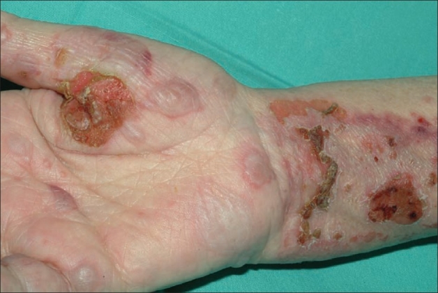
00:01 Welcome. With this talk we're going to discuss the major blistering lesions pemphigoid and pemphigus. 00:09 Now they seem like it's the same thing. 00:11 Well it is the same root word but they have very different outcomes as we'll talk about. 00:17 So pemphigoid and pemphigus are blistering autoimmune diseases. 00:21 Pemphigus is going to be the worst one. 00:23 And I'll emphasize that with some comments later on. 00:26 Pemphigoid is kind of like pemphigus hence the name, oid. So pemphigoid and pemphigus but they're both autoimmune. 00:34 They just attack different parts of the epidermis. 00:38 So first off the not so bad one bullous pemphigoid. 00:42 It is a nonlife-threatening . 00:44 Okay. That's important but it is chronic autoimmune disease. 00:48 What you get are subepithelial bullae. 00:51 Okay. Those words probably don't mean a lot right now. 00:54 In a moment when you see the images, they'll make all the sense in the world. 00:59 The location of where the blister is is important, because that also indicates which component of the epidermis has been affected, and in this case it's loss of adhesion molecules or breakdown. 01:14 Proteolytic catabolism of the adhesion molecules between the epidermis and the dermis. So the whole epidermis is coming up as part of the blister. 01:25 In comparison, pemphigus vulgaris, it is life-threatening . 01:29 This can be a lethal disease. 01:31 It is also chronic. It's also autoimmune and it's characterized by intraepithelial bullae. 01:37 So it's not a separation between the dermis and epidermis. 01:41 It's a separation within the epidermis. 01:44 And again the words don't mean so much. 01:46 But when you see the image it will make all the sense in the world. 01:49 In this case the antibodies that are responsible are causing loss of adhesion between keratinocytes. 01:57 So not between the keratinocytes and the basement membrane at the dermoepidermal junction, but rather between keratinocytes within the epidermis proper. 02:08 So let's discuss the epidemiology of both as you see in this chart. 02:13 Bullous pemphigoid and pemphigus vulgaris have very comparable incidences per year. 02:18 So this is not a major health crisis. 02:21 But nevertheless they do occur. 02:24 The age is slightly older for the bullous pemphigoid. 02:28 Women and men are equally affected in both cases. 02:32 There's no real ethnic or racial predilection for bullous pemphigoid. 02:37 We do see pemphigus vulgaris more commonly in certain populations as you see there. 02:43 The etiology in both are not well established. 02:48 And as in most diseases of the skin where we don't have a good cause, we invoke things like drugs and and particularly certain types of drugs that may drive an immune response. 03:03 Genetics certainly play a role, and particularly in pemphigus vulgaris, where certain HLA haplotypes are more prevalent in pemphigus. 03:12 That may also explain why we see pemphigus more commonly in the Southeastern Europe, Middle Eastern and Indian populations. 03:21 Radiation therapy and and other kind of causes can be invoked, but we don't know in most cases. 03:31 So the pathophysiology this we do understand. 03:35 But in order to understand this you also have to go back to basic cell biology. 03:39 So my apologies if this is giving you like tremors. 03:43 Um we have to understand where the autoantibodies are acting. 03:47 The epithelium is held together and is held to the basement membrane by a variety of adhesion molecules between individual epithelial cells are desmosomes. 03:59 These are junctional complexes. 04:01 They can they connect via transmembrane proteins between the cells. 04:06 It's a homotypic interaction. 04:08 Desmogleins. Desmocollin. 04:10 They have a variety of names. 04:12 These transmembrane proteins link one to another across the gap between epithelial cells. They also then connect with intermediate filaments keratin that sits within the cytoplasm. And they may link from side to side from desmosomes across the cell to the next desmosome to allow some structural integrity. 04:35 So that's one junctional complex we have to pay attention to. 04:39 The other one is the hemidesmosome, appropriately named because half of the structure is the same as the desmosomes. 04:45 So it's hemidesmosome. 04:47 This one anchors through a transmembrane protein to the extracellular matrix of the basement membrane. Now, instead of linking intracellularly to intermediate filaments, it links through integrins and then to intermediate filaments. 05:04 And those proteins are again provide structural integrity and allow the epithelium to be adherent to the basement membrane. 05:12 So recall desmosomes and hemidesmosomes. 05:15 And in the two diseases pemphigus and bullous pemphigoid we are going to see different antibodies attacking different structures. 05:24 Okay. Here's our normal skin. 05:26 Again kind of a refresher. 05:28 We have the dermis on top of which is a basement membrane on top of which is the basal layer of the epidermis. 05:35 And then we have all the keratinocytes in the stratum spinosum going all the way up to the granular layer. And the stratum corneum. 05:44 That's our normal structure. 05:47 So that was the normal skin. 05:48 Let's talk about our blistering lesions. 05:51 So the first one up is bullous pemphigoid. 05:53 In bullous pemphigoid we have an IgG autoantibody that attacks the hemidesmosome proteins. Those are the proteins that connect the basal layer to the underlying basement membrane. We know what the proteins are. 06:07 They have various names. You don't need to remember those. But we should know that when you have IgG binding there we're going to activate complement. 06:14 And we're going to also recruit other inflammatory cells which will release in turn proteases and other inflammatory mediators. 06:22 But it's the proteases in particular that break those connections between the hemidesmosome integrins and the underlying basement membrane. 06:31 And when that happens, the whole epidermis lifts off and then we have a blister underneath. 06:36 Importantly, the desmosomes that connect the keratinocytes one to another, they're fine okay. So there's no problem there. 06:44 And that gives us a fairly good hunk of epidermis that has normal integrity. 06:50 And that's why bullous pemphigoid is not as bad as pemphigus vulgaris that we'll talk about next. Here's where the blister is. 06:59 That basement membrane. 07:00 That's fine. But the blister is above that. 07:04 And then we have the whole layer of the epidermis the all of the stratum spinosum corneum granulosa all that stuff. 07:13 So it is a subepidermal blister okay. 07:18 All right. In comparison pemphigus vulgaris in this case the autoantibodies are against desmosomal proteins. So the ones that connect keratinocyte to keratinocyte. 07:28 And when they bind they don't activate complement so much. 07:32 But what they do do is that they turn on the epithelial cells to make a urokinase like plasminogen activator, a protease that will break those connections. 07:44 Oh, that's a problem, because now I have disruption of the connections between individual keratinocytes. 07:50 That whole layer of epidermis begins to fall apart. 07:54 And when that happens, I lose barrier function. 07:58 We get fragile flaccid bullae blisters that very easily rupture. 08:04 And this gives no barrier function. 08:07 You're very prone to loss of fluid. 08:10 So you need a hypervolemia. 08:12 But more importantly you're very susceptible to secondary bacterial infections. 08:16 And so this is just a schematic demonstrating what's going on there. 08:19 The basal layer is connected to the underlying basement membrane. 08:24 That's fine. But that's just one layer of cells. 08:27 Now all the other cells are dissociating. 08:30 They're dis cohesive one to another as they as they dissociate, they also die. But we end up with a blister that is intraepithelial, not subepithelial, but intraepithelial. 08:42 Okay, Hopefully now you have very clearly in your mind what's different between bullous pemphigoid and pemphigus vulgaris.
About the Lecture
The lecture Pemphigus and Bullous Pemphigoid: Pathophysiology by Richard Mitchell, MD, PhD is from the course Blistering Skin Disorders.
Included Quiz Questions
Which characteristic correctly distinguishes pemphigus vulgaris from bullous pemphigoid?
- Pemphigus vulgaris is potentially life-threatening, while bullous pemphigoid is not
- Pemphigus vulgaris affects only elderly patients
- Bullous pemphigoid has a higher annual incidence
- Bullous pemphigoid causes intraepidermal blisters
- Pemphigus vulgaris primarily affects the basement membrane
In bullous pemphigoid, where do blisters form in relation to the epidermis?
- Subepidermal (between epidermis and basement membrane)
- Within the epidermis
- Between keratinocytes
- Within the dermis
- Between the dermis and subcutaneous tissue
Which structural component is targeted by autoantibodies in pemphigus vulgaris?
- Desmosomes
- Hemidesmosomes
- Basement membrane
- Keratin filaments
- Integrins
What mechanism leads to blister formation in pemphigus vulgaris?
- Activation of urokinase-like plasminogen activator breaking keratinocyte connections
- Complement activation at the basement membrane
- Direct destruction of keratin filaments
- Inflammatory cell infiltration of the dermis
- Degradation of the basement membrane
Author of lecture Pemphigus and Bullous Pemphigoid: Pathophysiology

Richard Mitchell, MD, PhD
Customer reviews
5,0 of 5 stars
| 5 Stars |
|
5 |
| 4 Stars |
|
0 |
| 3 Stars |
|
0 |
| 2 Stars |
|
0 |
| 1 Star |
|
0 |
