Playlist
Show Playlist
Hide Playlist
Pemphigus and Bullous Pemphigoid: Diagnosis and Management

-
Reference List Pathology.pdf
-
Slides Pemphigus and Bullous Pemphigoid Diagnosis Management.pdf
-
Download Lecture Overview
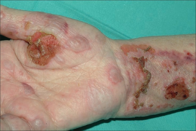
00:01 The clinical manifestations also very importantly depend on where those which kind of autoantibodies you have and where they are binding and causing their problems. 00:11 So bullous pemphigoid you it is an autoimmune disease. 00:15 There is a prodromal phase that lasts weeks to months before you may actually get visible blisters. Um, there's often moderate to severe pruritus. 00:25 It's itchy. And again this is probably because of local activation of pro-inflammatory markers. 00:30 But we haven't yet at that point elicited sufficient proteases to cause the blister. 00:36 And then the skin lesions may appear. 00:38 And they may initially be little tiny guys kind of papular. 00:41 Eventually they'll become plaques. 00:43 They can look like hives, urticaria, which is an entirely different disease that we'll talk about in a different talk. 00:51 Bullous pemphigoid gives you very classically the subepidermal bullae. 00:56 So the blisters again are below the epidermis between the epidermis and the underlying basement membrane. They're very tense. 01:03 They're not very easily ruptured. 01:05 And that's why they're not as bad as pemphigus vulgaris. 01:09 They can be quite large. 01:11 They contain clear fluid. 01:12 This is just edema. That is kind of because of the pro-inflammatory state we are causing vascular congestion. Increased vascular permeability in the fluid is getting into that blister there. They are typically throughout the body. 01:27 They can be quite widespread. 01:29 And they may appear on skin that is very red or skin that is not so inflamed. 01:34 And it just has to do with the degree of secondary inflammatory cell recruitment. 01:39 In bullous pemphigoid, you can get bullae that rupture and certainly in areas intertriginous areas where they may rub, you may get some some that rupture. 01:50 That's fine, but it's not typically life threatening. 01:53 They leave. As you might expect, underneath kind of a moist erosion. 01:57 In fact, when they when the blister comes off, you're looking at basement membrane and there's not much of a barrier. 02:03 So you're going to ooze fluid and you can get secondary infections there. 02:08 But in the absence of secondary infection you typically don't get scarring. 02:13 All right. Now let's compare that with pemphigus vulgaris. 02:17 These are characterized by bullae that have different properties. 02:23 So the blisters are coming up. 02:27 They're quite flaccid. 02:28 They rupture very easily because we've lost that separation. 02:32 We've lost the connection between the individual keratinocytes and they separate quite easily. This can be incredibly painful, usually as a result of secondary inflammation but also because of infection. 02:46 And they can appear depending again on the degree of inflammatory cell recruitment, either on normal-appearing skin or erythematous skin. 02:56 Pemphigus can occur anywhere on the body, but most commonly will involve other areas that we don't see involved with bullous pemphigoid. 03:05 Bullous pemphigoid doesn't involve the mouth. 03:08 On the other hand, pemphigus vulgaris typically does almost always present. 03:12 You can also get in other stratified squamous epithelium like the esophagus, the nasal mucosa, conjunctiva, etc. 03:20 you can get lesions as well. 03:22 And it's because the autoantibodies are binding to those epithelial cell desmosome connections and causing the same proteolytic activation. 03:33 When we see it, it can be anywhere on the body. 03:36 But in addition to the mouth, it's going to be face and scalp, trunk, groin and axilla. 03:42 Secondary infection is quite common. 03:44 We have lost barrier function. 03:47 And because of the secondary infection we're going to get recruitment of neutrophils. 03:51 And so we're going to get purulence. 03:52 It's going to be pussey. 03:54 And because of that because of the recruitment of neutrophils we're going to see a lot of erythema. Secondary inflammatory disease that is related to cause is related to vasodilation increased vascular permeability etc.. 04:10 The diagnosis. So Nikolsky's sign we'll talk about Nikolsky's sign on the next slide. 04:16 But we may want to do in fact most cases we'll want to do biopsies for histopathology and immunofluorescence. 04:24 Looking for the antibodies is going to be really important for us to make the appropriate diagnosis. 04:30 You may also do Elisa testing enzyme-linked immunosorbent assay testing to look for antibodies in the serum that are directed against the proteins that we know are associated either with pemphigoid or pemphigus. 04:44 Okay. As promised, Nikolsky's sign. 04:46 And this helps to determine whether we're dealing with a lesion that's at the dermal epidermal junction or one that is between keratinocytes. 04:56 So what you do is you apply scraping pressure. 04:58 You're not you're not trying to erode the skin, but you just kind of a firm pressure. 05:03 And you look for development of lesions. 05:07 So basically the the antibodies in pemphigus are everywhere. 05:12 Even though you may not have lesional skin, they're already bound there. If you rub that you will provoke the dissociation of that skin. 05:19 And so you will cause skin sloughing and rupture. 05:23 That means that you have antibodies against the desmosomes. 05:27 On the other hand, if you do that same Maneuver nikolsky's Maneuver and you get no skin sloughing or rupture, that means that the lesion is at the dermal epidermal junction. So with one Maneuver, you'll be able to reasonably specifically say whether something is pemphigus where you get the blistering or when you don't. 05:47 That it's pemphigoid. So the skin biopsies. 05:51 Yay. This is where pathology comes in. 05:53 The ideal biopsy site kind of depends on where the lesion is and which one you're dealing with. In bullous pemphigoid you want to go right into the middle of that blister. 06:02 Believe it or not. In pemphigus vulgaris you want to go to normal appearing peri lesional skin because you want to be able to see the antibodies around the individual keratinocytes. And I'll show you what that looks like on the next slide. 06:17 We can do routine histopathology to see where the blister is in the epithelium below the epithelium. And then we do immunofluorescence. 06:25 That's what the I-f stands for direct immunofluorescence testing. 06:30 Here's what the histology looks like on H and E hematoxylin and eosin. 06:36 The bullous pemphigoid slide shows that there's a separation between the dermis and the epidermis, and we will be able to say very characteristically, oh, this is this is very likely to be pemphigoid. 06:50 On the other hand, pemphigus vulgaris leaves the basal layer intact and the blister is up in the epithelium. We can also do, as I've mentioned, serum testing looking for circulating antibodies. 07:05 This can be a variety of assays and the Elisa assay. 07:08 The enzyme linked immunosorbent assay is probably the easiest where you have. 07:13 And the proteins that we know antibodies bind to. 07:17 And you look for the binding in the serum of those antibodies. 07:22 Immunofluorescence, however, is beautiful. 07:25 And this sort of image is often seen on national board exams because it's so classic. 07:33 And it allows us to distinguish the two entities. 07:36 So bullous pemphigoid remember the antibodies are against proteins at the dermal epidermal junction. So you get that nice linear staining at the base of the blister. 07:48 On the other hand pemphigus vulgaris that's those are antibodies that bind to the desmosomes around each one of the keratinocytes. 07:56 So now we're seeing within the epithelial layer this kind of reticular mesh like lace like binding of the autoantibodies all the way around individual keratinocytes. 08:07 And it looks very distinct from that linear dermoepidermal junction staining. 08:14 So how do we manage it. 08:15 Well in fact one is chronic and non-lethal. 08:19 One is chronic and potentially lethal. 08:21 So it's very important that we make the distinction. 08:24 And here's what we do once we've made that distinction. 08:26 So the report comes back from pathology. 08:28 Your patient has bullous pemphigoid. 08:30 Terrific. So we can give immunosuppression. 08:33 That's kind of the the first bullet out of the gun in terms of treatment, and you can give topical or systemic depending on the severity of the disease. 08:43 Other agents can be can be administered. 08:46 The tetracyclines are important for potentially limiting secondary infection, but that's not usually a big problem in bullous pemphigoid. 08:56 With really advanced or refractory disease, we can give other immunomodulatory immunosuppressive agents cyclosporin and the like. 09:04 And sometimes we can go to monoclonal antibodies that will limit B cell antibody production or B cell proliferation. 09:13 The prognosis is good overall. 09:16 The patients are walking around with blisters. 09:18 A long term remission is possible, especially after more systemic therapy involving B cell suppression. 09:26 On the other hand, pemphigus vulgaris. 09:28 Again, we want to pull out the bigger guns on this one because this is potentially lethal. 09:34 So you wouldn't just use topical steroids as a first step. 09:37 You'd go to systemic and you may then if the patient also doesn't tolerate that very well, go to other immunosuppressive agents, methotrexate, cyclosporin and other things. 09:49 In refractory disease, you can give intravenous immunoglobulin. 09:54 That's IV IgG which is globally immunosuppressive. 09:59 And you can also do cyclophosphamide and other chemotherapeutic agents. 10:03 The prognosis without treatment this is a bad disease. 10:07 Patients will die typically as a result of sepsis because they've lost their barrier function. So you really want to be aggressive once you make the diagnosis of pemphigus. 10:17 And with that we've talked about the major blistering lesions pemphigus pemphigoid. 10:24 And hopefully you now in your mind you have a very clear distinction between the two and what you do when you make the diagnosis.
About the Lecture
The lecture Pemphigus and Bullous Pemphigoid: Diagnosis and Management by Richard Mitchell, MD, PhD is from the course Blistering Skin Disorders.
Included Quiz Questions
Which characteristic describes the bullae in pemphigus vulgaris?
- Flaccid blisters that rupture easily
- Tense blisters that remain intact
- Subepidermal blisters with clear fluid
- Small papules that don't rupture
- Hemorrhagic blisters with blood
Which condition typically presents with oral mucosal involvement?
- Pemphigus vulgaris
- Bullous pemphigoid
- Both equally
- Neither condition
- Only in severe cases of both
What pattern is seen on direct immunofluorescence in bullous pemphigoid?
- Linear staining at the dermal-epidermal junction
- Reticular pattern around keratinocytes
- Granular deposits throughout epidermis
- Punctate staining in dermis
- Diffuse staining throughout all layers
What does a positive Nikolsky's sign indicate?
- Presence of antibodies against desmosomes
- Subepidermal blister formation
- Basement membrane destruction
- Normal skin integrity
- Secondary bacterial infection
Which treatment approach is most appropriate for pemphigus vulgaris?
- Immediate systemic immunosuppression
- Topical steroids alone
- Observation only
- Antibiotics alone
- Local wound care only
Author of lecture Pemphigus and Bullous Pemphigoid: Diagnosis and Management

Richard Mitchell, MD, PhD
Customer reviews
5,0 of 5 stars
| 5 Stars |
|
5 |
| 4 Stars |
|
0 |
| 3 Stars |
|
0 |
| 2 Stars |
|
0 |
| 1 Star |
|
0 |
