Playlist
Show Playlist
Hide Playlist
Panniculitis: Pathophysiology

-
Reference List Pathology.pdf
-
Slides Panniculitis Pathophysiology Dermatopathology.pdf
-
Download Lecture Overview
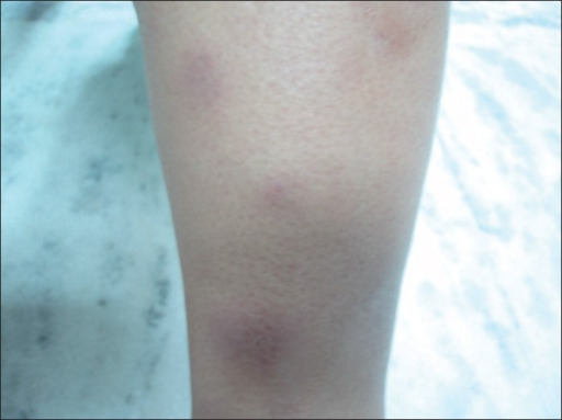
00:01 Welcome. In this talk, we're going to dive a little bit deeper into the skin. Most of the other talks have been talking about inflammation or disorders that involve the epithelial layer. 00:13 Well we're going to go below the epithelial layer and actually below the dermis into the hypodermis or the subcutaneous fat. 00:22 The topic is panniculitis. 00:24 So this is literally inflammation of the subcutaneous fat. 00:28 As we'll see there are a variety of causes. 00:32 But the manifestation that we'll see is more commonly on the surface of the skin. It will be an area of reddening and warmth due to inflammation that's kind of pushing up from below. 00:44 The epidemiology of panniculitis. 00:48 It's relatively uncommon. 00:50 So in your lifetime, depending on whether or not you do dermatology, you may or may not see it. 00:57 The age range is from teenagers into octogenarians. It is more common in women than in men. That may be because more autoimmune diseases are are more common in women than in men, and most of the causes for panniculitis are autoimmune. 01:18 So maybe that's the reason the legs overall are the most common location. And of the various kinds of panniculitis, erythema nodosum. Keep that word in mind, or that phrase in mind is going to be the most common. So the pathophysiology, as with a lot of inflammatory processes, there are kind of a broad list of things that can be causing this. 01:44 Inflammatory and autoimmune are going to be the major ones but infections trauma deposition disorders as we'll talk about very briefly, enzymatic destruction as we'll talk about very briefly. 01:57 And also malignancy can all present with inflammation of the subcutaneous fat. So the inflammatory lesions causing panniculitis. Erythema nodosum is the most common, but there are others that are listed here. 02:12 Again, because it's relatively uncommon overall I wouldn't worry about lipodermatosclerosis for example. 02:20 But erythema nodosum in a woman in her legs is actually going to be something that you may see. 02:27 Infections. So a variety of infections can cause inflammation that predominantly affects the subcutaneous fat, particularly if there has been an inoculation into that area by trauma or whatever. 02:41 Uh, cold panniculitis. 02:43 So there may be vasospasm that occurs deep associated with cold that may cause a relatively ischemic necrosis and factitious panniculitis where people inject things deliberately to get a sort of medical attention. 03:03 Deposition disorder. So calciphylaxis, if you have, uh, primary dysregulation of calcium, you can get deposition within vessels. And typically deep vessels within the subcutaneous fat will be affected. 03:15 And that may profoundly affect how well you perfuse the fat leading to ischemic necrosis and then panniculitis. 03:24 Gout. Uric acid crystallization can also occur deep within the fat and then systemic things. 03:31 So if you have pancreatitis that releases a variety of lipases and amylases things that can break down fat, and you can get a panniculitis, an inflammation and necrosis of the subcutaneous fat because of circulating pancreatic enzymes. Alpha -1 antitrypsin deficiency can also drive this by inhibiting or not allowing the inhibition , the alpha -1 antitrypsin of neutrophil activation. 04:01 So if you have that primary disorder because you're not making alpha -1 antitrypsin, then you may be more prone to having panniculitis. 04:10 So what's going on here. 04:11 We're going to basically describe what is happening with um with the more common erythema nodosum. 04:20 But you can kind of envision similar pathways going on with any of the other causes. 04:26 So in the subcutaneous adipose tissue again we are below the dermis clearly way below the epidermis. 04:34 We're down in the fat. 04:35 The fat is actually not just there as a big blob of lipid, but in fact is within adipocytes. 04:42 And the adipocytes are held together with fibrous connective tissue and investing blood vessels. 04:48 So there is a structure to the subcutaneous fat with septation throughout. 04:53 So kind of envision that. 04:55 And that's what's being demonstrated there. There's also clearly vasculature within the adipose or the subcutaneous adipose tissue. 05:03 So there's injury not otherwise specified. 05:05 But in the case of erythema nodosum, presumed to be autoimmune, directed specifically against the adipocytes and or the fibrous connective tissue septa. 05:15 As a result, we recruited inflammatory cells. 05:17 And what's demonstrated here is it's a mixture of neutrophils and macrophages and mononuclear lymphocytes for the most part. 05:26 So they are coming in and depending on the injury and the nature of that injury they're one or the other population may predominate. 05:34 They are going to then injure attack through their various cytokines, chemokines, proteases, reactive oxygen species, the typical things elaborated by inflammatory cells. 05:46 They are going to attack the adipocytes and we're going to get injury. We are also going to be attacking or attacking. 05:55 We're going to be interacting with the septum of the septal fibrous connective tissue. And overall you'll get destruction of the adipose tissue with inflammation. And you will get increased septal fibrosis because that's how extracellular matrix connective tissue responds to inflammation. 06:15 It lays down more connective tissue. 06:18 It can be septal fibrosis or it can be nodular. 06:21 And basically it's all on a spectrum. 06:24 So initially it's mainly going to be just accentuation of the septum with greater amounts of extracellular matrix deposited as part of the inflammatory healing. If you have much more extensive disease with lots of destruction of the adipocytes, then you can expect to see more nodular fibrosis. 06:42 But it's on a spectrum. 06:44 They are not distinct entities. 06:46 This is just what it looks like histologically. 06:48 So on the left hand side, identified as septal panniculitis, we have accentuation of the septum around the fibrous, around the fatty adipose tissue and in the lobular or nodular panniculitis. We have much greater areas of density and much less fat, because we've killed off the adipocytes and we've filled in the gap with that fibrous connective tissue. 07:13 Clearly, in both settings, what used to be kind of pliable, malleable, squishy fat is now going to be much more firm indurated non pliable.
About the Lecture
The lecture Panniculitis: Pathophysiology by Richard Mitchell, MD, PhD is from the course Inflammatory Lesions of the Skin.
Included Quiz Questions
Which characteristic is true about the epidemiology of panniculitis?
- More common in women than men
- More common in men than women
- Equal distribution between genders
- Only occurs in elderly patients
- Only affects children
Which area is most commonly affected by panniculitis?
- Legs
- Arms
- Face
- Trunk
- Hands
Which form of panniculitis is most common?
- Erythema nodosum
- Calciphylaxis
- Cold panniculitis
- Factitious panniculitis
- Lipodermatosclerosis
What distinguishes septal from lobular panniculitis?
- Septal shows accentuation around fat septae, while lobular shows dense areas with fat loss
- Septal affects superficial fat, while lobular affects deep fat
- Septal involves neutrophils, while lobular involves lymphocytes
- Septal is always infectious, while lobular is always autoimmune
- Septal affects women, while lobular affects men
Author of lecture Panniculitis: Pathophysiology

Richard Mitchell, MD, PhD
Customer reviews
5,0 of 5 stars
| 5 Stars |
|
5 |
| 4 Stars |
|
0 |
| 3 Stars |
|
0 |
| 2 Stars |
|
0 |
| 1 Star |
|
0 |
