Playlist
Show Playlist
Hide Playlist
Neurovascular Structures – Back Muscles
-
Slides 03 Abdominal Wall Canby.pdf
-
Download Lecture Overview
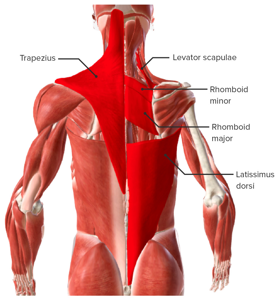
00:01 This slide depicts over all the neurovascular structures that can be observed within the suboccipital region, the suboccipital triangle. So, if we take a look here, here is our triangle in through this area here and you can see a lot of neurovascular structures in view. 00:22 The nerve of the triangle is your suboccipital nerve and we see that yellow nerve coming out here and if we look at some of the twigs of that nerve, we will see that they then penetrate the suboccipital muscles. And so, we do see some twigs of the suboccipital nerve, for example, going in to the rectus major. 00:45 This large nerve that we see entering the suboccipital region and ascending toward the midline of the cervical area in the skull is the greater occipital nerve and you can see it enters this region by passing just inferior to the inferior oblique that we see right in through here. This provides for sensory innervation of the skin associated with the posterior portion of your scalp. 01:17 We also have the third occipital nerve coming into play. This is a branch coming from the posterior ramus of the third cervical nerve. It will have a communicating branch into the greater occipital nerve and also, have a branch, not shown here, extends a course upwards and medial to your greater occipital nerve. 01:42 And then lastly, within the triangle and crossing on the superior portion of the posterior arch of C1, we have our vertebral artery and accompanying that artery would be the vertebral vein. 02:05 Some additional neurovascular structures that relate to the back that we have talked about previously would be the accessory nerve and the dorsal scapular nerve. And then we have a couple of arteries that we have not talked about yet, but want to introduce at this point that will be the superficial branch of the transverse cervical artery and the deep branch of the transverse cervical artery. 02:32 So, let’s take a look at what is depicted here. We will begin with this nerve that is crossing onto the deep surface of your trapezius muscle. The trapezius muscle is being reflected. 02:46 This is your accessory nerve that is providing motor innervation to your trapezius. The artery that accompanies it is seen branching off here and coming into the substance of the trapezius. This is the superficial branch of the transverse cervical artery. 03:11 We also have innervating the levator scapulae and the two rhomboids, the dorsal scapular nerve. That nerve is seen right along in through here, this yellow nerve and it’s running deep to the rhomboids after it supplies twigs to the levator scapulae, will run deep to the rhomboids in the area where they attach to the medial aspect or medial border of the scapula. 03:42 The artery that accompanies it is shown here. This is the deep branch of the transverse cervical artery. And then it will disappear under the rhomboid minor and then major along the medial border of the scapula. We also made reference to the thoracodorsal nerve as it innvervates the latissimus dorsi muscle. That nerve is seen in this particular illustration right in through here in yellow and it will penetrate and ramify or branch within the substance of the latissimus and we are just seeing the latissimus come up this way. The artery that is in here and travelling with the thoracodorsal nerve is your thoracodorsal artery and certainly, you will have a vein by the same name, thoracodorsal vein. 04:54 This is a more global look at the neural vasculature of the back. If we take a look here, we can see a deeper dissection on the left side of the image, more of a superficial view or dissection on the right side of the image. These branches that we see crossing along here with some of the terminal twigs going into the iliocostalis column of the erector spinae, these are the posterior rami of the spinal nerves in this general location. More superficially branches of these posterior rami will pierce through the back musculature and will then provide innervation to the skin that’s lying at the back more centrally. 05:48 We would also have, but not clearly in view, we would have some deep cervical arteries in the cervical area, they would provide for some branches that would cross posteriorly and help supply some of the musculature in the cervical area that we have discussed. 06:08 We have already mentioned the vertebral arteries as being members of the back region. And then we have posterior branches, intercostal vessels, so you see some of those branches here and there are some other branches up in through here as well, intercostal vessels travelling in the initial segment of the intercostal space. 06:33 There will be branches that will poke through the back musculature and will help supply the back muscles as well as the skin overlying those muscles. When you are in the lumbar area, as we would be down through in here, you see some penetrating arteries as well. 06:51 These are from segmental lumbar arteries helping to supply the lower back musculature in this area and the overlying skin that’s been removed.
About the Lecture
The lecture Neurovascular Structures – Back Muscles by Craig Canby, PhD is from the course Abdominal Wall with Dr. Canby.
Included Quiz Questions
What is the nerve supply of the suboccipital triangle?
- Suboccipital nerve
- 3rd cervical nerve
- 4th cervical nerve
- Subscapular nerve
- Trochlear nerve
What is the sensory innervation of the posterior surface of the scalp?
- Greater occipital nerve
- Zygomaticotemporal nerve
- Auriculotemporal nerve
- Greater auricular nerve
- Facial nerve
Which muscle is innervated by an accessory nerve?
- Trapezius
- Latissimus dorsi
- Levator scapulae
- Biceps
- Erector spinae
Customer reviews
5,0 of 5 stars
| 5 Stars |
|
5 |
| 4 Stars |
|
0 |
| 3 Stars |
|
0 |
| 2 Stars |
|
0 |
| 1 Star |
|
0 |


