Playlist
Show Playlist
Hide Playlist
Molluscum Contagiosum: Pathophysiology

-
Reference List Pathology.pdf
-
Slides Molluscum Contagiosum Pathophysiology.pdf
-
Download Lecture Overview
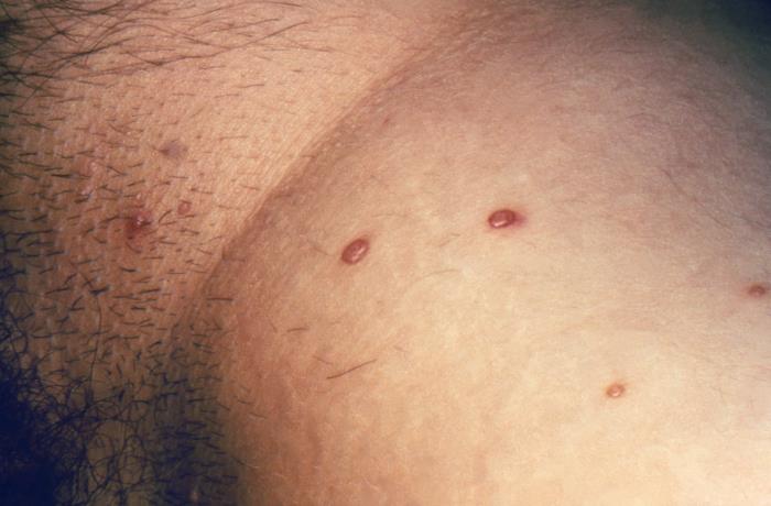
00:01 Welcome. In this talk, we're going to discuss an interesting entity called Molluscum Contagiosum. 00:07 And like the name suggests, this is an incredibly infectious cutaneous disorder. Molluscum contagiosum is caused by a pox virus infection. 00:18 Although you can have a lot of cutaneous manifestations. 00:22 And we'll see some great pictures of that. 00:24 Typically you don't have systemic manifestations. 00:26 These patients are not particularly sick. 00:29 And even when the immune response catches up with the virus and eliminates it, patients are still don't have much in the way of systemic symptoms like fever or joint pain, etc. The epidemiology of this is it's quite common. 00:44 It's 1% of all skin disorders, classically affects kids, don't usually see this in an older population unless they are immunosuppressed. 00:54 Boys get it more frequently than girls. 00:56 That may have something to do with boys roughhousing together. 00:59 And also kind of giving skin-to-skin transmission. 01:05 There's an increased incidence in hot, humid climates, and it's more common in patients who are immunocompromised, say, with HIV, or have inflammatory dermatoses such as eczema. 01:17 The pathophysiology of this entity. 01:19 Well, okay, so I already told you that it is a double-stranded DNA poxvirus in. 01:27 It's called Molluscum Contagiosum virus (MCV) good name because that's the name of the disease. The transmission of MCV is basically skin to skin contact. 01:38 You have exposure to infected individuals. 01:41 It can be sexually transmitted and can be transmitted at the time of birth from mom to baby. It is a reasonably persistent on fomites, so it can be on towels or toys or razors or whatever, and can be shared and transmitted in that way. 01:59 And because it is so darn infectious, you can do autoinoculation. 02:04 So if you have a lesion in one place, you kind of scratch it and then scratch yourself someplace else, or even just touch the skin someplace else, you can transmit it. The incubation period ranges from two weeks to six months. 02:17 It's usually closer to 2 to 4 weeks. 02:20 The virus only affects keratinocytes. 02:22 It's localized just to the epidermis. 02:25 And interestingly, one of its mechanisms of survival is that it makes a protein, the MC005 protein. Don't remember the name, but just remember it does this. 02:35 And that protein inhibits NF kappa B activation. 02:39 So that will suppress more broadly the innate immune system. 02:42 Despite that, the specific immune system CD4 and CD8 positive T cells will eventually eradicate it. So the clinical manifestations, great pictures coming, there usually no systemic symptoms we've already heard that. The dermatologic features include these dome-shaped , pearly papules that are flesh-colored . 03:03 They are up to kind of a half-centimeter in size. 03:07 They frequently have central areas of umbilicus or cavitation, and they are very rarely associated with itchiness or tenderness. 03:16 They just look like, wow, that's remarkable. 03:20 But they really don't have much in the way of other symptomatology. 03:25 On kids, they can pretty much be wherever face, trunk, axilla, antecubital, fossa, popliteal fossa, as opposed to kids where they may have lesions pretty much anywhere. 03:35 In adults, the lesions tend to be more limited in terms of where we find them, and very prominent in the anogenital area and in the abdomen and thighs. 03:46 Important to note that palms and soles are not ever involved. 03:50 The diagnosis, so it's a clinical diagnosis. 03:53 If you see a bunch of these and it's in the right distribution, that's usually going to be all you're going to need to make the diagnosis of molluscum. 04:02 And certainly don't touch them yourself without a glove. 04:06 The lesions if you see lesions in the inguinal region, that can be certain other sexually transmitted diseases. 04:13 So you may want to do a panel to rule out any sexually transmitted infections. 04:17 And if you absolutely want to and have to, you can do a biopsy to confirm. 04:22 The biopsy is beautiful. 04:24 From a pathologic perspective, you see kind of the changes of the virus. 04:28 So in the epithelial cells you get this expansion of the viral particles making new viruses. Those are the Henderson- Patterson bodies also called molluscum bodies. And they're intracellular cytoplasmic inclusion bodies that are bright bright pink. 04:46 When you do a hematoxylin and eosin stain that's what's being circled on the on the image that you're looking at. 04:52 But the viruses also driving epithelial proliferation and then expansion of the stratum corneum. And that is what is giving us those rays, pearly lesions that we saw so beautifully on the gross photographs. 05:10 Now that we've diagnosed it, what do we do about it? Well, in healthy individuals you're self-limiting. 05:16 So you just have them write it out and try not to kind of self inoculate themselves. 05:22 Rarely do you ever have to go to surgical removal. 05:25 That's you know, that is not usually necessary. 05:28 And they're usually so many lesions. 05:29 That's not really a remedy for individuals who are immunocompromised with HIV. 05:35 Then you may want to use antiretroviral therapies to help restore the immune response with a healthy immune response. 05:41 You clear these within a couple of weeks. 05:44 And with that, we've concluded our diagnosis of molluscum contagiosum. 05:49 Thanks.
About the Lecture
The lecture Molluscum Contagiosum: Pathophysiology by Richard Mitchell, MD, PhD is from the course Infection Conditions of the Skin.
Included Quiz Questions
Which population has the highest incidence of Molluscum contagiosum infection?
- Children, particularly boys
- Elderly adults
- Middle-aged women
- Teenage girls
- Adult men
What is the characteristic microscopic finding in Molluscum contagiosum lesions?
- Henderson Patterson bodies
- Koilocytes
- Cowdry type A inclusions
- Multinucleated giant cells
- Tzanck cells
Which anatomical location is never involved in Molluscum contagiosum infection?
- Palms and soles
- Face
- Trunk
- Axilla
- Antecubital fossa
Author of lecture Molluscum Contagiosum: Pathophysiology

Richard Mitchell, MD, PhD
Customer reviews
5,0 of 5 stars
| 5 Stars |
|
5 |
| 4 Stars |
|
0 |
| 3 Stars |
|
0 |
| 2 Stars |
|
0 |
| 1 Star |
|
0 |
