Playlist
Show Playlist
Hide Playlist
Melanocytes and Skin Color

-
Slides Melanocytes and Skin Color.pdf
-
Download Lecture Overview
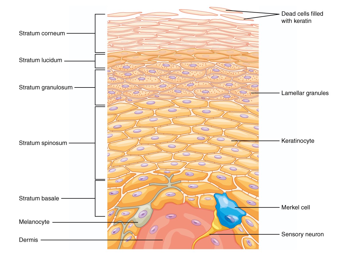
00:01 We are now going to talk about the melanocytes and we take a look at this particular cell. 00:07 Melanocytes contains melanin hence melanocytes. 00:11 So melanocytes are attached to the basal lamina via a certain channels called hemidesmosomes. They have long processes called dendrites which penetrate the stratum spinosum. These are long filaments that the melanocytes have, and they then communicate with the other cells. 00:35 Each melanocyte associates with keratinocytes via dendrites, forming the epidermal melanin unit. 00:42 What do you mean by that? What we mean is each melanocyte is attached to about 36 other keratinocytes, and this is how it is able to transfer the melanin to these cells. 00:56 It is important to note that the number of melanocytes is constant across all ethnic groups. What is the difference is the amount of melanin. 01:07 So if you differentiate between black skin and white skin, the number of melanocytes is the same. But it's the content the quantity of the melanin that is different. 01:21 Let's see how melanin is transferred from melanocytes to keratinocytes. 01:25 You can see the melanocytes there at the their base and attached to three keratinocytes through the dendrites. 01:33 So the melanin is stored in melanosomes, which are small pockets within the cytoplasm of the melanocyte. 01:40 It is then transported via the dendrites that I mentioned, like tentacles to the keratinocytes, and then they are taken from the extracellular space through a mechanism called phagocytosis, and then transferred into the stratum spinosum to all the cells based on the melanocytes keratinocytes unit that I mentioned to you. 02:07 So they then form a protective melanocytic umbrella which protects us against ultraviolet rays. 02:15 So what happens when there's a problem with melanocytes? We can get a condition called melanoma. 02:22 This condition is very common amongst patients who have light skin. 02:27 And it's a malignant cancer that affects the melanocytes. 02:34 Again another condition clinical example is congenital melanocytic nevus where you've got an aberrant increase in the number of end and replication of the melanocytes. 02:45 And as you can see those pictures there they are showing congenital melanocytic nevus.
About the Lecture
The lecture Melanocytes and Skin Color by Ncoza Dlova is from the course Introduction to Dermatology.
Included Quiz Questions
Which statement is correct regarding melanocytes?
- They have long processes that penetrate the stratum spinosum.
- They are attached to the basal lamina via dendrites.
- They compose a dermal-melanin unit.
- They compose a hypodermis-melanin unit.
- The number of melanocytes differs significantly between ethnic groups.
Author of lecture Melanocytes and Skin Color

Ncoza Dlova
Customer reviews
5,0 of 5 stars
| 5 Stars |
|
5 |
| 4 Stars |
|
0 |
| 3 Stars |
|
0 |
| 2 Stars |
|
0 |
| 1 Star |
|
0 |
