Playlist
Show Playlist
Hide Playlist
Introduction esp. Triangles of the Neck – Neck
-
Slides 25 Neck1 HeadNeckAnatomy.pdf
-
Download Lecture Overview
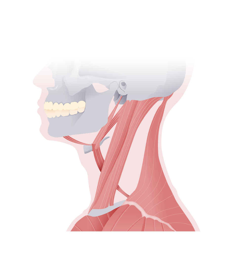
00:01 Welcome to this presentation on the neck, part I. 00:09 Here, we have the introduction to the lecture and two areas are highlighted. 00:15 And these happen to be the triangles of the neck. 00:17 So if you’ll continue on this journey with me, we’ll learn more about these geometric configurations. 00:25 So again, two colored areas here. We have the area in blue. This is one of the major triangles of the neck. And then the one located posteriorly is the second triangle of the neck. 00:42 Because of their anatomic relationships, the one in blue is referred to as the anterior cervical triangle. 00:56 The muscle that we see here as a major boundary is the sternocleidomastoid. 01:02 And it will separate this anterior triangle from the one that lies posterior. 01:10 That then leads us to this green-colored region which represents the posterior triangle of the neck. 01:19 Its posterior boundary is seen right along here. And that represents trapezius muscle. 01:29 Let's start with the anterior triangle, as this one is more complicated. We can see this region, just inferior to the mandible and bounded by the posterior and anterior belly of the digastric. This area represents your submandibular triangle. It contains the several structures. 01:48 The submandibular gland, some lymph nodes, and the facial artery and vein. Lying inferior to the anterior belly of the digastric muscle is a small region or triangle called the submental triangle. Its inferior boundary would be at the level of the hyoid bone that we see here and then another boundary would be the midline of this particular region. Here we can find the submental lymph nodes. Your third subdivision to the anterior triangle is your carotid triangle. 02:21 Here we can see the posterior belly for the digastric muscle and the superior belly of the omohyoid as well as the sternocleidomastoid forming its boundaries. The contents of the triangle are the common carotid artery, internal and external carotid arteries, internal jugular vein, vagus nerve and hypoglossal nerve. This is especially important as you can get access to the common carotid and internal carotid arteries. With a conventional carotid endarterectomy, you can surgically expose the common and internal carotid arteries to remove atherosclerotic lesions. With an access to the internal jugular vein, this triangle is also a site for insertion of a central line instead of the subclavian vein. The fourth and final subdivision of the anterior triangle is referred to as the muscular triangle. Its boundaries would be your superior belly of the omohyoid, the sternocleidomastoid and then it would run to the midline of the neck. The upper limit of this area would be your hyoid bone. 03:29 The important structures in this area are the infrahyoid muscles, the pharynx, the thyroid gland and parathyroid glands. The contents of the posterior triangle are the external jugular vein, the subclavian vein, the transverse cervical and suprascapular veins, the subclavian artery, the accessory nerve, the cervical plexus, the phrenic nerve and trunks of brachial plexus. Especially the external jugular vein, the subclavian vein and the cervical plexus have a high clinical relevance. 04:06 The superficial location of the external jugular vein makes it vulnerable to being severed. The lumen is held open by investing fascia. This resulting in an air embolism. 04:17 The subclavian vein is a site of access for a central line. 04:21 Two potential complications could be a puncture of a subclavian artery and a pneumothorax. Looking on the cervical plexus, you can use a nerve block here to anesthetize the neck region. 04:34 The anesthetic is injected over the posterior border of the sternocleidomastoid muscle at the junction of its superior and middle thirds. The posterior triangle is less complex with respect to its subdivisions. 04:49 Here, we see the occipital triangle, the posterior triangle identified. Its boundaries are the sternocleidomastoid. Here you see the inferior belly of your omohyoid. And then you see the trapezius. 05:08 The second and final subdivision of your posterior triangle is the omoclavicular triangle, this small area here. 05:19 And you can see it’s bounded by the inferior belly of the omohyoid, hence omo in the prefix. 05:26 This boundary here is the clavicle, hence clavicular as the suffix. 05:32 And then the third and final boundary to this triangle is going to be your sternocleidomastoid.
About the Lecture
The lecture Introduction esp. Triangles of the Neck – Neck by Craig Canby, PhD is from the course Head and Neck Anatomy with Dr. Canby.
Included Quiz Questions
Which of the following muscles separates the anterior neck triangle from the posterior neck triangle?
- Sternocleidomastoid muscle
- Scalene muscle
- Sternothyroid muscle
- Omohyoid muscle
- Trapezius muscle
Which of the following is NOT a subdivision of the anterior triangle of the neck?
- Occipital triangle
- Submandibular triangle
- Muscular triangle
- Carotid triangle
- Submental triangle
Which of the following is the name for the triangle located inferiorly to the mandible and bounded by the anterior and posterior bellies of the digastric muscle?
- Submandibular
- Carotid
- Sublingual
- Submental
- Muscular
Customer reviews
4,9 of 5 stars
| 5 Stars |
|
22 |
| 4 Stars |
|
2 |
| 3 Stars |
|
0 |
| 2 Stars |
|
0 |
| 1 Star |
|
0 |
it is self explanatory and so understandable , i like this a lot
amazing and well-thought lecture. explains extremely long pages of gray's anatomy within minutes. it is so much easier to read anatomy and go through studying it and memorizing instead of taking precious time to understand what the author wanted to say...
because the lecture is short and brief it is also not time consuming
Le he dado 5 estrellas a esta conferencia porque es una conferencia que realmente se lo merece, explica muy bien el tema y he aprendido bastante acerca de este tema muchas gracias profesor
1 user review without text


