Playlist
Show Playlist
Hide Playlist
Impetigo: Pathophysiology

-
Reference List Pathology.pdf
-
Slides Impetigo Pathophysiology.pdf
-
Download Lecture Overview
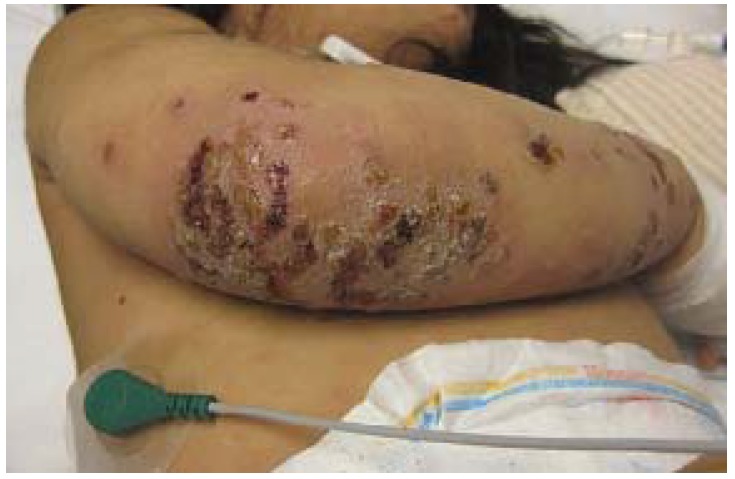
00:01 Welcome. In this talk, we're going to discuss another very infectious cutaneous disorder that's called impetigo. 00:09 So impetigo as I've already said, is highly contagious. 00:12 It's a superficial bacterial infection. 00:15 It's typically caused by staphylococcus but can also be caused by Streptococcus pyogenes. 00:21 So the epidemiology typically occurs in little kids. 00:25 By the time that you get to be teenaged into older adult ages, you've already developed a fairly good humoral immunity to those agents, so they tend not to be able to get a foothold. 00:36 But kids haven't yet developed that. 00:39 It's really infectious. 00:42 So there can be epidemics or outbreaks in schools. 00:45 It's easily spread among individuals. 00:47 And kids put each other's fingers in their mouths and they touch a variety of things. 00:52 It's very contagious. Spread by direct contact with lesions. 00:58 Even though they may not be grossly apparent, the bacteria can easily be spread. 01:03 The pathophysiology has already said the most common entity that drives this is staph aureus Staphylococcus aureus, a normal skin commensal. 01:12 But Streptococcus pyogenes can also be an etiology for this. 01:16 The routes of entry are primarily through the intact skin. 01:21 It doesn't take any breaks in the skin to make this happen, although breaks in the skin will allow deeper infections as we'll talk about. 01:29 When there are minor injuries like a cut or abrasion or eczema, where there are breaks in the barrier function that allows a deeper invasion and more readily seeds the bacteria once it makes contact. 01:43 The clinical presentation. 01:44 So there are three kind of general varieties that we recognize. 01:48 It's all the same disease. 01:49 It just has to be. It has to do with the extent and the deepness of infection. 01:55 So there's nonbullous impetigo which means it's not really forming big bully. 02:00 There is bullous impetigo where we get kind of larger blisters and then there's Ecthyma. 02:07 You may also hear it as Ecthyma gangrenosum. 02:10 And that is a very deep seated impetigo. 02:14 So let's talk about nonbullous impetigo first. 02:17 It's the most common variant that we're going to see. 02:20 It's not really a type. It's just it's more superficial and not as extensive. 02:25 70% of cases are going to be this the nonbullous variety. 02:28 And previously it got got the nomenclature, contagious impetigo. 02:34 Kids as again as I said previously are going to have this when they have it, it's going to be typically classically around the nose or mouth or on the hands, although it can be in other areas as well. 02:47 So the lesions of nonbullous impetigo are going to be very superficial. 02:52 They're basically basically just going to be in the epithelium. 02:55 So the initial lesion will be a proliferation of bacteria that will drive an innate inflammatory response. 03:02 So we will see some vascular dilation that will be kind of a rash. 03:06 It will be red a little bit warm with a central papule. 03:09 As we get more and more bacteria in there, we'll start recruiting more and more acute inflammatory cells, neutrophils. 03:17 So we will turn this initial lesion, the papule, into a vesicle, which has a lot of fluid within it that represents kind of edema in the tissue, but also a very large and increasing collection of neutrophils and bacteria. There will be a surrounding rim of erythema again reflecting vasodilation as part of the inflammatory response. 03:38 This will then evolve into a formal pustule. 03:41 There will be so many neutrophils that we'll be able to see the yellow necrotic debris and neutrophils that are there. 03:48 And at this point it's capped only by a very thin layer of stratum corneum. 03:53 So that will rupture. The pustule will rupture and will ooze and exudate pus and serous fluid and necrotic debris and bacteria over the surface that dries. 04:02 That gives rise to the classic honey colored crust. 04:06 It's non-tender. It can be itchy again because of mediators made by the inflammatory cells, but it's important to note that this is very superficial. 04:15 It's only in the epithelium, so it will typically not scar. 04:20 What's shown on the right hand side is kind of an actual image of this, with the honey crusting and the surrounding erythema. 04:27 Perfect. All right. That was nonbullous impetigo. 04:30 Let's talk about bullous impetigo, which is essentially the same process. 04:35 Just a little more so. 04:37 So 30% of cases overall are going to be bullous. 04:40 The lesions occur on the trunk. 04:42 And that the reason that the trunk is more commonly affected with this is that there's looser connective tissue. 04:48 So you may be able to get more extensive spread of the bacteria before we kind of corral it. It's the same sequence of events, just larger lesions, occasionally with systemic manifestations. 05:00 So the patient may have fever, may have malaise, may have myalgias. Again these are superficial lesions for the most part. 05:07 So they won't scar. The systemic manifestations include fatigue and fever general malaise. You may actually have nausea and vomiting as well. 05:16 So that's bullous impetigo. 05:18 And then there's ecthyma or ecthyma. 05:20 Gangrenosum. And ecthyma is kind of very infrequent. 05:24 But it may happen if there's a deep seated infection, if it occurs at an area where there has been prior kind of breaching of the epithelial barrier or a very, uh, a very aggressive bug. 05:40 The exact incidence is kind of unknown, but it is relatively rare, fortunately. It affects the deeper layers of the dermis. 05:48 So it is going into the dermis as and can also get into the subcutaneous fat or the hypodermis. The lesions in this case are usually on the extremities, legs and arms with ecthyma gangrenosum. 06:04 This is a deeper seated infection. 06:07 And what we'll have are coin size anywhere from kind of penny to quarter size punched out lesions that go very deep. 06:15 They will have thick yellow to gray scabs over the surface. 06:20 Okay. Trigger warning. 06:22 The next image may be a little distressing to you. 06:26 What we're seeing are multiple gross images of ecthyma gangrenosum. 06:31 You can appreciate that these appear to be more indurated. 06:34 They are deeper diving. 06:35 There is more injury. The histologic image that's shown side by side is showing that we have transmural necrosis with inflammation. 06:46 If we look there would be bacteria from top to bottom getting down into the dermis in many cases into the subcutaneous fat. 06:53 Because we are getting beyond the epidermis, these lesions, when they heal with appropriate antibiotic treatment, will end up being scarred. 07:02 And so there will be some residual scarring in the dermis and into the subcutaneous fat. 07:09 Diagnosis. It's clinical. 07:11 I mean the classic honey crusting, that sort of appearance, little kids contact with daycare, all of that sort of stuff. 07:19 But you can also, and you probably do want to do a gram stain and culture of the exudate to know which bacteria you're dealing with and what might be their antibiotic sensitivity. 07:29 Management. So for the nonbullous impetigo, the very noninvasive topical antibiotics are more than adequate cleaning and then topical antibiotics. 07:39 With the bullous impetigo and the more severe forms you probably want to give, in fact, you do want to give oral antibiotics. 07:49 And if you have been cultured, if you have cultured out methicillin resistant Staphylococcus aureus, that's something that requires special consideration. And in some locales will involve connecting with the the local authorities to let them know that we have an MRSA or MRSA infection. 08:08 And with that, we've covered a very common topic, and one that if you if and when you have children, you will face head-on because they'll get it. 08:18 Thanks.
About the Lecture
The lecture Impetigo: Pathophysiology by Richard Mitchell, MD, PhD is from the course Infection Conditions of the Skin.
Included Quiz Questions
Which bacteria is the most common cause of impetigo?
- Staphylococcus aureus
- Streptococcus pneumoniae
- Pseudomonas aeruginosa
- Escherichia coli
- Haemophilus influenzae
Which variant of impetigo accounts for approximately 70% of cases?
- Nonbullous impetigo
- Bullous impetigo
- Ecthyma gangrenosum
- Impetigo contagiosa
- Secondary impetigo
Why do bullous impetigo lesions commonly occur on the trunk?
- Due to looser connective tissue allowing bacterial spread
- Due to higher concentration of sweat glands
- Due to increased sun exposure
- Due to friction from clothing
- Due to higher bacterial colonization
Which type of impetigo is most likely to result in scarring?
- Ecthyma gangrenosum
- Nonbullous impetigo
- Bullous impetigo
- Superficial impetigo
- Crusted impetigo
Author of lecture Impetigo: Pathophysiology

Richard Mitchell, MD, PhD
Customer reviews
5,0 of 5 stars
| 5 Stars |
|
5 |
| 4 Stars |
|
0 |
| 3 Stars |
|
0 |
| 2 Stars |
|
0 |
| 1 Star |
|
0 |
