Playlist
Show Playlist
Hide Playlist
Imaging in Dermatology: Wood's Lamp and Dermoscopy

-
Slides Imaging in Dermatology Wood's Lamp and Dermoscopy.pdf
-
Download Lecture Overview
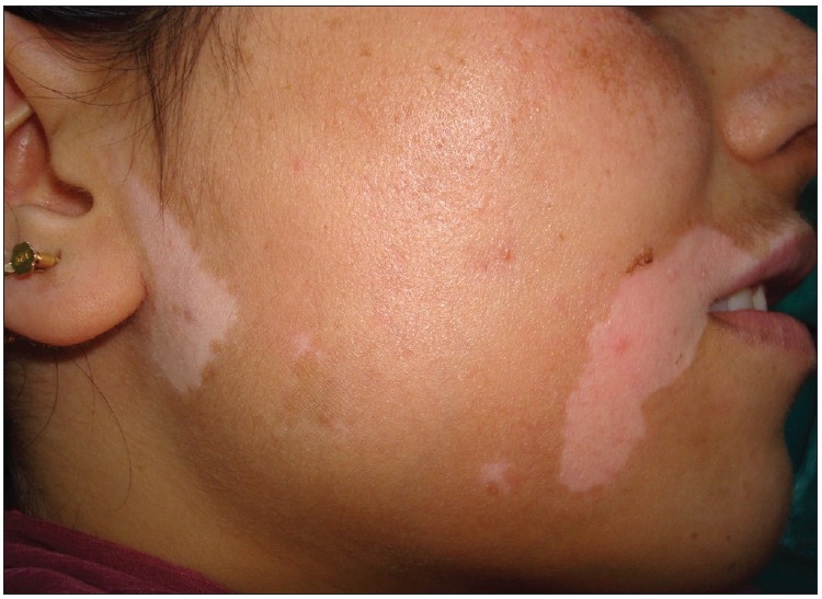
00:01 We are now going to talk about imaging in dermatology. 00:06 So one of the instruments we use are is what we call the Wood's lamp for skin examination. 00:13 It is a diagnostic test in which skin or hair is examined while exposed to the black light emitted by a Wood's lamp. 00:21 The black light is invisible to the naked eye, and it is found on the ultraviolet spectrum. The lamp also emits light in the violet part of the electromagnetic spectrum, resulting in a violet glow. 00:41 So this is how the Wood's lamp is used. 00:46 It is turned on to warm for about a minute. 00:52 The room has to be dark, and the lamp is held at 10 to 30cm away from the skin. 01:01 So what do we use the Wood's lamp for? We use it for the following conditions. 01:06 Bacterial infections, viral infections, porphyria, as well as differentiating between vitiligo and conditions like tinea versicolor, which we will talk about later. 01:18 So here are the examples of positive results on a Wood's lamp examination. 01:25 This is a patient with a pigmentary disorder, which is more obvious when you use the Wood's lamp. 01:36 This second picture shows a positive result in a patient with tinea pedis. 01:44 This patient has got a condition called tinea pedis, which is caused by Trichophyton rubrum. That is the name of the fungus rubrum, meaning red. 01:54 And you can see that it fluoresces red on the Wood's lamp. 02:00 The last imaging equipment that we're going to talk about is a dermoscopy. 02:05 Now, in recent years, dermoscopy has become indispensable for a dermatologist. 02:11 It's almost like a stethoscope for a cardiologist. 02:15 So the examination of the skin using surface microscopy is what we do when we use the dermatoscope. It is mainly used for evaluation of pigmented skin and hair disorders and other skin conditions. 02:33 This is a benign melanocytic nevus nevus on dermoscopy. 02:41 You can see that there's brown pigmentation and some laser pattern which is regular. 02:46 So this is probably a normal nevus or birthmark. 02:53 One needs training to be able to detect the different variations on using Dermoscopy. 02:58 It's not something that you do overnight. 03:00 It takes time and training and skill and experience. 03:04 If you look on the right hand side, this is a white patient with melanoma and you can see the difference between the two. 03:11 The one is darker irregular pigmentation Asian, as well as ulceration and a grayish lace pattern, so to the trained eye of a dermatologist. 03:22 That signifies that is melanoma. 03:27 We do also use dermoscopy for hair conditions, for example patients with alopecia areata. 03:34 You see what we call exclamation marks. 03:37 You can see on this picture with the arrow, the exclamation mark, which is typical of alopecia areata. 03:44 However, we do see that even in other hair conditions. 03:49 And you also see yellow dots, which are typical of alopecia areata. 03:55 So it is important that one is trained on using dermoscopy. 04:03 It takes time.
About the Lecture
The lecture Imaging in Dermatology: Wood's Lamp and Dermoscopy by Ncoza Dlova is from the course Introduction to Dermatology.
Included Quiz Questions
What is a Wood's lamp examination used for?
- It uses black light to detect bacterial or fungal infections on the skin.
- It uses infrared light to assess skin cancer.
- It uses X-rays to diagnose bone fractures.
- It uses visible light to examine hair follicles.
- It uses ultrasound to detect deep tissue abnormalities.
What is NOT typically tested on Wood's lamp examination?
- Malignancy
- Bacterial infections
- Fungal infections
- Porphyria
- Alterations in skin pigmentation
Author of lecture Imaging in Dermatology: Wood's Lamp and Dermoscopy

Ncoza Dlova
Customer reviews
5,0 of 5 stars
| 5 Stars |
|
5 |
| 4 Stars |
|
0 |
| 3 Stars |
|
0 |
| 2 Stars |
|
0 |
| 1 Star |
|
0 |
