Playlist
Show Playlist
Hide Playlist
Ichthyosis Vulgaris: Diagnosis and Management

-
Reference List Pathology.pdf
-
Slides Ichthyosis Vulgaris Diagnosis Management Dermatopathology.pdf
-
Download Lecture Overview
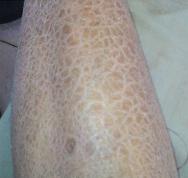
00:01 So the clinical presentation, you see this diffuse, dry, scaly skin. The plaques tend to be on extensor surfaces, and they are kind of firmly semi adherent to the underlying skin. 00:18 This has been likened to fish scales hence the name of that ichthyosis. 00:23 So where do we find ichthyosis vulgaris. 00:27 It tends to have a fairly characteristic distribution where you will see it on the trunk. You also tend to see it on extensor surfaces of the extremities, the flexor surfaces in comparison, or the intertriginous areas in the groin and the axilla tend to be relatively spared. 00:43 It is also quite characteristically seen on the palmar and plantar surfaces of the hand and foot, as you see here, with very impressive kind of accentuation of the creases due to this hyperkeratosis. 00:59 The hereditary form, those lesions, because it is hereditary, happened very early. They begin to appear in infancy or early childhood. 01:07 It may improve in warm, sunny weather. 01:12 And that may be because as you sweat or you have increased production of of of a variety of materials that are coming out of the various glands that may provide kind of an emollient surface, it may lubricate better. 01:28 So it may be better in that environment as compared to cold, dry weather. It will gradually improve with adolescence. 01:35 This hereditary form. And that's because as you get as you go through adolescence, you now have apocrine secretion and you will have increased sebaceous gland secretion associated with the kind of hormone spurts that happen with puberty. 01:49 So that may also improve some of the clinical manifestations. 01:55 And it uh, in patients who have very severe ichthyosis, you may have a functional vitamin D deficiency. 02:03 Why? Well, that's because in fact, the skin the stratum corneum layer is much thicker. And so UV penetration is much less. 02:11 And they don't get the metabolism of the of the vitamin D precursor into active vitamin D. How do we diagnose this. 02:18 Well it's kind of by inspection also by clinical history. 02:22 If a mom or dad had it, it's very likely that when you have scales, that's what you have. Again, you can also do formal genetic testing. 02:28 We know many of the mutations in the Filaggrin gene that will give rise to ichthyosis. We can do a biopsy. 02:36 I always like seeing a biopsy as a pathologist, but it probably in most cases is not absolutely necessary. 02:42 But if you do a biopsy, what you're going to see is acanthosis. 02:47 So a very thickening of the epidermis the epithelial layer you will see hyperkeratosis. 02:55 So a markedly thickened stratum corneum. 02:58 You will see parakeratosis which is just another way of saying nucleated keratinocytes in the stratum corneum which you don't normally see. 03:09 You will also see that there is a very thin granular layer. 03:11 So the maturation from the granular layer up is abnormal. 03:17 And that's what we're kind of seeing reflected here. You may also see secondary effects due to the abnormal keratinization that will give rise to cytoplasmic vacuolization those kind of bubbles in the very superficial layers of the keratinocytes. 03:33 How do you manage this? Well, a lot of it is just treating the symptoms, the very dry, flaky skin and then you. 03:40 So you would give emollients moisturizers. 03:42 You can give topical retinoids. 03:45 That reduces the the abnormal keratinocytes that are at the very superficial layer. 03:50 It suppresses keratin synthesis. 03:52 So you don't get that really thick layer on top of the epithelium that is to say the stratum corneum. You can do keratolytic which are basically exfoliation. 04:02 You basically wipe away the excess dead layer on top in the stratum corneum. 04:08 And if there's any underlying disease you manage that. 04:11 So if there's malignancy you manage that. 04:13 If there is the inflammatory disorder like lupus or celiac disease, you manage that. If it's drug related, you take them off the drug. 04:20 If it's hypothyroidism, you treat their hypothyroidism. 04:23 Et cetera. So that kind of kind of all makes sense with that. 04:27 You are now experts in the world of ichthyosis vulgaris. 04:31 And again, not that uncommon. 04:34 And you'll be able to give your patients some understanding of why this happens. 04:38 So thanks.
About the Lecture
The lecture Ichthyosis Vulgaris: Diagnosis and Management by Richard Mitchell, MD, PhD is from the course Disorders of Maturation and Pigmentation of the Skin.
Included Quiz Questions
Which area is typically spared in the distribution pattern of ichthyosis vulgaris?
- Intertriginous areas of the groin and axilla
- Extensor surfaces of extremities
- Palmar surfaces of hands
- Plantar surfaces of feet
- Trunk
Which topical treatment for ichthyosis vulgaris works by suppressing keratin synthesis?
- Topical retinoids
- Emollients
- Keratolytics
- Moisturizers
- Vitamin D supplements
Author of lecture Ichthyosis Vulgaris: Diagnosis and Management

Richard Mitchell, MD, PhD
Customer reviews
5,0 of 5 stars
| 5 Stars |
|
5 |
| 4 Stars |
|
0 |
| 3 Stars |
|
0 |
| 2 Stars |
|
0 |
| 1 Star |
|
0 |
