Playlist
Show Playlist
Hide Playlist
Diaphragmatic Hiatus

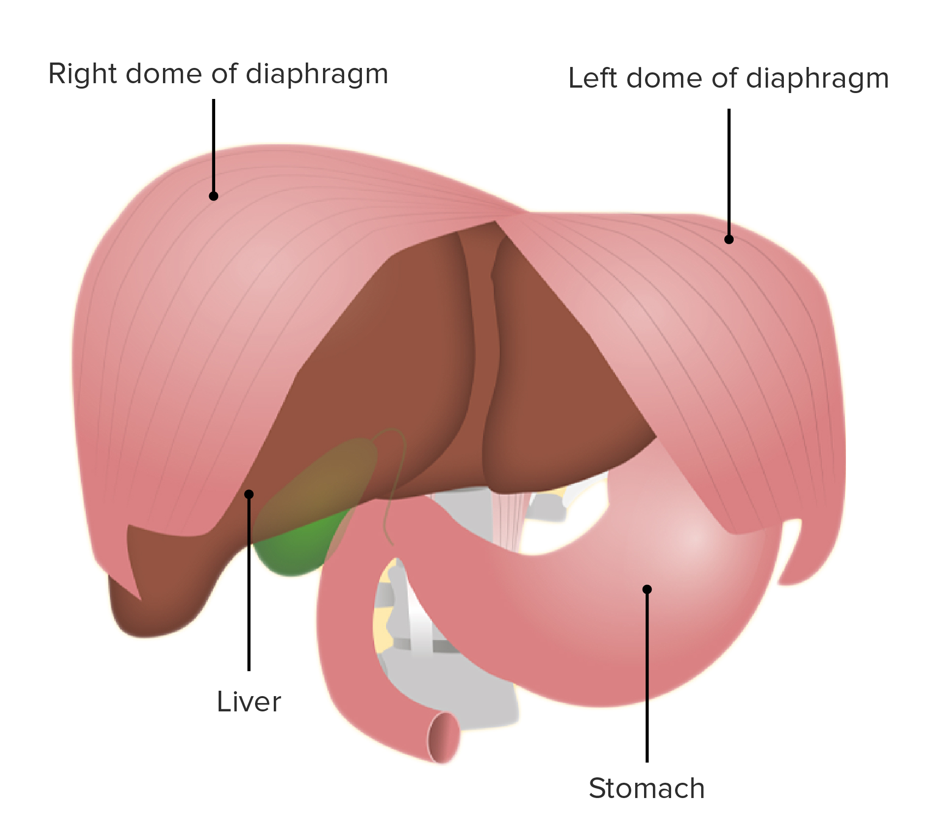
00:00 What we have done so far is up the diaphragm. 00:04 We reached up to the diaphragm. 00:06 Nerve supply, we did phrenic nerve. 00:11 So, this is pretty much the diaphragm. 00:18 Blood supply, we touched upon. 00:21 Your internal mammary artery goes down here. 00:26 That's your superior epigastric then you have the musculophrenic. 00:30 That's the main artery to the diaphragm. 00:32 But then you also have the superior phrenic and the inferior phrenic to the diaphragm as well. 00:39 As I said, the three blood vessels supply the diaphragm. 00:43 One is the musculophrenic coming off the internal mammary, superior phrenic coming off the thoracic aorta, and inferior phrenic coming off the abdominal aorta. 00:55 So, that is blood supply to the diaphragm. 00:57 Diaphragmatic hiatus, quite important. 01:00 We have discussed this during the day. 01:03 So, that's your dome. 01:06 The first hiatus is T8. 01:09 You have esophagus. 01:14 You have the inferior vena cava and the right phrenic nerve. 01:19 Left phrenic nerve has its own hiatus. 01:22 Come two levels below, T10. 01:25 That is when you have the esophagus and the right vagus. 01:29 Sorry, the anterior vagus and the left vagus. 01:32 I'm not sure whether I covered this before. 01:34 Your left vagus runs close to the hilum here and then becomes the anterior vagus. 01:44 The right vagus continues as the posterior vagus. 01:49 So, the left is anterior, right is posterior. 01:53 Then you come vertebral level lower down is T12, abdominal aorta, thoracic duct, and the azygos vein. 02:01 So, these are the important structures coming off the diaphragm, coming through the hiatus. 02:07 Then the long, expendable transpyloric plane: The transpyloric plane, as the name says, it travels the pylorus of the stomach. 02:16 It corresponds to the lower border of T. 02:20 Just turn around for a minute, Johnny. Thank you. 02:23 It corresponds to the lower border of L1 vertebra. 02:31 Transpyloric plane. 02:37 In other words, it is midway between the suprasternal notch and the pubic symphysis. 02:48 That's the transpyloric plane, lower border of L1. 02:51 Now, if you draw two lines, one in the right midaxillary line and then one on the left midaxillary line, and then you identify the highest point of the iliac crest which is called the intertubercular, the tubercular area there or the tubercular crest. 03:18 If you draw this line, this essentially divides your abdomen to 9 regions. 03:26 Now of this, clearly, all the regions are important in its own unique way, but for the purpose of your exam, L1 is probably most important. 03:36 If you start from the right to the left, what are the important structures you will see in a CT scan? If you slice it across L1 vertebra, what is the first thing? You see the hepatic flexure of the colon here. 03:56 Then what else do you see? You go then, the liver is slightly at fundus of the gallbladder. 04:05 They're at the fundus of the gallbladder. 04:09 Here you have the hepatic flexure, fundus of gallbladder As you go more medially, sort of more towards the midline, what do you see? Second part of the duodenum? Go on. You're right. 04:26 Neck of the pancreas? Portal vein, where does the portal vein come from? Splenic vein, very good. 04:37 What else? We have covered quite a few things. 04:46 Hilum of the right lung.
About the Lecture
The lecture Diaphragmatic Hiatus by Stuart Enoch, PhD is from the course Upper Part of the Body Anatomy.
Author of lecture Diaphragmatic Hiatus

Stuart Enoch, PhD
Customer reviews
5,0 of 5 stars
| 5 Stars |
|
5 |
| 4 Stars |
|
0 |
| 3 Stars |
|
0 |
| 2 Stars |
|
0 |
| 1 Star |
|
0 |
