Playlist
Show Playlist
Hide Playlist
Autonomic Innervation of the Hindgut, Kidneys, and Adrenal Glands

-
Slides Autonomic Innervation of the Hindgut Kidneys and Adrenal Glands.pdf
-
Reference List Anatomy.pdf
-
Download Lecture Overview
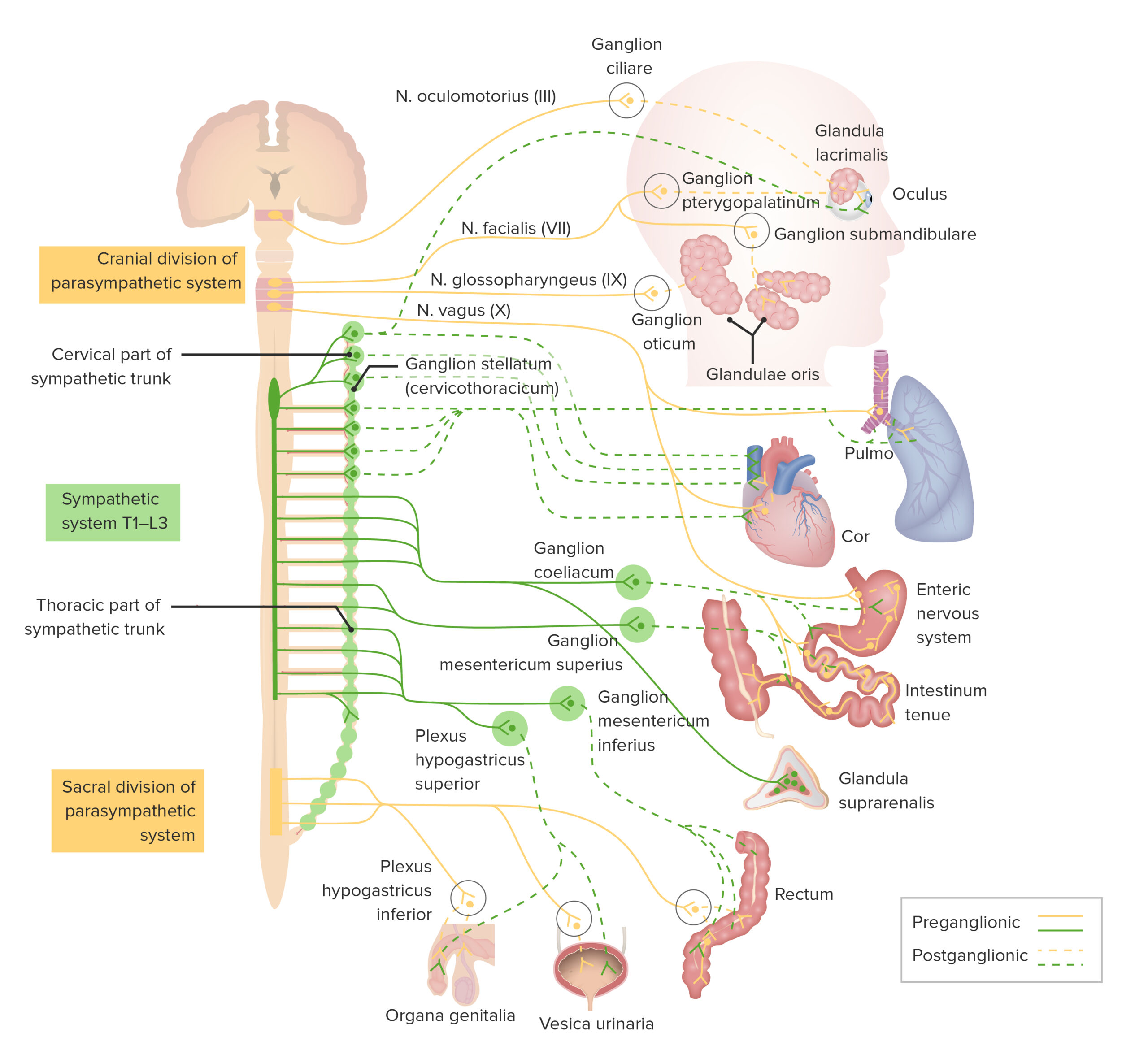
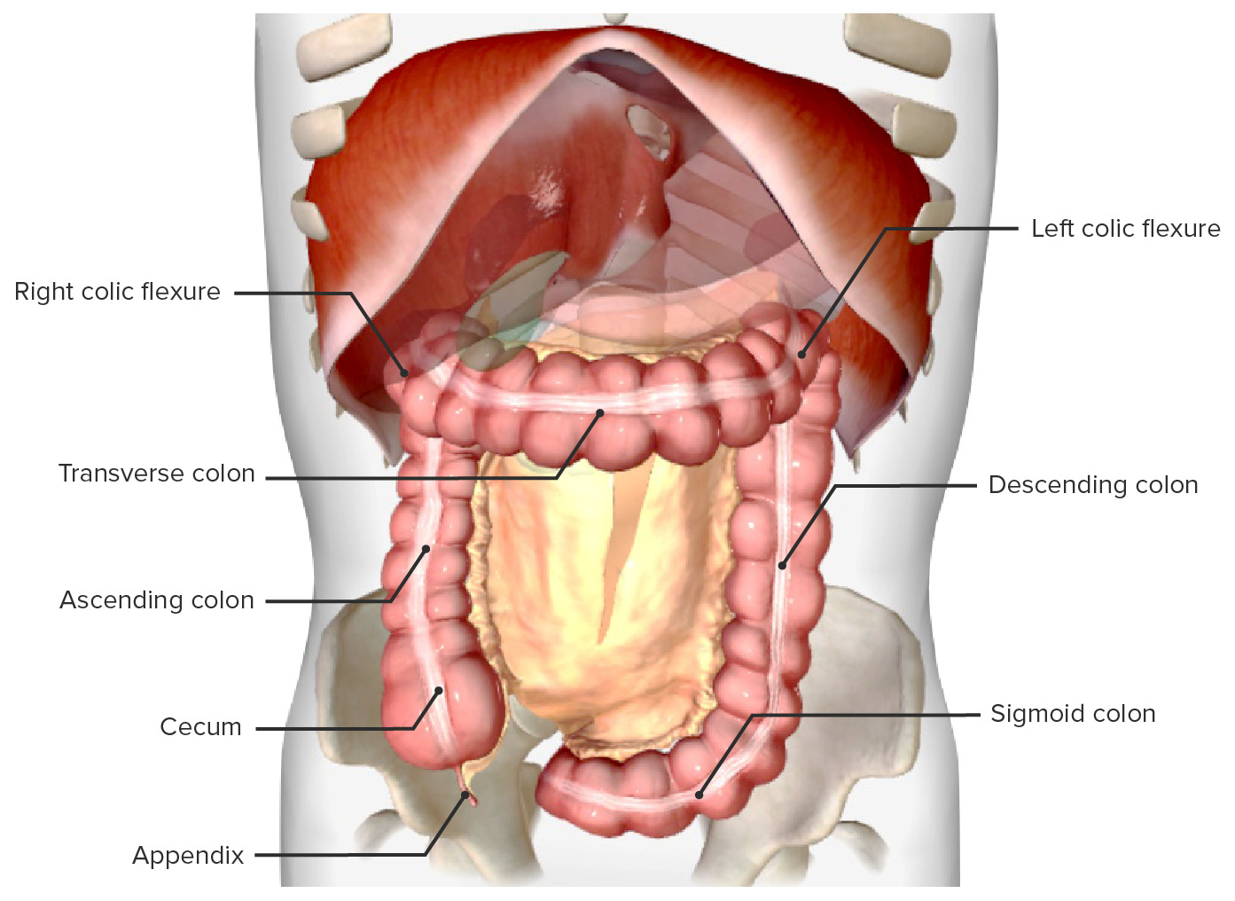
00:01 Now, let's move on to the hindgut. 00:03 Here we're going to have a look at the innovation of the hindgut. 00:07 We can see that the descending colon, the sigmoid colon, and the rectum. 00:11 These are going to be supplied by the inferior mesenteric plexus. 00:15 So, here we have the inferior mesenteric ganglion, formed by the lumbar splanchnic nerves. 00:21 We can see contributing here in green. 00:23 And here we can see the pelvic splanchnic nerves. 00:26 Remember, these pelvic splanchnic nerves are coming up from the inferior hypogastric plexus. 00:32 So these fibers are coming up from the inferior hypogastric plexus so then merge with the inferior mesenteric plexus to form these periarterial plexuses. 00:43 These are going to supply the descending colon, sigmoid colon, and rectum. 00:48 So, here we can see the inferior mesenteric ganglion contributed to via the lumbar splanchnic nerves for sympathetic. 00:56 Pelvic splanchnic nerves coming up via the inferior hypogastric plexus going towards it to form the inferior mesenteric plexus. 01:06 Now for the first time we can see this superior hypogastric plexus. 01:10 So remember, the inferior hypogastric plexus is lying on the lateral wall of the pelvis. 01:17 It's got parasympathetic and sympathetic nerve fibers within it. 01:21 These pelvic splanchnic nerves are leaving the inferior hypogastric plexus and passing towards the superior hypogastric plexus via those two hypogastric nerves. 01:33 Once they've got here, they form the inferior mesenteric plexus which goes to supply the descending colon, the sigmoid colon and the superior parts of the rectum. 01:43 Next, they will have a little bit of a sideways step and look at the kidneys and adrenal glands. 01:48 So here we can see the greater splanchnic nerve is actually contributing to the adrenal medulla. We can see here. 01:55 And it actually is slightly different. 01:57 In that it does have a very long preganglionic fibers. 02:02 This really is an exception to that sympathetic. 02:05 It doesn't synapse within the celiac ganglion like it did previously. 02:09 It's similar to the parasympathetic. 02:12 So here we can see the posterior vagal trunk is now going towards the kidney alongside the lesser splanchnic nerve. 02:19 But importantly, see how the lesser splanchnic nerve does now synapse in the aorticorenal ganglion. 02:26 So the only exception here is going into the adrenal medulla. 02:30 That is because embryologically, the adrenal gland is derived from the sympathetic nervous system. 02:36 So really is an exception that helps us to characterize the parasympathetic and sympathetic long or short preganglionic fibers. 02:45 Here we can see for the actual kidney how they've reverted to type. 02:51 Long preganglionic fiber for parasympathetic. 02:55 Short preganglionic for sympathetic synapses in the aorticorenal ganglion. 03:02 We can see the lesser splanchnic nerve also has a contribution to the renal ganglion here, And again, a short pre, and a long post. 03:10 The first lumbar spinal nerve will also have a contribution. 03:13 We can see here. 03:14 These together form the renal plexus that takes autonomic nerves towards the kidney.
About the Lecture
The lecture Autonomic Innervation of the Hindgut, Kidneys, and Adrenal Glands by James Pickering, PhD is from the course Lymphatics and Nerves of Abdominopelvic Region.
Included Quiz Questions
Which region contributes to the lumbar splanchnic nerves?
- L1 - L3
- L2 - L4
- L3 - L5
- L2 - L5
- L4 - L5
Which region contributes to the pelvic splanchnic nerves?
- S2 - S4
- S3 - S5
- S1 - S3
- S1 - S4
- S4 - S5
The greater splanchnic nerves are associated with which ganglion?
- Celiac ganglion
- Aorticorenal ganglion
- Superior hypogastric ganglion
- Inferior mesenteric ganglion
- Superior mesenteric ganglion
Author of lecture Autonomic Innervation of the Hindgut, Kidneys, and Adrenal Glands

James Pickering, PhD
Customer reviews
5,0 of 5 stars
| 5 Stars |
|
5 |
| 4 Stars |
|
0 |
| 3 Stars |
|
0 |
| 2 Stars |
|
0 |
| 1 Star |
|
0 |
