Playlist
Show Playlist
Hide Playlist
Articulations – Vertebral Column
-
Slides 02 Abdominal Wall Canby.pdf
-
Download Lecture Overview
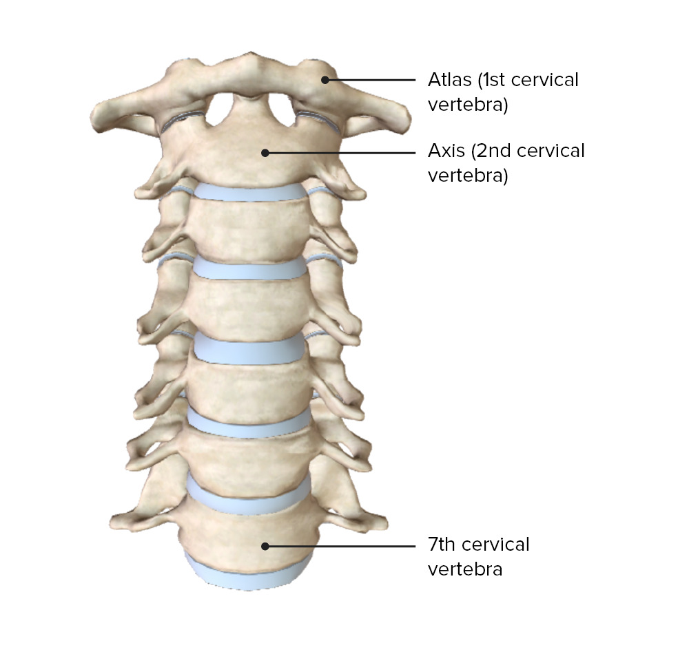
00:01 Now, let's understand the various types of articulations that we have within the vertebral column, and the first type of joint or articulation you wanna understand is that it exists between the articular processes superior to inferior, and these are refered to as zygapophyseal articulations or joints. 00:30 And if we take a look here we have one, two, three, four, five vertebrae, represented. 00:38 We see coastal facets here on the bodies of these vertebrae, so these are thoracic and then we get down to this level, we no longer see coastal facets so we know that we are looking at lumbar vertebra 1 and 2, but what we wanna focus in on is this area here or here for example, here we have the inferior articular process of this vertebra above and then the vertebra below here has it's superior articular process and then as result we form this type of joint. 01:18 There are segmental differences in the orientation of the zygapophyseal joints that will confer different ranges of motion from one segment to another, so if you look at the cervical region, the orientation of these articulations would be somewhat oblique in nature are allowing for or conferring for greater range of motion within the thoracic area, the orientation of these joints is more on anterior/posterior direction that will help to limit to some degree the range of motion. 01:57 In addition at the thoracic level we have super imposed here the ribs and that will further restrict the range of motion, and then within the lumbar segment, the greatest restriction in movement imposed because of the more lateral orientation of these particular articulations. 02:20 This particular slide is demonstrating a type of joint refered to as a symphysis, the plural of that is symphyses and simply an anatomic symphysis is shown in through here, this is a fibro-cartilaginous joint so this type of joint is found between our vertebral bodies and the structural component of this type of joint is gonna be the intervertebral disc. 02:49 This region here represents the intervertebral disc, the vertebral body above, vertebral body below. 02:59 Twenty three of these are present within the vertebral column. 03:05 You do not have one between C1 and C2 nor do you have intervertebral disc arranged within the sacral region nor the coccygeal region so you have 23. 03:21 An intervertebral disc has an outer ring called the annulus fibrosis and the tissue that constitutes the annulus fibrosis is fibrocartilage and then the central component which is kinda hard to really demonstrate here becomes less distinct as one ages in this more central gelatinous component of an intervertebral disc would represent the nucleus pulposus which is a remnant of the nodachord. 03:58 There are also end plates here on the inferior aspect of the superior vertebral body and on the superior aspect of the inferior vertebral body and these end plates are composed of hyaline as well as fibrocartilage.
About the Lecture
The lecture Articulations – Vertebral Column by Craig Canby, PhD is from the course Abdominal Wall with Dr. Canby.
Included Quiz Questions
How many intervertebral discs are present in a normal human spine?
- 23
- 21
- 33
- 27
- 30
What is the gelatinous material of the intervertebral disc called?
- Nucleus pulposus
- Annulus fibrosis
- Hyaline cartilage
- Fibrous cartilage
- End plates
Customer reviews
5,0 of 5 stars
| 5 Stars |
|
5 |
| 4 Stars |
|
0 |
| 3 Stars |
|
0 |
| 2 Stars |
|
0 |
| 1 Star |
|
0 |


