Playlist
Show Playlist
Hide Playlist
Anatomy of the Vertebral Column

-
Slides Anatomy of the Vertebral Column.pdf
-
Reference List Anatomy.pdf
-
Download Lecture Overview
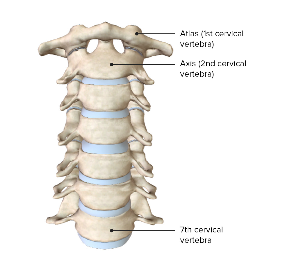
00:01 Now we're going to start talking about the anatomy of the back. 00:05 We're going to start by talking about the back bone or what we actually call the vertebral column. 00:12 The vertebral column is collection of bones that we group according to their location. 00:18 We have the cervical area which has seven portions. 00:23 We have the thoracic area, which has 12. 00:28 And we have the lumbar that has five. 00:31 By the time we get to the inferior most portion, we have the sacrum, which is actually a single bone formed by the fusion of five different bones. 00:41 And then the tiniest bit at the end, also called the tailbone, is the coccyx formed by between three and five fused bones. 00:51 If we look at the vertebral column from an anterior view, the first thing we'll see is a typical what we say vertebra. 01:00 And each vertebra is separated from the adjacent one by an intervertebral disc. 01:08 And this is going to be a description of a what we call basic vertebra. 01:12 Later on, we'll talk about regional variations that make for example, the cervical vertebra slightly different from the lumbar vertebra. 01:20 But for right now, we'll start with a superior view of the basic features of a vertebra. 01:27 In anteriorly, the majority of the vertebra overall is made up of the body. 01:34 Posteriorly, we have a little bit more complicated structure here and we call this collectively the vertebral arch. 01:42 We see that laterally we have these processes called transverse processes. 01:49 And then although it's hard to tell in a superior view, but these are pointing superiorly as the superior articular facets and articular means referring to a joints. 01:59 We know that that's going to be participating in a joint. 02:04 And then posteriorly, we have a single midline structure called the spinous process. 02:10 The spinous process is connected to the rest of the vertebral arch by lamina. 02:17 And the vertebral arch connects to the body by short pedicles. 02:23 Let's look at a basic vertebra from an anterior point of view. 02:28 From this point of view, mostly what we see is the vertebral body. 02:33 But laterally we can see a bit of the transverse processes. 02:36 Just a little bit of the superior and inferior articular facets on either side. 02:45 In a lateral view, we can still see a lot of the vertebral body. 02:49 A little bit of the transverse process. 02:51 And from this view, we can really see the superior and inferior articular facets as well as the spinous process. 03:00 From a posterior point of view, now the body is kind of out of our viewpoint here we can barely see it. 03:06 But we can still see the transverse processes, the superior and inferior articular facets. 03:12 And absolutely the spinous process. 03:17 Let's look at a cross section of an intervertebral disk now. 03:21 There intervertebral disk basically has two components, a tough outer portion called the annulus fibrosus, and a softer squishier inner portion called the nucleus pulposus. 03:36 And together these intervertebral discs form fibrocartilaginous joints. 03:42 These aren't the highly movable joints are used to in other parts of the body. 03:46 They're really there to provide strength and support and only a limited degree of movement. 03:52 Because after all, we do have the spinal cord running through the vertebral column so we don't want to have too much movement. 04:01 Now over time, the intervertebral disc can become degenerated. 04:06 And that can lead to all sorts of problems. 04:10 For example, if the annulus fibrosis becomes weak, it may bulge outward from its normal area and cause what's called a bulging disc. 04:20 If it gets worn down to the point that there's a defect or crack in the annulus fibrosis, then the nucleus pulposus can squeeze out through that opening causing a herniated disc because it's herniating through an opening in the annulus fibrosis. 04:38 And over time, the disc can basically dehydrate and get thin. 04:43 And when it does that the adjacent vertebral bodies can rub up against each other and cause osteophyte formation in a form of osteoarthritis. 04:55 So let's look at a superior view of what happens with a herniated disc. 05:01 So with a normal disc, the spinal nerve has a free pathway out beyond the vertebral column. 05:09 With a herniated disc that nucleus pulposus can squeeze out and impinge upon the nerve root at that area. 05:19 And unfortunately what that means is there will be pain and numbness in any of the areas that are innervated by that compressed nerve.
About the Lecture
The lecture Anatomy of the Vertebral Column by Darren Salmi, MD, MS is from the course Back Anatomy.
Included Quiz Questions
What separates the vertebrae?
- Intervertebral discs
- Facets
- Vertebral bodies
- Transverse processes
- Pedicles
Which structure is NOT part of the vertebral arch?
- Body
- Facet
- Lamina
- Spinous process
- Transverse process
What pathology leads to osteophyte formation in the vertebral column?
- Disc degeneration
- Herniated disc
- Vertebral fracture
- Hematoma
- Abscess
Author of lecture Anatomy of the Vertebral Column

Darren Salmi, MD, MS
Customer reviews
5,0 of 5 stars
| 5 Stars |
|
5 |
| 4 Stars |
|
0 |
| 3 Stars |
|
0 |
| 2 Stars |
|
0 |
| 1 Star |
|
0 |
