Playlist
Show Playlist
Hide Playlist
Anatomy of the Bladder and Urethra

-
Slides Anatomy of the Bladder and Urethra.pdf
-
Reference List Anatomy.pdf
-
Download Lecture Overview
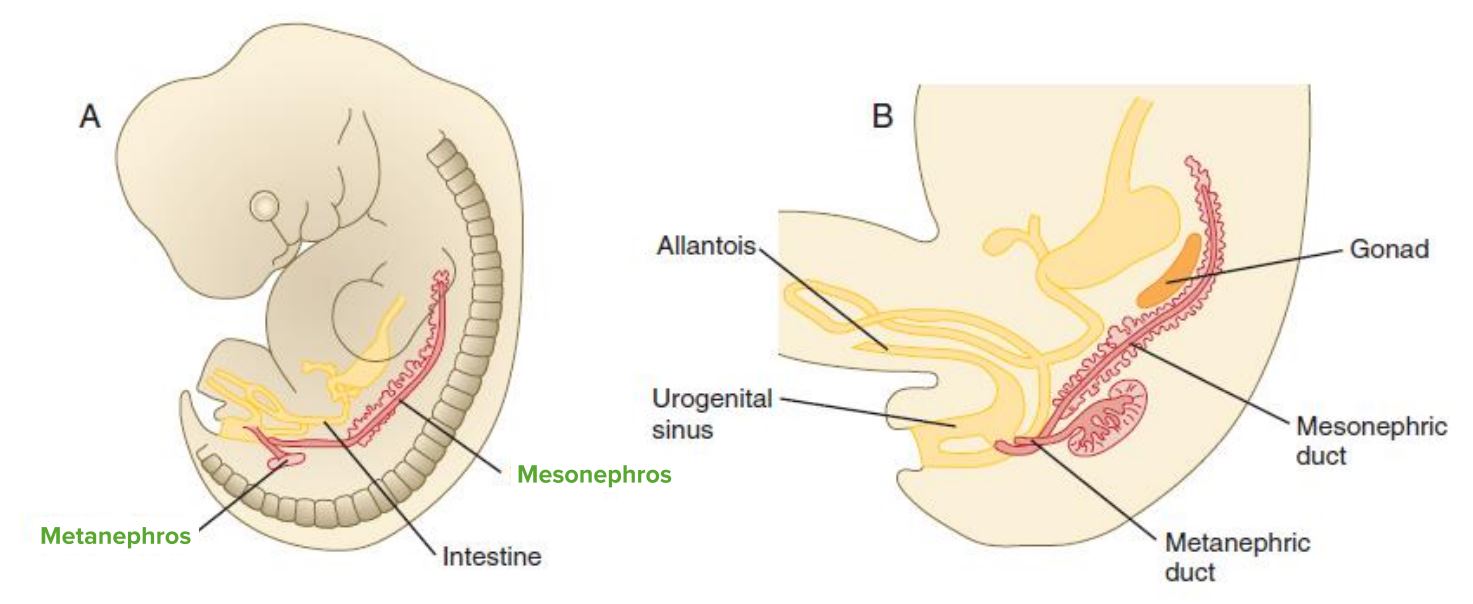
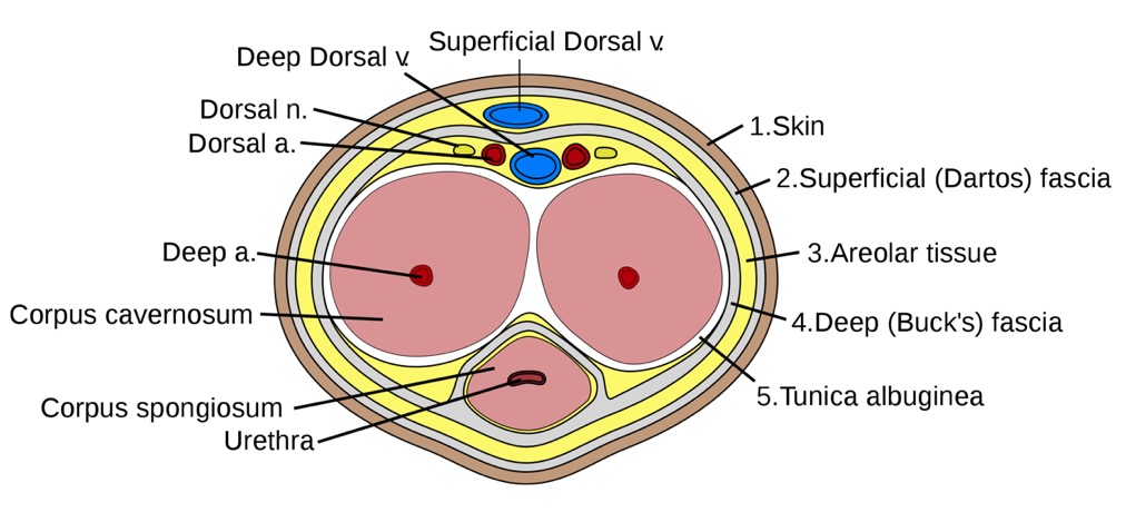
00:01 Now, let's look at the bladder and the urethra. 00:06 So here we're going to start looking at the urinary system really. 00:09 So, let's have a reminder of that urinary system. 00:11 Obviously, it's associated with a number of important structures: the kidneys, the ureters, the bladder, and the single urethra. 00:19 So let's remind ourselves of the position of the two kidneys. 00:22 We can see here, either side of the vertebral column. 00:25 And these lie retroperitoneal on the posterior abdominal wall. 00:30 Emerging from the renal hilum of each of these kidneys, we find a pair of ureters. 00:36 And these descend down the posterior abdominal wall, either side again of the vertebral column to enter into the pelvis by passing through the pelvic inlet. 00:45 So, the two ureters then pass down and they pass into the lateral aspects of the bladder. 00:51 So urine produced by the kidneys is passed down into the bladder via the ureters, where it's then stored before micturition allows that urine to escape. 01:02 So, here we have the bladder collecting and storing the urine produced by the kidneys, and then passing away from the bladder, we have the urethra that allows you to leave the body. 01:13 Here we can see the lateral aspect of the bladder. 01:16 Anteriorly we have the apex of the bladder, which we can see highlighted on the screen at the moment. 01:21 And then running posteriorly from the apex, we find this superior surface of the bladder, which we can see here. 01:29 And that's that surface that that layer of peritoneum will lie across as it runs down from the posterior aspect of the anterior abdominal wall. 01:38 Coming from the apex of the bladder, we have a remnant of embryo logical development, we have the median umbilical ligaments and this is a remnant of the urachus which was an important developmental structure. 01:50 So we have the apex, the superior surface, and then we have two inferolateral surfaces and we can see the left one here. 01:59 Obviously, the right one will be on the opposite side of the screen. 02:03 Then most posteriorly we have the base of the bladder. 02:06 And really you can imagine the shape of the bladder as the front of a ship. 02:10 So, it's very kind of pointed at the front as an a ship would pass through the breaking waves. 02:15 And then those two inferolateral surfaces then combine at the base of the posterior aspect of the bladder. 02:23 Here we can see passing into this connection, this kind of junction between the base and the inferolateral surfaces, we have the ureters, which have passed down in the posterior abdominal wall, entered into the pelvis via the pelvic inlet, and are now merging with the muscular layer of the pelvis. 02:42 So, now let's turn our attention to the internal surface of the bladder. 02:46 So, here we can see we have the bladder. 02:48 It's been sectioned in coronal section, and we can see that we have a nice thick muscular layer around the bladder. 02:55 This is known as detrusor muscle and that surrounds the bladder. 02:59 Really forming the muscle wall of the bladder. 03:02 We can see on the internal surface here we have elevations of that muscle and that is known as ruggae. 03:07 That helps to increase the surface area of the bladder as it fills with urine and allowing it to expand. 03:13 much like we had within the stomach. 03:16 As we then move down towards the urethra, the urethral opening, we have a transition in muscle fiber type, or at least how those muscles behave. 03:24 And these muscle fibers within the Trigone, they don't contract under the same influence as those muscles more superiorly. 03:31 And this makes sense as we need to allow urine that's within the bladder to pass from the bladder to the urethra. 03:38 And if all of these muscle fibers contracted under the same influence, then all what had happened is the urine would be compressed. 03:45 So, there's differentiation in how those muscle fibers contract due to different neurotransmitter signaling and the neural innovation. 03:52 That means that the muscles at the lower aspects of the bladder will remain relaxed as those superior muscle fibers contract, and that will maintain the opening of the bladder allowing urine to pass into the urethra. 04:05 So we're going to see the Trigone is demarcated here. 04:08 We can see importantly, that the ureters open up into the trigone aspects of the bladder via the right and left ureteric of orifices, respectively. 04:17 So we can see those two openings there where the ureters pass urine into the bladder. 04:22 It then tapers all the way down towards the urethral opening. 04:26 And here in the male we can see the prostate has been added. 04:28 And this is the neck of the bladder that then tapers down into the internal urethral orifice. 04:34 And this internal urethral orifice is then the beginning of the urethra, which as I said a moment ago is in this case passing down through the prostate in the male. 04:43 In the female without the prostate, the urethra would just pass through the perineum and exit the body as we'll see later on. 04:52 So now let's have a look at the bladder in a bit more detail and its relations to other structures within the pelvis. 04:58 So again, let's orientate ourselves. 05:00 Anteriorly, we have the pubic symphysis and then behind the posterior to the pubic symphysis we can see in red, we have the bladder, and then inferior to it we have the prostate. 05:11 And you can see the urethra passing through it here. 05:14 Posterior to the bladder, we find we have the rectovesical pouch which we mentioned before that folding of peritoneum. 05:21 And then posteriorly we find we have the rectum. 05:24 These two structures form the posterior relations of the bladder. 05:29 Superiorly, remember we'll have that layer of peritoneum that's lying over the body or the superior surface of the bladder, which we looked at previously. 05:37 And if we then transition into the female and introduce the uterus into this space, again, anteriorly we find the pubic symphysis. 05:46 But now posteriorly, with the introduction of the uterus and the vagina, we find we have that layer of peritoneum forming the vesicouterine pouch. 05:56 We then have the cervix of the uterus that junction between the uterus and the vagina. 06:00 And here we can see the vagina introduced most posteriorly to the base of the bladder. 06:06 We can see now that the mediate relation of the bladder is not the rectum as it was in the male, because we have the uterus and the vagina forming that posterior boundary within the female. 06:17 Here we can see the uterus and then again laying over those structures, we have that layer of peritoneum. 06:24 These two structures in combination from the superior relation as the body of the uterus, we can see arches over the bladder. 06:32 But importantly, this isn't the case in all female pelvis. 06:36 Sometimes uterus will actually be retroverted or retroflexed away from the bladder, and we're going to have a look at that in a moment or two. 06:44 Inferiorly, so below the bladder, we have the urogenital diaphragm. 06:49 And this is an important structure that allows the urethra, the vagina to pass through from the pelvis into the perineum. 06:56 But again, we'll come back to that in a moment or two. 07:00 Let's have a look at the urethra specifically within the female. 07:04 We looked at it with in the male a moment or two ago. 07:07 So here we can see that the urethra is much shorter than in the male. 07:10 Obviously, because you don't have the length of the penis in the female, it's much shorter, and it passes from the internal urethral orifice. 07:18 And it then runs that approximately 4cm course, to have that external opening, which is the external urethral orifice. 07:26 As it does so, it passes through that urogenital diaphragm we mentioned a moment ago. 07:32 Immediately posterior to the urethra, we have the vagina. 07:35 So anterior to posterior, we have the urethra, the vaginal opening, and then the rectum and it's opening the anus. 07:44 In that anterior to posterior order. 07:47 If we then go back and have a look at the male, there's a couple of important structures associated with the male urethra here. 07:55 Here we can see the urethra passing down from the bladder through the prostate into the length of the penis. 08:01 We have the intramural part, which is that part passing through the muscle wall of the bladder, where we have the internal urethral orifice. 08:10 The muscle that surrounds that orifice is the internal urethral sphincter. 08:14 And that's an important muscle under autonomic nervous control that helps to regulate the flow of urine from the bladder into the urethra. 08:24 So, we have the intramural parts of the urethra running through the muscle wall of the bladder. 08:28 It then runs through the prostate, so we have the prostatic urethra. 08:33 It then runs through a membranous layer that sits underneath the prostate. 08:37 We'll come back to that when we look at the perineum. 08:39 But this is the membranous urethra. 08:41 It then forms the external urethral sphincter around the urethra, and this is controlled by somatic nerves. 08:49 So this is how you can control when you go to the toilet. 08:53 This urethra passing through the membranous urethra then passes through the bulb of the penis. 08:58 So we have the bulbous portion which we can see here. 09:01 And then it runs through into the penile portion, and these bits are running through the spongy aspects of the penis. 09:07 Again, we'll look at the various parts of the penis in a later lecture. But here, we can see the urethra running down through the substance of the penis through the spongy urethra. 09:17 It then expands before enters into the external urethral opening. 09:22 And here we have the navicular fossa and expansion of the urethra before it exits the body via the tip of the penis, via the external urethral orifice, which we can see here. 09:33 So various parts intramural, prostatic, membranous, and spongy. 09:38 Four important parts of the male urethra.
About the Lecture
The lecture Anatomy of the Bladder and Urethra by James Pickering, PhD is from the course Anatomy of the Urinary System and Suprarenal Glands.
Included Quiz Questions
What is the origin of the median umbilical ligament?
- Urachus
- Cloaca
- Amniotic sac
- Membranous urethra
- Prostatic urethra
What is the most inferior part of the bladder?
- Neck of the bladder
- Trigone
- Apex
- Left ureteric orifice
- Prostatic urethra
In females, what organ is most immediately superior to the bladder?
- Uterus
- Pubic symphysis
- Rectum
- Cervix
- Vagina
Which statement is true about the urethra?
- It is shorter in females.
- It is 4 cm in length in males.
- The most superior part is the external ureteral orifice.
- It passes behind the urogenital diaphragm.
- It passes anterior to the prostate.
In males, where is the external urethral sphincter located?
- Membranous urethra
- Prostatic urethra
- Intramural part
- Detrusor muscle
- Corpus cavernosum
Author of lecture Anatomy of the Bladder and Urethra

James Pickering, PhD
Customer reviews
5,0 of 5 stars
| 5 Stars |
|
5 |
| 4 Stars |
|
0 |
| 3 Stars |
|
0 |
| 2 Stars |
|
0 |
| 1 Star |
|
0 |
