Playlist
Show Playlist
Hide Playlist
Acquired Melanocytic Nevi

-
Reference List Pathology.pdf
-
Slides Acquired Melanocytic Nevi Dermatopathology.pdf
-
Download Lecture Overview
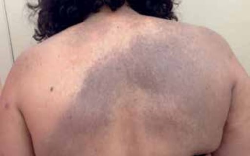
00:01 Welcome. In this talk, we're going to discuss acquired melanocytic nevi to be compared and contrasted with congenital melanocytic nevi, which were done in a previous talk. 00:13 Acquired melanocytic nevi are skin lesions. 00:17 We can call them moles because that's another name for them that arise from the proliferation after birth of melanocytes that have been present since birth. 00:27 So at some point there are acquired mutations that drive a proliferation of those cells. 00:33 Let's discuss the epidemiology. 00:36 It's an incredibly common lesion. 00:38 Acquired melanocytic nevi occur pretty frequently across the entire population. 00:45 The mean number in the white adolescent population, say in America and in Europe, about 15 to 30 such lesions. 00:54 They reach a peak count typically in the second to third decade. 00:57 And then you will see slow regression for the vast majority of them. 01:02 How are these happening? So there is a genetic component. 01:06 There can be activating mutations in Braf, which is a tyrosine kinase, or Nras, which is a intracellular protein that drives the activation and proliferation of cells. Notably, these genetic mutations also underlie congenital melanocytic nevi and can also be driver mutations that occur in melanoma. 01:28 Just again, to alleviate any concern that you may have, since you have all of these nevi on your body, the conversion from a benign acquired melanocytic nevus to anything malignant, such as a melanoma, is exceptionally rare. 01:42 Clearly, sun exposure drives some of this as well. 01:45 That may drive the proliferation and certainly the expression of melanin by the melanocytes. So what's happening that gives rise to these lesions. 01:55 Fundamentally melanocytes are proliferating. 01:58 So we can get a little nodule of them that we can recognize. 02:01 And then there are also migrating from their normal resting spot, which would be at the dermal epidermal junction into the dermis. 02:09 So melanocytes, typically in a normal skin, are scattered, you know, separately along the dermal epidermal junction. 02:16 Single cells in the beginning of a, say, a junctional nevus. 02:22 Then you will get proliferation at that dermoepidermal junction. 02:25 So you'll get a whole a recognizable little nodule of proliferating melanocytes. 02:31 With time, those may migrate and will migrate into the underlying dermis. 02:37 So we'll get a compound nevus where we have proliferated melanocytes both at the dermal epidermal junction and deep within the dermis. 02:47 With time, they tend to accumulate all within the dermis. 02:51 We lose that collection at the dermal epidermal junction, and that will give us a dermal nevus. 02:58 And then remember that these are neural crest derived cells with time. 03:03 And we had also talked about the possibility that these would regress with time as you age that those collections within the dermis, they undergo neuron ization so they will become more neural like and less pigmented. 03:16 And that's how you can have regression of an acquired melanocytic nevi. 03:21 The images, the histologic images are just showing you what a junctional nevus looks like with proliferation of pigmented cells at the dermal epidermal junction. 03:30 And the compound nevus is showing a collection between cells that are present within the dermal epidermal junction and also deeper within the dermis. 03:38 And you can easily pick them out even at this low power because they're pigmented. 03:43 What's the clinical presentation? Well, basically these are small, less than a centimeter in diameter. 03:51 They generally have an even pigmentation, as we'll talk about later when we do melanoma in a different talk. Uneven or kind of kind of a mosaic of pigmentation is typically more suggestive of malignancy. 04:08 So usually the acquired melanocytic nevi are uniformly one color. 04:13 They tend to be round or oval. 04:14 This is because they grew from a single cell that proliferated. 04:18 So they tend to have a very sharply demarcated round or oval shape, and they do tend to be more obvious in sun exposed areas. 04:28 And that's only because in sun exposed areas they become they develop and accumulate more melanin. So they may be more apparent. 04:35 But you can also clearly get the acquired melanocytic nevi anywhere on the body, even in areas not exposed to the sun. 04:44 A typical neoi are a variation on this theme. 04:47 They are often associated with other mutations. 04:50 They will be the topic of topic of another talk later on. 04:54 These are benign for the most part. 04:57 The vast majority never turn into anything more malignant. 05:00 They are associated with an increased number. 05:03 They may have a little bit of more of irregular border. 05:06 That's why they're called atypical. 05:08 And histologically they have some different features. 05:10 So this is a different set of mutations associated with a slightly different sort of lesion, but can look very similar to the more common acquired melanocytic nevus. 05:22 A blue nevus is actually the same entity. 05:26 But because the melanocytes are deep within the dermis the there is scattering of the color. So they go to shorter wavelengths of light because they're deep within the dermis. 05:38 And so they tend to have this bluish coloration. 05:41 But it's the same lesion just deeper. 05:44 They're benign. They're solitary. 05:46 They're uniformly blue black and dome shaped. 05:49 And they typically occur in certain areas, so the acquired melanocytic nevi tend to become more dermal on the scalp, dorsum of the hands and feet, and in the coccygeal area as well as buttocks. 06:05 Spitz nevi are an interesting variant. 06:08 Yet again, these are benign melanocytic proliferations. 06:12 Histologically they can look awful. 06:15 They can resemble melanoma, but spitz nevi epidemiologically occur in childhood, and it's very important that the dermatopathologist be able to distinguish a spitz, which will have a benign behavior, versus a melanoma, which clearly has a very malignant behavior. 06:34 These nevi occurring in kids are commonly located on the face lower extremities, and the melanocytes look bad. 06:43 And we are starting to learn some of the specific genetic variations that occur in occur in a Spitz versus your typical acquired melanocytic nevus, but we don't yet completely understand why these behaves so well when they look so terrible. The diagnosis. 06:59 So this is made largely clinically. 07:02 In most cases. We would never biopsy it. 07:05 But if there's a question of potentially something being malignant then we will do biopsy and excision. And I'm just showing you on the slide on the upper right hand side what a junctional nevus looks like with proliferation of melanocytes both at the dermoepidermal junction and then the slide below where the dermal nevus, where there's all these pigmented melanocytes sitting deep within the dermis. 07:29 And grossly this would have looked like a blue nevus. 07:32 How do we manage this? Mostly we let them be. 07:37 There's clinical observation to make sure that we're not missing malignant transformation, or a malignant cell or a malignant lesion that Spontaneously arose. 07:49 As I had already said previously, the risk of melanoma. 07:52 Is very, very, very low. 07:54 And so, um, we're mainly looking for the. 07:58 For new lesions that arise that might have malignant potential, but not from past lesions. 08:03 When we are worried about them or they're cosmetically undesirable, we can surgically excise them. 08:10 And they're clearly amenable to laser therapy, which basically zaps the proliferating melanocytes. 08:17 So that brings us to the end of our discussion of acquired melanocytic nevi. 08:21 And if you want, go back and compare them with congenital melanocytic nevi, you'll see that there are some common features. 08:29 There are some overlaps, but there is a difference between something you're born with and something you acquire later on. 08:35 In both cases, they're really mostly almost entirely benign. 08:41 And that's the major take home message.
About the Lecture
The lecture Acquired Melanocytic Nevi by Richard Mitchell, MD, PhD is from the course Disorders of Maturation and Pigmentation of the Skin.
Included Quiz Questions
Which sequence correctly describes the progression of acquired melanocytic nevi based on melanocyte migration?
- Junctional nevus → Compound nevus → Dermal nevus
- Dermal nevus → Compound nevus → Junctional nevus
- Compound nevus → Junctional nevus → Dermal nevus
- Dermal nevus → Junctional nevus → Compound nevus
- Junctional nevus → Dermal nevus → Compound nevus
During which age period do acquired melanocytic nevi typically reach their peak count?
- Second to third decade
- First decade
- Fourth to fifth decade
- Sixth to seventh decade
- At birth
Why do blue nevi appear blue-black in color?
- Due to melanocytes being located deep within the dermis causing light scattering
- Due to the presence of blue melanin pigment
- Due to increased blood vessel formation
- Due to inflammation in the dermis
- Due to oxidation of melanin
Which characteristic best describes Spitz nevi?
- Benign childhood lesions that can histologically resemble melanoma
- Malignant lesions that only occur in adults
- Blue-black lesions found on the scalp
- Congenital lesions present at birth
- Acquired lesions that never require biopsy
Author of lecture Acquired Melanocytic Nevi

Richard Mitchell, MD, PhD
Customer reviews
5,0 of 5 stars
| 5 Stars |
|
5 |
| 4 Stars |
|
0 |
| 3 Stars |
|
0 |
| 2 Stars |
|
0 |
| 1 Star |
|
0 |
