Advertisement

Advertisement

Advertisement

Advertisement

Urologic Cancer

Urologic cancer is a broad term that involves cancer of the male and female urinary tracts and male reproductive organs. Risk factors for urologic cancer are smoking Smoking Willful or deliberate act of inhaling and exhaling smoke from burning substances or agents held by hand. Interstitial Lung Diseases; exposure to chemicals such as benzidine and beta-naphthylamine, and arsenic Arsenic A shiny gray element with atomic symbol as, atomic number 33, and atomic weight 75. It occurs throughout the universe, mostly in the form of metallic arsenides. Most forms are toxic. According to the fourth annual report on carcinogens, arsenic and certain arsenic compounds have been listed as known carcinogens. Metal Poisoning (Lead, Arsenic, Iron); genetic predisposition; and chronic irritation of the urinary system. Clinical presentation includes painless hematuria Hematuria Presence of blood in the urine. Renal Cell Carcinoma, flank and/or suprapubic pain Pain An unpleasant sensation induced by noxious stimuli which are detected by nerve endings of nociceptive neurons. Pain: Types and Pathways, dysuria Dysuria Painful urination. It is often associated with infections of the lower urinary tract. Urinary Tract Infections (UTIs), and unexplained significant weight loss Weight loss Decrease in existing body weight. Bariatric Surgery. The gold standard for diagnosis is endoscopy Endoscopy Procedures of applying endoscopes for disease diagnosis and treatment. Endoscopy involves passing an optical instrument through a small incision in the skin i.e., percutaneous; or through a natural orifice and along natural body pathways such as the digestive tract; and/or through an incision in the wall of a tubular structure or organ, i.e. Transluminal, to examine or perform surgery on the interior parts of the body. Gastroesophageal Reflux Disease (GERD) of the urologic structures (cystoscopy, cystourethroscopy, ureteropyeloscopy) with biopsy Biopsy Removal and pathologic examination of specimens from the living body. Ewing Sarcoma. Additional studies include radiologic imaging, which gives information about the tumor Tumor Inflammation invasion and spread of the disease to other sites or organs. Management includes surgery, chemotherapy Chemotherapy Osteosarcoma, radiotherapy, and supportive treatment, depending on the location, extent, and histology.

Last updated: May 17, 2024

Editorial responsibility: Stanley Oiseth, Lindsay Jones, Evelin Maza

Advertisement

Advertisement

Advertisement

Advertisement

Advertisement

Advertisement

Overview

Definition

Growth of abnormal cells from the lining of organs of the male and female urinary tracts and the male reproductive organs.
Epidemiology

  • Bladder Bladder A musculomembranous sac along the urinary tract. Urine flows from the kidneys into the bladder via the ureters, and is held there until urination. Pyelonephritis and Perinephric Abscess cancer is the most common urologic cancer.
  • Incidence Incidence The number of new cases of a given disease during a given period in a specified population. It also is used for the rate at which new events occur in a defined population. It is differentiated from prevalence, which refers to all cases in the population at a given time. Measures of Disease Frequency in men is 4x more than that in women.
  • The incidence Incidence The number of new cases of a given disease during a given period in a specified population. It also is used for the rate at which new events occur in a defined population. It is differentiated from prevalence, which refers to all cases in the population at a given time. Measures of Disease Frequency of bladder Bladder A musculomembranous sac along the urinary tract. Urine flows from the kidneys into the bladder via the ureters, and is held there until urination. Pyelonephritis and Perinephric Abscess cancer increases with age, with a median age at diagnosis of 73 years.

Risk factors

  • Smoking Smoking Willful or deliberate act of inhaling and exhaling smoke from burning substances or agents held by hand. Interstitial Lung Diseases tobacco (most common risk factor for urothelial bladder Bladder A musculomembranous sac along the urinary tract. Urine flows from the kidneys into the bladder via the ureters, and is held there until urination. Pyelonephritis and Perinephric Abscess cancer in the United States)
  • Long-term exposure to chemicals (benzidine, heavy metals, aniline dye, and beta-naphthylamine) 
  • Age > 55 years
  • Chronic urinary tract Urinary tract The urinary tract is located in the abdomen and pelvis and consists of the kidneys, ureters, urinary bladder, and urethra. The structures permit the excretion of urine from the body. Urine flows from the kidneys through the ureters to the urinary bladder and out through the urethra. Urinary Tract: Anatomy inflammation Inflammation Inflammation is a complex set of responses to infection and injury involving leukocytes as the principal cellular mediators in the body’s defense against pathogenic organisms. Inflammation is also seen as a response to tissue injury in the process of wound healing. The 5 cardinal signs of inflammation are pain, heat, redness, swelling, and loss of function. Inflammation due to:
    • Schistosomiasis Schistosomiasis Infection with flukes (trematodes) of the genus schistosoma. Three species produce the most frequent clinical diseases: Schistosoma haematobium (endemic in Africa and the Middle East), Schistosoma Mansoni (in Egypt, northern and southern Africa, some West Indies islands, northern 2/3 of South america), and Schistosoma japonicum (in Japan, China, the Philippines, Celebes, Thailand, Laos). S. mansoni is often seen in Puerto Ricans living in the United States. Schistosoma/Schistosomiasis infection of the bladder Bladder A musculomembranous sac along the urinary tract. Urine flows from the kidneys into the bladder via the ureters, and is held there until urination. Pyelonephritis and Perinephric Abscess
    • Recurrent or chronic urinary tract Urinary tract The urinary tract is located in the abdomen and pelvis and consists of the kidneys, ureters, urinary bladder, and urethra. The structures permit the excretion of urine from the body. Urine flows from the kidneys through the ureters to the urinary bladder and out through the urethra. Urinary Tract: Anatomy infections Infections Invasion of the host organism by microorganisms or their toxins or by parasites that can cause pathological conditions or diseases. Chronic Granulomatous Disease
    • Renal fibrosis Fibrosis Any pathological condition where fibrous connective tissue invades any organ, usually as a consequence of inflammation or other injury. Bronchiolitis Obliterans from Chinese herbs (especially those that contain aristolochic acid), nephropathy
    • Balkan endemic nephropathy (inflammatory process of the renal interstitium noted in the Balkan region)
  • History of exposure to radiation Radiation Emission or propagation of acoustic waves (sound), electromagnetic energy waves (such as light; radio waves; gamma rays; or x-rays), or a stream of subatomic particles (such as electrons; neutrons; protons; or alpha particles). Osteosarcoma in the pelvic area 
  • Previous treatment with cyclophosphamide Cyclophosphamide Precursor of an alkylating nitrogen mustard antineoplastic and immunosuppressive agent that must be activated in the liver to form the active aldophosphamide. It has been used in the treatment of lymphoma and leukemia. Its side effect, alopecia, has been used for defleecing sheep. Cyclophosphamide may also cause sterility, birth defects, mutations, and cancer. Immunosuppressants 
  • Use of phenacetin (analgesic)
  • Arsenic Arsenic A shiny gray element with atomic symbol as, atomic number 33, and atomic weight 75. It occurs throughout the universe, mostly in the form of metallic arsenides. Most forms are toxic. According to the fourth annual report on carcinogens, arsenic and certain arsenic compounds have been listed as known carcinogens. Metal Poisoning (Lead, Arsenic, Iron) in drinking water
  • Genetic predisposition:
    • Mutations in the RB1, TP53, or PTEN gene Gene A category of nucleic acid sequences that function as units of heredity and which code for the basic instructions for the development, reproduction, and maintenance of organisms. Basic Terms of Genetics
    • Lynch syndrome Lynch syndrome Lynch syndrome, also called hereditary non-polyposis colorectal cancer (HNPCC), is the most common inherited colon cancer syndrome, and carries a significantly increased risk for endometrial cancer and other malignancies. Lynch syndrome has an autosomal dominant inheritance pattern involving pathogenic variants in one of the mismatch repair (MMR) genes or epithelial cell adhesion molecule (EpCAM). Lynch syndrome (hereditary nonpolyposis colorectal cancer Colorectal cancer Colorectal cancer (CRC) is the 2nd leading cause of cancer-related deaths in the United States. Colorectal cancer is a heterogeneous disease that arises from genetic and epigenetic abnormalities, with influence from environmental factors. Colorectal Cancer ( HNPCC HNPCC Lynch syndrome, also called hereditary non-polyposis colorectal cancer (HNPCC), is the most common inherited colon cancer syndrome, and carries a significantly increased risk for endometrial cancer and other malignancies. Lynch syndrome has an autosomal dominant inheritance pattern involving pathogenic variants in one of the mismatch repair (MMR) genes or epithelial cell adhesion molecule (EpCAM). Lynch syndrome))
  • Exposure to human papillomavirus Human papillomavirus Human papillomavirus (HPV) is a nonenveloped, circular, double-stranded DNA virus belonging to the Papillomaviridae family. Humans are the only reservoir, and transmission occurs through close skin-to-skin or sexual contact. Human papillomaviruses infect basal epithelial cells and can affect cell-regulatory proteins to result in cell proliferation. Papillomavirus (HPV) (HPV-16)

Types

  • Urinary bladder Urinary Bladder A musculomembranous sac along the urinary tract. Urine flows from the kidneys into the bladder via the ureters (ureter), and is held there until urination. Urinary Tract: Anatomy cancer
  • Renal cell carcinoma Renal cell carcinoma Renal cell carcinoma (RCC) is a tumor that arises from the lining of the renal tubular system within the renal cortex. Renal cell carcinoma is responsible for 80%-85% of all primary renal neoplasms. Most RCCs arise sporadically, but smoking, hypertension, and obesity are linked to its development. Renal Cell Carcinoma 
  • Testicular cancer Testicular cancer Testicular cancer is the most common solid malignancy affecting men 15-35 years of age. Most of the testicular cancers are of the germ cell tumor type, and they can be classified as seminomas and nonseminomas. The most common presentation of testicular cancer is a painless testicular mass. Testicular Cancer
  • Penile cancer Penile cancer Malignant lesions of the penis arise from the squamous epithelium of the glans, prepuce, or penile shaft. Penile cancer is rare in the United States, but there is a higher prevalence in lower socioeconomic regions. The most common histologic subtype is squamous cell carcinoma. Penile Cancer 
  • Urethral cancer 
  • Ureteral cancer 
  • Prostate Prostate The prostate is a gland in the male reproductive system. The gland surrounds the bladder neck and a portion of the urethra. The prostate is an exocrine gland that produces a weakly acidic secretion, which accounts for roughly 20% of the seminal fluid. cancer

Bladder Cancer

Histologic types

  • Urothelial carcinoma:
    • Previously known as transitional cell carcinoma
    • Urothelial cells physiologically expand and contract when the bladder Bladder A musculomembranous sac along the urinary tract. Urine flows from the kidneys into the bladder via the ureters, and is held there until urination. Pyelonephritis and Perinephric Abscess is full and empty, respectively.
    • Most common type of bladder Bladder A musculomembranous sac along the urinary tract. Urine flows from the kidneys into the bladder via the ureters, and is held there until urination. Pyelonephritis and Perinephric Abscess cancer in the United States
  • Squamous cell carcinoma Squamous cell carcinoma Cutaneous squamous cell carcinoma (cSCC) is caused by malignant proliferation of atypical keratinocytes. This condition is the 2nd most common skin malignancy and usually affects sun-exposed areas of fair-skinned patients. The cancer presents as a firm, erythematous, keratotic plaque or papule. Squamous Cell Carcinoma (SCC):
    • Associated with chronic bladder Bladder A musculomembranous sac along the urinary tract. Urine flows from the kidneys into the bladder via the ureters, and is held there until urination. Pyelonephritis and Perinephric Abscess irritation
    • Commonly occurs in countries where the incidence Incidence The number of new cases of a given disease during a given period in a specified population. It also is used for the rate at which new events occur in a defined population. It is differentiated from prevalence, which refers to all cases in the population at a given time. Measures of Disease Frequency of schistosomiasis Schistosomiasis Infection with flukes (trematodes) of the genus schistosoma. Three species produce the most frequent clinical diseases: Schistosoma haematobium (endemic in Africa and the Middle East), Schistosoma Mansoni (in Egypt, northern and southern Africa, some West Indies islands, northern 2/3 of South america), and Schistosoma japonicum (in Japan, China, the Philippines, Celebes, Thailand, Laos). S. mansoni is often seen in Puerto Ricans living in the United States. Schistosoma/Schistosomiasis is high 
  • Adenocarcinoma:
    • Occurs in mucus-secreting cells of the bladder Bladder A musculomembranous sac along the urinary tract. Urine flows from the kidneys into the bladder via the ureters, and is held there until urination. Pyelonephritis and Perinephric Abscess
    • Rare type

Clinical presentation

  • Painless gross hematuria Hematuria Presence of blood in the urine. Renal Cell Carcinoma (classic presentation), which can differ depending on site of lesion(s):
    • Terminal hematuria Hematuria Presence of blood in the urine. Renal Cell Carcinoma:
    • Blood in the beginning of urination: typically from the urethra Urethra A tube that transports urine from the urinary bladder to the outside of the body in both the sexes. It also has a reproductive function in the male by providing a passage for sperm. Urinary Tract: Anatomy
    • Hematuria Hematuria Presence of blood in the urine. Renal Cell Carcinoma throughout urination: can be from the kidneys Kidneys The kidneys are a pair of bean-shaped organs located retroperitoneally against the posterior wall of the abdomen on either side of the spine. As part of the urinary tract, the kidneys are responsible for blood filtration and excretion of water-soluble waste in the urine. Kidneys: Anatomy, ureter, and/or bladder Bladder A musculomembranous sac along the urinary tract. Urine flows from the kidneys into the bladder via the ureters, and is held there until urination. Pyelonephritis and Perinephric Abscess
  • Dysuria Dysuria Painful urination. It is often associated with infections of the lower urinary tract. Urinary Tract Infections (UTIs) 
  • Urinary frequency
  • Urge incontinence Urge incontinence Involuntary discharge of urine that is associated with an abrupt and strong desire to void. It is usually related to the involuntary contractions of the detrusor muscle of the bladder (detrusor hyperreflexia or detrusor instability). Urinary Incontinence 
  • Back pain Pain An unpleasant sensation induced by noxious stimuli which are detected by nerve endings of nociceptive neurons. Pain: Types and Pathways
  • Flank pain Flank pain Pain emanating from below the ribs and above the ilium. Renal Cell Carcinoma 
  • Suprapubic pain Pain An unpleasant sensation induced by noxious stimuli which are detected by nerve endings of nociceptive neurons. Pain: Types and Pathways 
  • Fatigue Fatigue The state of weariness following a period of exertion, mental or physical, characterized by a decreased capacity for work and reduced efficiency to respond to stimuli. Fibromyalgia 
  • Unexplained significant weight loss Weight loss Decrease in existing body weight. Bariatric Surgery

Diagnosis

  • History and physical examination:
    • Solid pelvic mass Mass Three-dimensional lesion that occupies a space within the breast Imaging of the Breast on abdominal examination Abdominal examination The abdominal examination is the portion of the physical exam evaluating the abdomen for signs of disease. The abdominal examination consists of inspection, auscultation, percussion, and palpation. Abdominal Examination (late sign)
    • Enlarged lymph nodes Lymph Nodes They are oval or bean shaped bodies (1 – 30 mm in diameter) located along the lymphatic system. Lymphatic Drainage System: Anatomy on examination
    • History of chronic bladder Bladder A musculomembranous sac along the urinary tract. Urine flows from the kidneys into the bladder via the ureters, and is held there until urination. Pyelonephritis and Perinephric Abscess irritation
  • Urinalysis Urinalysis Examination of urine by chemical, physical, or microscopic means. Routine urinalysis usually includes performing chemical screening tests, determining specific gravity, observing any unusual color or odor, screening for bacteriuria, and examining the sediment microscopically. Urinary Tract Infections (UTIs) in Children:
    • Indicated for any presentation of hematuria Hematuria Presence of blood in the urine. Renal Cell Carcinoma: considered significant if > 3 RBCs RBCs Erythrocytes, or red blood cells (RBCs), are the most abundant cells in the blood. While erythrocytes in the fetus are initially produced in the yolk sac then the liver, the bone marrow eventually becomes the main site of production. Erythrocytes: Histology per high-power field (HPF)
    • RBC morphology:
      • Dysmorphic or formed in casts (cylindrical or tubular particles): likely intrinsic renal disease
      • Normally shaped RBCs RBCs Erythrocytes, or red blood cells (RBCs), are the most abundant cells in the blood. While erythrocytes in the fetus are initially produced in the yolk sac then the liver, the bone marrow eventually becomes the main site of production. Erythrocytes: Histology: infection, tumor Tumor Inflammation, calculi, or obstruction
  • Urine cytology:
    • A urine sample is collected and observed under a microscope to detect cancer cells. 
    • Has poor sensitivity 
  • Cystoscopy + biopsy Biopsy Removal and pathologic examination of specimens from the living body. Ewing Sarcoma:
    • Gold standard for initial diagnosis and staging Staging Methods which attempt to express in replicable terms the extent of the neoplasm in the patient. Grading, Staging, and Metastasis of bladder Bladder A musculomembranous sac along the urinary tract. Urine flows from the kidneys into the bladder via the ureters, and is held there until urination. Pyelonephritis and Perinephric Abscess cancer 
    • Visible tumors or suspicious lesions are biopsied or resected transurethrally. 
    • Fluorescence cystoscopy: photoactive protoporphyrin accumulates in the neoplastic tissue.  
  • Urine-based tumor Tumor Inflammation markers:
    • A noninvasive diagnostic tool
    • Based on expressed tumor-related proteins Proteins Linear polypeptides that are synthesized on ribosomes and may be further modified, crosslinked, cleaved, or assembled into complex proteins with several subunits. The specific sequence of amino acids determines the shape the polypeptide will take, during protein folding, and the function of the protein. Energy Homeostasis, DNA DNA A deoxyribonucleotide polymer that is the primary genetic material of all cells. Eukaryotic and prokaryotic organisms normally contain DNA in a double-stranded state, yet several important biological processes transiently involve single-stranded regions. DNA, which consists of a polysugar-phosphate backbone possessing projections of purines (adenine and guanine) and pyrimidines (thymine and cytosine), forms a double helix that is held together by hydrogen bonds between these purines and pyrimidines (adenine to thymine and guanine to cytosine). DNA Types and Structure, RNA RNA A polynucleotide consisting essentially of chains with a repeating backbone of phosphate and ribose units to which nitrogenous bases are attached. RNA is unique among biological macromolecules in that it can encode genetic information, serve as an abundant structural component of cells, and also possesses catalytic activity. RNA Types and Structure, methylation Methylation Addition of methyl groups. In histo-chemistry methylation is used to esterify carboxyl groups and remove sulfate groups by treating tissue sections with hot methanol in the presence of hydrochloric acid. . Glucocorticoids changes, or cellular markers
    • More sensitive than urine cytology, but not sensitive enough to replace cystoscopy
  • Imaging:
  • For metastasis Metastasis The transfer of a neoplasm from one organ or part of the body to another remote from the primary site. Grading, Staging, and Metastasis screening Screening Preoperative Care:
    • Chest x-ray X-ray Penetrating electromagnetic radiation emitted when the inner orbital electrons of an atom are excited and release radiant energy. X-ray wavelengths range from 1 pm to 10 nm. Hard x-rays are the higher energy, shorter wavelength x-rays. Soft x-rays or grenz rays are less energetic and longer in wavelength. The short wavelength end of the x-ray spectrum overlaps the gamma rays wavelength range. The distinction between gamma rays and x-rays is based on their radiation source. Pulmonary Function Tests: sensitive for lesions ≥ 1 cm 
    • CT is more sensitive than chest x-ray X-ray Penetrating electromagnetic radiation emitted when the inner orbital electrons of an atom are excited and release radiant energy. X-ray wavelengths range from 1 pm to 10 nm. Hard x-rays are the higher energy, shorter wavelength x-rays. Soft x-rays or grenz rays are less energetic and longer in wavelength. The short wavelength end of the x-ray spectrum overlaps the gamma rays wavelength range. The distinction between gamma rays and x-rays is based on their radiation source. Pulmonary Function Tests
    • Bone scan Bone Scan Osteosarcoma:
      • Used to detect bone Bone Bone is a compact type of hardened connective tissue composed of bone cells, membranes, an extracellular mineralized matrix, and central bone marrow. The 2 primary types of bone are compact and spongy. Bones: Structure and Types metastases 
      • Recommended in cases with elevated alkaline phosphatase Alkaline Phosphatase An enzyme that catalyzes the conversion of an orthophosphoric monoester and water to an alcohol and orthophosphate. Osteosarcoma

Stages of bladder Bladder A musculomembranous sac along the urinary tract. Urine flows from the kidneys into the bladder via the ureters, and is held there until urination. Pyelonephritis and Perinephric Abscess cancer

Bladder Bladder A musculomembranous sac along the urinary tract. Urine flows from the kidneys into the bladder via the ureters, and is held there until urination. Pyelonephritis and Perinephric Abscess cancer pathologic stages are based on the TNM staging system TNM staging system Grading, Staging, and Metastasis.

Table: Primary tumor Tumor Inflammation
Tumor Tumor Inflammation (T)
category
Description
Tx Primary tumor Tumor Inflammation cannot be assessed.
T0 No evidence of primary tumor Tumor Inflammation
Ta TA Thyrotoxicosis and Hyperthyroidism Noninvasive papillary or exophytic Exophytic Retinoblastoma lesions
Tis Carcinoma in situ (“flat tumor Tumor Inflammation”)
T1 Tumor Tumor Inflammation invades lamina propria Lamina propria Whipple’s Disease (or submucosa).
T2 Tumor Tumor Inflammation invades muscularis propria.
T2a: Tumor Tumor Inflammation invades superficial muscularis propria (inner half).
T2b: Tumor Tumor Inflammation invades deep muscularis propria (outer half).
T3 T3 A T3 thyroid hormone normally synthesized and secreted by the thyroid gland in much smaller quantities than thyroxine (T4). Most T3 is derived from peripheral monodeiodination of T4 at the 5′ position of the outer ring of the iodothyronine nucleus. The hormone finally delivered and used by the tissues is mainly t3. Thyroid Hormones Tumor Tumor Inflammation invades perivesical fat.
T3a: Microscopic invasion
T3b: Macroscopic (extravesical mass Mass Three-dimensional lesion that occupies a space within the breast Imaging of the Breast) invasion
T4 T4 The major hormone derived from the thyroid gland. Thyroxine is synthesized via the iodination of tyrosines (monoiodotyrosine) and the coupling of iodotyrosines (diiodotyrosine) in the thyroglobulin. Thyroxine is released from thyroglobulin by proteolysis and secreted into the blood. Thyroxine is peripherally deiodinated to form triiodothyronine which exerts a broad spectrum of stimulatory effects on cell metabolism. Thyroid Hormones Tumor Tumor Inflammation invades 1 of the following: prostatic stroma, seminal vesicles Vesicles Female Genitourinary Examination, uterus Uterus The uterus, cervix, and fallopian tubes are part of the internal female reproductive system. The uterus has a thick wall made of smooth muscle (the myometrium) and an inner mucosal layer (the endometrium). The most inferior portion of the uterus is the cervix, which connects the uterine cavity to the vagina. Uterus, Cervix, and Fallopian Tubes: Anatomy, vagina Vagina The vagina is the female genital canal, extending from the vulva externally to the cervix uteri internally. The structures have sexual, reproductive, and urinary functions and a rich blood supply, mainly arising from the internal iliac artery. Vagina, Vulva, and Pelvic Floor: Anatomy, pelvic wall, abdominal wall Abdominal wall The outer margins of the abdomen, extending from the osteocartilaginous thoracic cage to the pelvis. Though its major part is muscular, the abdominal wall consists of at least seven layers: the skin, subcutaneous fat, deep fascia; abdominal muscles, transversalis fascia, extraperitoneal fat, and the parietal peritoneum. Surgical Anatomy of the Abdomen.
T4a: Tumor Tumor Inflammation invades prostatic stroma, uterus Uterus The uterus, cervix, and fallopian tubes are part of the internal female reproductive system. The uterus has a thick wall made of smooth muscle (the myometrium) and an inner mucosal layer (the endometrium). The most inferior portion of the uterus is the cervix, which connects the uterine cavity to the vagina. Uterus, Cervix, and Fallopian Tubes: Anatomy, vagina Vagina The vagina is the female genital canal, extending from the vulva externally to the cervix uteri internally. The structures have sexual, reproductive, and urinary functions and a rich blood supply, mainly arising from the internal iliac artery. Vagina, Vulva, and Pelvic Floor: Anatomy (adjacent organs).
T4b: Tumor Tumor Inflammation invades pelvic wall, abdominal wall Abdominal wall The outer margins of the abdomen, extending from the osteocartilaginous thoracic cage to the pelvis. Though its major part is muscular, the abdominal wall consists of at least seven layers: the skin, subcutaneous fat, deep fascia; abdominal muscles, transversalis fascia, extraperitoneal fat, and the parietal peritoneum. Surgical Anatomy of the Abdomen, or other organs.
Table: Regional lymph nodes Lymph Nodes They are oval or bean shaped bodies (1 – 30 mm in diameter) located along the lymphatic system. Lymphatic Drainage System: Anatomy
Node (N) category Description
Nx Lymph nodes Lymph Nodes They are oval or bean shaped bodies (1 – 30 mm in diameter) located along the lymphatic system. Lymphatic Drainage System: Anatomy cannot be assessed.
N0 No lymph Lymph The interstitial fluid that is in the lymphatic system. Secondary Lymphatic Organs node metastasis Metastasis The transfer of a neoplasm from one organ or part of the body to another remote from the primary site. Grading, Staging, and Metastasis
N1 Single regional lymph Lymph The interstitial fluid that is in the lymphatic system. Secondary Lymphatic Organs node metastasis Metastasis The transfer of a neoplasm from one organ or part of the body to another remote from the primary site. Grading, Staging, and Metastasis in the true pelvis Pelvis The pelvis consists of the bony pelvic girdle, the muscular and ligamentous pelvic floor, and the pelvic cavity, which contains viscera, vessels, and multiple nerves and muscles. The pelvic girdle, composed of 2 “hip” bones and the sacrum, is a ring-like bony structure of the axial skeleton that links the vertebral column with the lower extremities. Pelvis: Anatomy (perivesical, obturator, internal and external iliac, or sacral lymph nodes Lymph Nodes They are oval or bean shaped bodies (1 – 30 mm in diameter) located along the lymphatic system. Lymphatic Drainage System: Anatomy)
N2 Multiple regional lymph Lymph The interstitial fluid that is in the lymphatic system. Secondary Lymphatic Organs node metastases in the true pelvis Pelvis The pelvis consists of the bony pelvic girdle, the muscular and ligamentous pelvic floor, and the pelvic cavity, which contains viscera, vessels, and multiple nerves and muscles. The pelvic girdle, composed of 2 “hip” bones and the sacrum, is a ring-like bony structure of the axial skeleton that links the vertebral column with the lower extremities. Pelvis: Anatomy (perivesical, obturator, internal and external iliac, or sacral lymph Lymph The interstitial fluid that is in the lymphatic system. Secondary Lymphatic Organs node metastasis Metastasis The transfer of a neoplasm from one organ or part of the body to another remote from the primary site. Grading, Staging, and Metastasis)
N3 Metastasis Metastasis The transfer of a neoplasm from one organ or part of the body to another remote from the primary site. Grading, Staging, and Metastasis to the common iliac lymph nodes Lymph Nodes They are oval or bean shaped bodies (1 – 30 mm in diameter) located along the lymphatic system. Lymphatic Drainage System: Anatomy
Table: Distant metastasis Metastasis The transfer of a neoplasm from one organ or part of the body to another remote from the primary site. Grading, Staging, and Metastasis
Metastasis Metastasis The transfer of a neoplasm from one organ or part of the body to another remote from the primary site. Grading, Staging, and Metastasis (M) category Description
M0 No distant metastasis Metastasis The transfer of a neoplasm from one organ or part of the body to another remote from the primary site. Grading, Staging, and Metastasis
M1 Distant metastasis Metastasis The transfer of a neoplasm from one organ or part of the body to another remote from the primary site. Grading, Staging, and Metastasis
M1a: distant metastasis Metastasis The transfer of a neoplasm from one organ or part of the body to another remote from the primary site. Grading, Staging, and Metastasis limited to lymph nodes Lymph Nodes They are oval or bean shaped bodies (1 – 30 mm in diameter) located along the lymphatic system. Lymphatic Drainage System: Anatomy beyond the common iliac nodes
M1b: non– lymph Lymph The interstitial fluid that is in the lymphatic system. Secondary Lymphatic Organs node distant metastasis Metastasis The transfer of a neoplasm from one organ or part of the body to another remote from the primary site. Grading, Staging, and Metastasis
Table: Stage/prognostic groups
Stage T N M
Stage 0a Ta TA Thyrotoxicosis and Hyperthyroidism N0 M0
Stage 0is Tis N0 M0
Stage I T1 N0 M0
Stage II T2a N0 M0
T2b N0 M0
Stage IIIA T3a, T3b, T4a N0 M0
Stage IIIA T1–T4a N1 M0
Stage IIIB T1–T4a N2, N3 M0
Stage IVA T4b Any N M0
Stage IVA Any T Any N M1a
Stage IVB Any T Any N M1b

Management

  • Lifestyle changes:
    • Cessation of smoking Smoking Willful or deliberate act of inhaling and exhaling smoke from burning substances or agents held by hand. Interstitial Lung Diseases
    • Increased intake of water 
    • Multivitamin supplementation 
    • Adequate exercise and dietary changes 
  • Treatment for bladder Bladder A musculomembranous sac along the urinary tract. Urine flows from the kidneys into the bladder via the ureters, and is held there until urination. Pyelonephritis and Perinephric Abscess cancer depends on the stage of the tumor Tumor Inflammation:
  • Transurethral resection of bladder Bladder A musculomembranous sac along the urinary tract. Urine flows from the kidneys into the bladder via the ureters, and is held there until urination. Pyelonephritis and Perinephric Abscess tumor Tumor Inflammation
  • Potential adjuvant Adjuvant Substances that augment, stimulate, activate, potentiate, or modulate the immune response at either the cellular or humoral level. The classical agents (freund’s adjuvant, bcg, corynebacterium parvum, et al.) contain bacterial antigens. Some are endogenous (e.g., histamine, interferon, transfer factor, tuftsin, interleukin-1). Their mode of action is either non-specific, resulting in increased immune responsiveness to a wide variety of antigens, or antigen-specific, i.e., affecting a restricted type of immune response to a narrow group of antigens. The therapeutic efficacy of many biological response modifiers is related to their antigen-specific immunoadjuvanticity. Vaccination intravesical therapy to decrease risk of recurrence (dependent on risk)
  • Intravesical bacille Calmette-Guérin ( BCG BCG An active immunizing agent and a viable avirulent attenuated strain of Mycobacterium bovis, which confers immunity to mycobacterial infections. It is used also in immunotherapy of neoplasms due to its stimulation of antibodies and non-specific immunity. Cancer Immunotherapy; live attenuated form of Mycobacterium bovis Mycobacterium bovis The bovine variety of the tubercle Bacillus. It is called also Mycobacterium tuberculosis var. Bovis. Mycobacterium) is used for high-risk disease.
  • Other intravesical agents: gemcitabine Gemcitabine Antimetabolite Chemotherapy, mitomycin Mitomycin A group of methylazirinopyrroloindolediones obtained from certain streptomyces strains. They are very toxic antibiotics used as antineoplastic agents in some solid tumors. Porfiromycin and mitomycin are the most useful members of the group. Antitumor Antibiotics, epirubicin Epirubicin An anthracycline which is the 4′-epi-isomer of doxorubicin. The compound exerts its antitumor effects by interference with the synthesis and function of DNA. Antitumor Antibiotics
  • Muscle invasive disease (any lymph Lymph The interstitial fluid that is in the lymphatic system. Secondary Lymphatic Organs node status but no metastasis Metastasis The transfer of a neoplasm from one organ or part of the body to another remote from the primary site. Grading, Staging, and Metastasis (M0)):
    • Radical cystectomy (requires urinary diversion): removes the bladder Bladder A musculomembranous sac along the urinary tract. Urine flows from the kidneys into the bladder via the ureters, and is held there until urination. Pyelonephritis and Perinephric Abscess, adjacent organs ( prostate Prostate The prostate is a gland in the male reproductive system. The gland surrounds the bladder neck and a portion of the urethra. The prostate is an exocrine gland that produces a weakly acidic secretion, which accounts for roughly 20% of the seminal fluid. , seminal vesicles Vesicles Female Genitourinary Examination, uterus Uterus The uterus, cervix, and fallopian tubes are part of the internal female reproductive system. The uterus has a thick wall made of smooth muscle (the myometrium) and an inner mucosal layer (the endometrium). The most inferior portion of the uterus is the cervix, which connects the uterine cavity to the vagina. Uterus, Cervix, and Fallopian Tubes: Anatomy, ovaries Ovaries Ovaries are the paired gonads of the female reproductive system that contain haploid gametes known as oocytes. The ovaries are located intraperitoneally in the pelvis, just posterior to the broad ligament, and are connected to the pelvic sidewall and to the uterus by ligaments. These organs function to secrete hormones (estrogen and progesterone) and to produce the female germ cells (oocytes). Ovaries: Anatomy) and regional lymph nodes Lymph Nodes They are oval or bean shaped bodies (1 – 30 mm in diameter) located along the lymphatic system. Lymphatic Drainage System: Anatomy
    • Neoadjuvant chemotherapy Neoadjuvant Chemotherapy Osteosarcoma
    • Trimodal therapy (transurethral resection of bladder Bladder A musculomembranous sac along the urinary tract. Urine flows from the kidneys into the bladder via the ureters, and is held there until urination. Pyelonephritis and Perinephric Abscess tumor Tumor Inflammation, radiation Radiation Emission or propagation of acoustic waves (sound), electromagnetic energy waves (such as light; radio waves; gamma rays; or x-rays), or a stream of subatomic particles (such as electrons; neutrons; protons; or alpha particles). Osteosarcoma, and chemotherapy Chemotherapy Osteosarcoma): preserves the bladder Bladder A musculomembranous sac along the urinary tract. Urine flows from the kidneys into the bladder via the ureters, and is held there until urination. Pyelonephritis and Perinephric Abscess for those who are not candidates for cystectomy
  • Metastasis Metastasis The transfer of a neoplasm from one organ or part of the body to another remote from the primary site. Grading, Staging, and Metastasis present (M1):
    • Chemotherapy Chemotherapy Osteosarcoma (platinum-based)
    • Checkpoint inhibitor immunotherapy targeting programmed cell death-1 protein ( PD-1 PD-1 An inhibitory t-lymphocyte receptor that has specificity for CD274 antigen and programmed cell death 1 ligand 2 protein. Signaling by the receptor limits T cell proliferation and interferon gamma synthesis. The receptor also may play an essential role in the regulatory pathway that induces peripheral tolerance. T cells: Types and Functions) or its ligand (PD-L1)
    • Palliative care
  • Surveillance Surveillance Developmental Milestones and Normal Growth:
    • Second primary tumors can arise anywhere in the genitourinary tract.
    • Cystoscopy and urine cytology performed every 3 months in the 1st 2 years.
    • Frequency subsequently decreases.

Prognosis Prognosis A prediction of the probable outcome of a disease based on a individual’s condition and the usual course of the disease as seen in similar situations. Non-Hodgkin Lymphomas

  • The recurrence rate for urothelial carcinoma is high, with 80% of cases having ≥ 1 recurrence. 
  • The 5-year survival rate:
    • CIS CIS Multiple Sclerosis/minimal muscle invasion (T1): 82%–100%
    • Muscle invasion ( T3 T3 A T3 thyroid hormone normally synthesized and secreted by the thyroid gland in much smaller quantities than thyroxine (T4). Most T3 is derived from peripheral monodeiodination of T4 at the 5′ position of the outer ring of the iodothyronine nucleus. The hormone finally delivered and used by the tissues is mainly t3. Thyroid Hormones onward): up to 71%
    • T4 T4 The major hormone derived from the thyroid gland. Thyroxine is synthesized via the iodination of tyrosines (monoiodotyrosine) and the coupling of iodotyrosines (diiodotyrosine) in the thyroglobulin. Thyroxine is released from thyroglobulin by proteolysis and secreted into the blood. Thyroxine is peripherally deiodinated to form triiodothyronine which exerts a broad spectrum of stimulatory effects on cell metabolism. Thyroid Hormones: up to 22%
  • Risk factors for recurrence and progression are:
    • Female sex Sex The totality of characteristics of reproductive structure, functions, phenotype, and genotype, differentiating the male from the female organism. Gender Dysphoria
    • Large tumor Tumor Inflammation size
    • Multifocality 
    • Advanced stage 
    • Presence of CIS CIS Multiple Sclerosis

Ureteral Cancer

Definition

Abnormal growth of cells in the inner lining of the ureter/ ureters Ureters One of a pair of thick-walled tubes that transports urine from the kidney pelvis to the urinary bladder. Urinary Tract: Anatomy, of which > 90% are urothelial carcinomas.
Clinical presentation

  • Ureteral cancer is usually asymptomatic in the initial stages, with clinical presentation as follows:
  • On presentation, up to 17% have bladder Bladder A musculomembranous sac along the urinary tract. Urine flows from the kidneys into the bladder via the ureters, and is held there until urination. Pyelonephritis and Perinephric Abscess cancer as well.
  • Because the entire urothelial surface is exposed to carcinogenic alteration ( field cancerization Field Cancerization Actinic Keratosis), multifocality can be seen.

Diagnosis

  • History and physical examination:
    • Presenting signs and symptoms
    • Exposure history
    • History of cancer or genetic predisposition 
    • Generally, exam is unremarkable, but in rare cases, a flank mass Mass Three-dimensional lesion that occupies a space within the breast Imaging of the Breast may be found. 
  • Urinalysis Urinalysis Examination of urine by chemical, physical, or microscopic means. Routine urinalysis usually includes performing chemical screening tests, determining specific gravity, observing any unusual color or odor, screening for bacteriuria, and examining the sediment microscopically. Urinary Tract Infections (UTIs) in Children: evaluate hematuria Hematuria Presence of blood in the urine. Renal Cell Carcinoma
  • Urine cytology:
    • Generally negative in low-grade urothelial cancers of the upper tract
    • ↑ Chance of positive cytology in higher-grade lesions
    • Positive cytology + negative cystoscopy/ biopsy Biopsy Removal and pathologic examination of specimens from the living body. Ewing Sarcoma → must evaluate the upper tracts and prostatic urethra Prostatic urethra
  • Radiologic imaging:
    • Imaging is used to identify the tumor Tumor Inflammation (usually seen as a filling defect) and to assess the extent of the cancer.
    • CT urography provides better visualization than IVP. 
    • MRI is an alternative (but gadolinium Gadolinium An element of the rare earth family of metals. It has the atomic symbol gd, atomic number 64, and atomic weight 157. 25. Its oxide is used in the control rods of some nuclear reactors. Magnetic Resonance Imaging (MRI) is contraindicated in impaired renal function, which carries the risk of development of nephrogenic systemic fibrosis Fibrosis Any pathological condition where fibrous connective tissue invades any organ, usually as a consequence of inflammation or other injury. Bronchiolitis Obliterans).
  • Ureteropyeloscopy with cystoscopy:
    • Visualization of the entire collecting system with a camera
    • The scope passes the urethra Urethra A tube that transports urine from the urinary bladder to the outside of the body in both the sexes. It also has a reproductive function in the male by providing a passage for sperm. Urinary Tract: Anatomy and bladder Bladder A musculomembranous sac along the urinary tract. Urine flows from the kidneys into the bladder via the ureters, and is held there until urination. Pyelonephritis and Perinephric Abscess into the ureters Ureters One of a pair of thick-walled tubes that transports urine from the kidney pelvis to the urinary bladder. Urinary Tract: Anatomy
    • Biopsy Biopsy Removal and pathologic examination of specimens from the living body. Ewing Sarcoma of the mass Mass Three-dimensional lesion that occupies a space within the breast Imaging of the Breast can be taken.
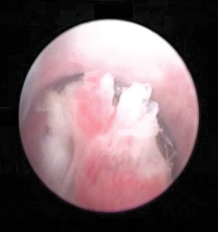
Ureteral tumor

Ureteral tumor noted on endoscopic examination of the ureter.

Image: “Ureteral tumour with elective indication for endoscopic treatment” by Niţă G, Georgescu D, Mulţescu R, Draguţescu M, Mihai B, Geavlete B, Persu C, Geavlete P. License: CC BY 2.0

Stages of ureteral cancer

Staging Staging Methods which attempt to express in replicable terms the extent of the neoplasm in the patient. Grading, Staging, and Metastasis applies to cancer involving the renal pelvis Renal pelvis Kidneys: Anatomy and ureter.

Table: Primary tumor Tumor Inflammation
T category Description
TX Primary tumor Tumor Inflammation cannot be assessed.
T0 No evidence of primary tumor Tumor Inflammation
Ta TA Thyrotoxicosis and Hyperthyroidism Noninvasive papillary or exophytic Exophytic Retinoblastoma lesions
Tis CIS CIS Multiple Sclerosis
T1 Tumor Tumor Inflammation invasion of subepithelial Subepithelial Membranoproliferative Glomerulonephritis connective tissue Connective tissue Connective tissues originate from embryonic mesenchyme and are present throughout the body except inside the brain and spinal cord. The main function of connective tissues is to provide structural support to organs. Connective tissues consist of cells and an extracellular matrix. Connective Tissue: Histology
T2 Tumor Tumor Inflammation invasion of muscularis
T3 T3 A T3 thyroid hormone normally synthesized and secreted by the thyroid gland in much smaller quantities than thyroxine (T4). Most T3 is derived from peripheral monodeiodination of T4 at the 5′ position of the outer ring of the iodothyronine nucleus. The hormone finally delivered and used by the tissues is mainly t3. Thyroid Hormones For ureter only: invasion beyond muscularis into periureteric fat. For renal pelvis Renal pelvis Kidneys: Anatomy only: invasion beyond muscularis into peripelvic fat or into the renal parenchyma
T4 T4 The major hormone derived from the thyroid gland. Thyroxine is synthesized via the iodination of tyrosines (monoiodotyrosine) and the coupling of iodotyrosines (diiodotyrosine) in the thyroglobulin. Thyroxine is released from thyroglobulin by proteolysis and secreted into the blood. Thyroxine is peripherally deiodinated to form triiodothyronine which exerts a broad spectrum of stimulatory effects on cell metabolism. Thyroid Hormones Tumor Tumor Inflammation invasion of adjacent organs or the kidney into the perinephric fat
Table: Regional lymph nodes Lymph Nodes They are oval or bean shaped bodies (1 – 30 mm in diameter) located along the lymphatic system. Lymphatic Drainage System: Anatomy
Node category Description
NX Regional lymph nodes Lymph Nodes They are oval or bean shaped bodies (1 – 30 mm in diameter) located along the lymphatic system. Lymphatic Drainage System: Anatomy cannot be assessed.
N0 No lymph Lymph The interstitial fluid that is in the lymphatic system. Secondary Lymphatic Organs node metastasis Metastasis The transfer of a neoplasm from one organ or part of the body to another remote from the primary site. Grading, Staging, and Metastasis
N1 Metastasis Metastasis The transfer of a neoplasm from one organ or part of the body to another remote from the primary site. Grading, Staging, and Metastasis ≤ 2 cm (single lymph Lymph The interstitial fluid that is in the lymphatic system. Secondary Lymphatic Organs node)
N2 Metastasis Metastasis The transfer of a neoplasm from one organ or part of the body to another remote from the primary site. Grading, Staging, and Metastasis > 2 cm (single lymph Lymph The interstitial fluid that is in the lymphatic system. Secondary Lymphatic Organs node or multiple lymph nodes Lymph Nodes They are oval or bean shaped bodies (1 – 30 mm in diameter) located along the lymphatic system. Lymphatic Drainage System: Anatomy)
Table: Metastasis Metastasis The transfer of a neoplasm from one organ or part of the body to another remote from the primary site. Grading, Staging, and Metastasis
M category Description
M0 No distant metastasis Metastasis The transfer of a neoplasm from one organ or part of the body to another remote from the primary site. Grading, Staging, and Metastasis
M1 Distant metastasis Metastasis The transfer of a neoplasm from one organ or part of the body to another remote from the primary site. Grading, Staging, and Metastasis
Table: Stage/prognostic groups
Stage T N M
Stage 0a Ta TA Thyrotoxicosis and Hyperthyroidism N0 M0
Stage 0is Tis N0 M0
Stage I T1 N0 M0
Stage II T2 N0 M0
Stage III T3 T3 A T3 thyroid hormone normally synthesized and secreted by the thyroid gland in much smaller quantities than thyroxine (T4). Most T3 is derived from peripheral monodeiodination of T4 at the 5′ position of the outer ring of the iodothyronine nucleus. The hormone finally delivered and used by the tissues is mainly t3. Thyroid Hormones N0 M0
Stage IV T4 T4 The major hormone derived from the thyroid gland. Thyroxine is synthesized via the iodination of tyrosines (monoiodotyrosine) and the coupling of iodotyrosines (diiodotyrosine) in the thyroglobulin. Thyroxine is released from thyroglobulin by proteolysis and secreted into the blood. Thyroxine is peripherally deiodinated to form triiodothyronine which exerts a broad spectrum of stimulatory effects on cell metabolism. Thyroid Hormones NX, N0 M0
Any T N1, N2 M0
Any T Any N M1

Management

Treatment of ureteral cancer depends on the site, size, and extent of cancer, with surgery providing curative treatment. 
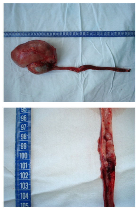
  • Localized disease:
    • Nephroureterectomy (laparoscopic or open)
      • Removal of the ureter and kidney reduces risk of local recurrence.
      • Surgery includes excision of the cuff of the bladder Bladder A musculomembranous sac along the urinary tract. Urine flows from the kidneys into the bladder via the ureters, and is held there until urination. Pyelonephritis and Perinephric Abscess and bladder Bladder A musculomembranous sac along the urinary tract. Urine flows from the kidneys into the bladder via the ureters, and is held there until urination. Pyelonephritis and Perinephric Abscess mucosa.
      • Lymph Lymph The interstitial fluid that is in the lymphatic system. Secondary Lymphatic Organs node dissection is performed in high-grade lesions.
    • Kidney-sparing surgery:
      • Considered in low-risk lesions
      • Also a consideration in solitary kidney or impaired renal function
    • Chemotherapy Chemotherapy Osteosarcoma:
      • Adjuvant Adjuvant Substances that augment, stimulate, activate, potentiate, or modulate the immune response at either the cellular or humoral level. The classical agents (freund’s adjuvant, bcg, corynebacterium parvum, et al.) contain bacterial antigens. Some are endogenous (e.g., histamine, interferon, transfer factor, tuftsin, interleukin-1). Their mode of action is either non-specific, resulting in increased immune responsiveness to a wide variety of antigens, or antigen-specific, i.e., affecting a restricted type of immune response to a narrow group of antigens. The therapeutic efficacy of many biological response modifiers is related to their antigen-specific immunoadjuvanticity. Vaccination treatment given with muscle invasion
      • Gemcitabine Gemcitabine Antimetabolite Chemotherapy + platinum-based chemotherapy Chemotherapy Osteosarcoma
      • Intravesical treatment (pirarubicin or mitomycin Mitomycin A group of methylazirinopyrroloindolediones obtained from certain streptomyces strains. They are very toxic antibiotics used as antineoplastic agents in some solid tumors. Porfiromycin and mitomycin are the most useful members of the group. Antitumor Antibiotics) decreases bladder Bladder A musculomembranous sac along the urinary tract. Urine flows from the kidneys into the bladder via the ureters, and is held there until urination. Pyelonephritis and Perinephric Abscess recurrence.
  • Advanced disease:
Nephroureterectomy in ureteral cancer

Nephroureterectomy in ureteral cancer

Image: “Nephroureterectomy (with endoscopic desinsertion) for ureteral tumours” by Niţă G, Georgescu D, Mulţescu R, Draguţescu M, Mihai B, Geavlete B, Persu C, Geavlete P. License: CC BY 2.0

Prognosis Prognosis A prediction of the probable outcome of a disease based on a individual’s condition and the usual course of the disease as seen in similar situations. Non-Hodgkin Lymphomas

  • The 5-year survival rate for ureteral cancer overall is 71%, with prognosis Prognosis A prediction of the probable outcome of a disease based on a individual’s condition and the usual course of the disease as seen in similar situations. Non-Hodgkin Lymphomas worsening with advancing stage. 
  • Upper urinary tract Urinary tract The urinary tract is located in the abdomen and pelvis and consists of the kidneys, ureters, urinary bladder, and urethra. The structures permit the excretion of urine from the body. Urine flows from the kidneys through the ureters to the urinary bladder and out through the urethra. Urinary Tract: Anatomy urothelial cancer has a risk of recurrence in the bladder Bladder A musculomembranous sac along the urinary tract. Urine flows from the kidneys into the bladder via the ureters, and is held there until urination. Pyelonephritis and Perinephric Abscess (seen in about 44%).

Urethral Cancer

Definition

Urethral cancer is an extremely rare malignancy Malignancy Hemothorax (< 1% of all genitourinary malignancies) that involves the abnormal growth of cells in the lining of the urethra Urethra A tube that transports urine from the urinary bladder to the outside of the body in both the sexes. It also has a reproductive function in the male by providing a passage for sperm. Urinary Tract: Anatomy.

Clinical presentation

Urethral cancer has an insidious onset and can remain asymptomatic for a long time. Symptoms vary between men and women:
Diagnosis

  • History and physical examination
    • History of:
      • Recurrent urethral infections Infections Invasion of the host organism by microorganisms or their toxins or by parasites that can cause pathological conditions or diseases. Chronic Granulomatous Disease
      • Urethral stricture Stricture Primary Sclerosing Cholangitis 
      • STIs STIs Sexually transmitted infections (STIs) or sexually transmitted diseases (STDs) are infections that spread either by vaginal intercourse, anal sex, or oral sex. Symptoms and signs may include vaginal discharge, penile discharge, dysuria, skin lesions (e.g., warts, ulcers) on or around the genitals, and pelvic pain. Some infections can lead to infertility and chronic debilitating disease. Sexually Transmitted Infections (STIs) 
    • Physical examination for urethral lesions and lymphadenopathy Lymphadenopathy Lymphadenopathy is lymph node enlargement (> 1 cm) and is benign and self-limited in most patients. Etiologies include malignancy, infection, and autoimmune disorders, as well as iatrogenic causes such as the use of certain medications. Generalized lymphadenopathy often indicates underlying systemic disease. Lymphadenopathy
    • Bimanual exam in women
  • Cystourethroscopy:
    • A cystoscope is used to view the urethra Urethra A tube that transports urine from the urinary bladder to the outside of the body in both the sexes. It also has a reproductive function in the male by providing a passage for sperm. Urinary Tract: Anatomy and the bladder Bladder A musculomembranous sac along the urinary tract. Urine flows from the kidneys into the bladder via the ureters, and is held there until urination. Pyelonephritis and Perinephric Abscess.
    • A tissue biopsy Biopsy Removal and pathologic examination of specimens from the living body. Ewing Sarcoma is taken, if necessary. 
    • Views the extent of spread of the cancer 
  • Imaging:
    • CT or MRI
    • MRI is sensitive and shows the location, size, and spread of the tumor Tumor Inflammation
    • Imaging should include the chest to check for metastasis Metastasis The transfer of a neoplasm from one organ or part of the body to another remote from the primary site. Grading, Staging, and Metastasis.
    • Bone scan Bone Scan Osteosarcoma is added if with elevated alkaline phosphatase Alkaline Phosphatase An enzyme that catalyzes the conversion of an orthophosphoric monoester and water to an alcohol and orthophosphate. Osteosarcoma, related symptoms, or imaging findings.
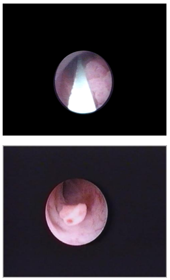
Papillary tumor

Video urethrocystoscopy showing a papillary tumor in the bulbar urethra

Image: “Video urethrocystoscopy showing papillary tumor at the level of bulbar urethra.” by Journal of Endourology Case Reports. License: CC BY 4.0

Staging Staging Methods which attempt to express in replicable terms the extent of the neoplasm in the patient. Grading, Staging, and Metastasis

Urethral cancer is staged according to the TNM criteria.

Table: Primary tumor Tumor Inflammation (female urethra Urethra A tube that transports urine from the urinary bladder to the outside of the body in both the sexes. It also has a reproductive function in the male by providing a passage for sperm. Urinary Tract: Anatomy and male penile urethra Penile urethra Urinary Tract: Anatomy)
T category Description
Tx Primary tumor Tumor Inflammation cannot be assessed.
T0 No evidence of primary tumor Tumor Inflammation
Ta TA Thyrotoxicosis and Hyperthyroidism Noninvasive papillary carcinoma
Tis Carcinoma in situ Carcinoma in situ A lesion with cytological characteristics associated with invasive carcinoma but the tumor cells are confined to the epithelium of origin, without invasion of the basement membrane. Leukoplakia
T1 Tumor Tumor Inflammation invasion of subepithelial Subepithelial Membranoproliferative Glomerulonephritis connective tissue Connective tissue Connective tissues originate from embryonic mesenchyme and are present throughout the body except inside the brain and spinal cord. The main function of connective tissues is to provide structural support to organs. Connective tissues consist of cells and an extracellular matrix. Connective Tissue: Histology
T2 Tumor Tumor Inflammation invasion of the corpus spongiosum Corpus spongiosum Penis: Anatomy or periurethral muscle
T3 T3 A T3 thyroid hormone normally synthesized and secreted by the thyroid gland in much smaller quantities than thyroxine (T4). Most T3 is derived from peripheral monodeiodination of T4 at the 5′ position of the outer ring of the iodothyronine nucleus. The hormone finally delivered and used by the tissues is mainly t3. Thyroid Hormones Tumor Tumor Inflammation invasion of the corpus cavernosum or anterior vagina Vagina The vagina is the female genital canal, extending from the vulva externally to the cervix uteri internally. The structures have sexual, reproductive, and urinary functions and a rich blood supply, mainly arising from the internal iliac artery. Vagina, Vulva, and Pelvic Floor: Anatomy
T4 T4 The major hormone derived from the thyroid gland. Thyroxine is synthesized via the iodination of tyrosines (monoiodotyrosine) and the coupling of iodotyrosines (diiodotyrosine) in the thyroglobulin. Thyroxine is released from thyroglobulin by proteolysis and secreted into the blood. Thyroxine is peripherally deiodinated to form triiodothyronine which exerts a broad spectrum of stimulatory effects on cell metabolism. Thyroid Hormones Tumor Tumor Inflammation invasion of other adjacent organs
Table: Primary tumor Tumor Inflammation ( prostatic urethra Prostatic urethra )
T category Description
Tx Primary tumor Tumor Inflammation cannot be assessed.
T0 No evidence of primary tumor Tumor Inflammation
Ta TA Thyrotoxicosis and Hyperthyroidism Noninvasive papillary carcinoma
Tis Carcinoma in situ (involves the prostatic urethra Prostatic urethra or periurethral or prostatic ducts Prostatic ducts without stromal invasion)
T1 Tumor Tumor Inflammation invasion of the subepithelial Subepithelial Membranoproliferative Glomerulonephritis connective tissue Connective tissue Connective tissues originate from embryonic mesenchyme and are present throughout the body except inside the brain and spinal cord. The main function of connective tissues is to provide structural support to organs. Connective tissues consist of cells and an extracellular matrix. Connective Tissue: Histology immediately underlying the urothelium Urothelium The epithelial lining of the urinary tract. Urinary Tract: Anatomy
T2 Tumor Tumor Inflammation invasion of the prostatic stroma surrounding ducts (by directly extending from the urothelial surface or by invading from prostatic ducts Prostatic ducts )
T3 T3 A T3 thyroid hormone normally synthesized and secreted by the thyroid gland in much smaller quantities than thyroxine (T4). Most T3 is derived from peripheral monodeiodination of T4 at the 5′ position of the outer ring of the iodothyronine nucleus. The hormone finally delivered and used by the tissues is mainly t3. Thyroid Hormones Tumor Tumor Inflammation invasion of the periprostatic fat
T4 T4 The major hormone derived from the thyroid gland. Thyroxine is synthesized via the iodination of tyrosines (monoiodotyrosine) and the coupling of iodotyrosines (diiodotyrosine) in the thyroglobulin. Thyroxine is released from thyroglobulin by proteolysis and secreted into the blood. Thyroxine is peripherally deiodinated to form triiodothyronine which exerts a broad spectrum of stimulatory effects on cell metabolism. Thyroid Hormones Tumor Tumor Inflammation invasion of the adjacent organs
Table: Regional lymph nodes Lymph Nodes They are oval or bean shaped bodies (1 – 30 mm in diameter) located along the lymphatic system. Lymphatic Drainage System: Anatomy
N category Description
Nx Regional lymph nodes Lymph Nodes They are oval or bean shaped bodies (1 – 30 mm in diameter) located along the lymphatic system. Lymphatic Drainage System: Anatomy cannot be assessed.
N0 Regional lymph Lymph The interstitial fluid that is in the lymphatic system. Secondary Lymphatic Organs node metastasis Metastasis The transfer of a neoplasm from one organ or part of the body to another remote from the primary site. Grading, Staging, and Metastasis is not present.
N1 Single regional lymph Lymph The interstitial fluid that is in the lymphatic system. Secondary Lymphatic Organs node metastasis Metastasis The transfer of a neoplasm from one organ or part of the body to another remote from the primary site. Grading, Staging, and Metastasis in the inguinal region Inguinal region Anterior Abdominal Wall: Anatomy or true pelvis Pelvis The pelvis consists of the bony pelvic girdle, the muscular and ligamentous pelvic floor, and the pelvic cavity, which contains viscera, vessels, and multiple nerves and muscles. The pelvic girdle, composed of 2 “hip” bones and the sacrum, is a ring-like bony structure of the axial skeleton that links the vertebral column with the lower extremities. Pelvis: Anatomy or presacral lymph Lymph The interstitial fluid that is in the lymphatic system. Secondary Lymphatic Organs node
N2 Multiple lymph Lymph The interstitial fluid that is in the lymphatic system. Secondary Lymphatic Organs node metastases in the inguinal region Inguinal region Anterior Abdominal Wall: Anatomy or true pelvis Pelvis The pelvis consists of the bony pelvic girdle, the muscular and ligamentous pelvic floor, and the pelvic cavity, which contains viscera, vessels, and multiple nerves and muscles. The pelvic girdle, composed of 2 “hip” bones and the sacrum, is a ring-like bony structure of the axial skeleton that links the vertebral column with the lower extremities. Pelvis: Anatomy or presacral lymph Lymph The interstitial fluid that is in the lymphatic system. Secondary Lymphatic Organs node
Table: Distant metastasis Metastasis The transfer of a neoplasm from one organ or part of the body to another remote from the primary site. Grading, Staging, and Metastasis
M category Description
M0 No distant metastasis Metastasis The transfer of a neoplasm from one organ or part of the body to another remote from the primary site. Grading, Staging, and Metastasis
M1 Distant metastasis Metastasis The transfer of a neoplasm from one organ or part of the body to another remote from the primary site. Grading, Staging, and Metastasis
Table: Staging Staging Methods which attempt to express in replicable terms the extent of the neoplasm in the patient. Grading, Staging, and Metastasis of urethral cancer
Stage T N M
Stage 0a Ta TA Thyrotoxicosis and Hyperthyroidism N0 M0
Stage 0is Tis N0 M0
Stage I T1 N0 M0
Stage II T2 M0
Stage III T1, T2 N1 M0
T3 T3 A T3 thyroid hormone normally synthesized and secreted by the thyroid gland in much smaller quantities than thyroxine (T4). Most T3 is derived from peripheral monodeiodination of T4 at the 5′ position of the outer ring of the iodothyronine nucleus. The hormone finally delivered and used by the tissues is mainly t3. Thyroid Hormones N0, N1 M0
Stage IV T4 T4 The major hormone derived from the thyroid gland. Thyroxine is synthesized via the iodination of tyrosines (monoiodotyrosine) and the coupling of iodotyrosines (diiodotyrosine) in the thyroglobulin. Thyroxine is released from thyroglobulin by proteolysis and secreted into the blood. Thyroxine is peripherally deiodinated to form triiodothyronine which exerts a broad spectrum of stimulatory effects on cell metabolism. Thyroid Hormones N0, N1 M0
Any T N2 M0
Any T Any N M1

Management

Localized disease (up to T2) is generally treated with surgery, while locally advanced conditions are treated with multimodal therapy.

  • In men, the surgical options for urethral cancer are:
    • Low-grade or small high-grade noninvasive tumors: endoscopic resection
    • High-grade, noninvasive tumors: segmental resection
    • Distal tumors: distal urethrectomy, creating hypospadias Hypospadias A birth defect due to malformation of the urethra in which the urethral opening is below its normal location. In the male, the malformed urethra generally opens on the ventral surface of the penis or on the perineum. In the female, the malformed urethral opening is in the vagina. Penile Anomalies and Conditions
    • More extensive tumors (T2): subtotal urethrectomy
    • Urothelial carcinoma invading the prostatic stroma: neoadjuvant chemotherapy Neoadjuvant Chemotherapy Osteosarcoma, radical cystoprostatectomy with urinary diversion 
    • More advanced disease ( T3 T3 A T3 thyroid hormone normally synthesized and secreted by the thyroid gland in much smaller quantities than thyroxine (T4). Most T3 is derived from peripheral monodeiodination of T4 at the 5′ position of the outer ring of the iodothyronine nucleus. The hormone finally delivered and used by the tissues is mainly t3. Thyroid Hormones or T4 T4 The major hormone derived from the thyroid gland. Thyroxine is synthesized via the iodination of tyrosines (monoiodotyrosine) and the coupling of iodotyrosines (diiodotyrosine) in the thyroglobulin. Thyroxine is released from thyroglobulin by proteolysis and secreted into the blood. Thyroxine is peripherally deiodinated to form triiodothyronine which exerts a broad spectrum of stimulatory effects on cell metabolism. Thyroid Hormones) requires extensive surgical resection ( urethra Urethra A tube that transports urine from the urinary bladder to the outside of the body in both the sexes. It also has a reproductive function in the male by providing a passage for sperm. Urinary Tract: Anatomy, prostate Prostate The prostate is a gland in the male reproductive system. The gland surrounds the bladder neck and a portion of the urethra. The prostate is an exocrine gland that produces a weakly acidic secretion, which accounts for roughly 20% of the seminal fluid. , bladder Bladder A musculomembranous sac along the urinary tract. Urine flows from the kidneys into the bladder via the ureters, and is held there until urination. Pyelonephritis and Perinephric Abscess with penectomy).
  • In women:
    • Low-grade or small high-grade noninvasive tumors: endoscopic resection
    • Extensive disease: urethrectomy, bladder Bladder A musculomembranous sac along the urinary tract. Urine flows from the kidneys into the bladder via the ureters, and is held there until urination. Pyelonephritis and Perinephric Abscess neck Neck The part of a human or animal body connecting the head to the rest of the body. Peritonsillar Abscess closure, and urinary diversion
    • Advanced disease ( T3 T3 A T3 thyroid hormone normally synthesized and secreted by the thyroid gland in much smaller quantities than thyroxine (T4). Most T3 is derived from peripheral monodeiodination of T4 at the 5′ position of the outer ring of the iodothyronine nucleus. The hormone finally delivered and used by the tissues is mainly t3. Thyroid Hormones or T4 T4 The major hormone derived from the thyroid gland. Thyroxine is synthesized via the iodination of tyrosines (monoiodotyrosine) and the coupling of iodotyrosines (diiodotyrosine) in the thyroglobulin. Thyroxine is released from thyroglobulin by proteolysis and secreted into the blood. Thyroxine is peripherally deiodinated to form triiodothyronine which exerts a broad spectrum of stimulatory effects on cell metabolism. Thyroid Hormones): neoadjuvant chemotherapy Neoadjuvant Chemotherapy Osteosarcoma with extensive surgical resection ( urethra Urethra A tube that transports urine from the urinary bladder to the outside of the body in both the sexes. It also has a reproductive function in the male by providing a passage for sperm. Urinary Tract: Anatomy, anterior vagina Vagina The vagina is the female genital canal, extending from the vulva externally to the cervix uteri internally. The structures have sexual, reproductive, and urinary functions and a rich blood supply, mainly arising from the internal iliac artery. Vagina, Vulva, and Pelvic Floor: Anatomy, bladder Bladder A musculomembranous sac along the urinary tract. Urine flows from the kidneys into the bladder via the ureters, and is held there until urination. Pyelonephritis and Perinephric Abscess with possible pubectomy)
  • Chemotherapy Chemotherapy Osteosarcoma:
    • Regimen differs by histology 
    • Urothelial carcinoma can be treated with:
      • Cisplatin Cisplatin An inorganic and water-soluble platinum complex. After undergoing hydrolysis, it reacts with DNA to produce both intra and interstrand crosslinks. These cross links appear to impair replication and transcription of DNA. The cytotoxicity of cisplatin correlates with cellular arrest in the g2 phase of the cell cycle. Alkylating Agents and Platinum, gemcitabine Gemcitabine Antimetabolite Chemotherapy, ifosfamide Ifosfamide Positional isomer of cyclophosphamide which is active as an alkylating agent and an immunosuppressive agent. Alkylating Agents and Platinum (CGI)
      • Ifosfamide Ifosfamide Positional isomer of cyclophosphamide which is active as an alkylating agent and an immunosuppressive agent. Alkylating Agents and Platinum, paclitaxel Paclitaxel A cyclodecane isolated from the bark of the pacific yew tree, taxus brevifolia. It stabilizes microtubules in their polymerized form leading to cell death. Microtubule and Topoisomerase Inhibitors, cisplatin Cisplatin An inorganic and water-soluble platinum complex. After undergoing hydrolysis, it reacts with DNA to produce both intra and interstrand crosslinks. These cross links appear to impair replication and transcription of DNA. The cytotoxicity of cisplatin correlates with cellular arrest in the g2 phase of the cell cycle. Alkylating Agents and Platinum
      • Methotrexate Methotrexate An antineoplastic antimetabolite with immunosuppressant properties. It is an inhibitor of tetrahydrofolate dehydrogenase and prevents the formation of tetrahydrofolate, necessary for synthesis of thymidylate, an essential component of DNA. Antimetabolite Chemotherapy, vinblastine Vinblastine Antitumor alkaloid isolated from vinca rosea. Microtubule and Topoisomerase Inhibitors, doxorubicin, cisplatin Cisplatin An inorganic and water-soluble platinum complex. After undergoing hydrolysis, it reacts with DNA to produce both intra and interstrand crosslinks. These cross links appear to impair replication and transcription of DNA. The cytotoxicity of cisplatin correlates with cellular arrest in the g2 phase of the cell cycle. Alkylating Agents and Platinum
  • Radiation Radiation Emission or propagation of acoustic waves (sound), electromagnetic energy waves (such as light; radio waves; gamma rays; or x-rays), or a stream of subatomic particles (such as electrons; neutrons; protons; or alpha particles). Osteosarcoma therapy:
    • Used alone (localized tumors) or coupled with chemotherapy Chemotherapy Osteosarcoma especially in those who are not candidates for surgery
    • May be used for palliation 
    • Side effects:
      • Bladder Bladder A musculomembranous sac along the urinary tract. Urine flows from the kidneys into the bladder via the ureters, and is held there until urination. Pyelonephritis and Perinephric Abscess pain Pain An unpleasant sensation induced by noxious stimuli which are detected by nerve endings of nociceptive neurons. Pain: Types and Pathways 
      • Incontinence
      • Bleeding 
      • Rectal pain Pain An unpleasant sensation induced by noxious stimuli which are detected by nerve endings of nociceptive neurons. Pain: Types and Pathways 
      • Rectal bleeding
      • Vaginal pain Pain An unpleasant sensation induced by noxious stimuli which are detected by nerve endings of nociceptive neurons. Pain: Types and Pathways and/or bleeding, in women 
      • Narrowing of the urethra Urethra A tube that transports urine from the urinary bladder to the outside of the body in both the sexes. It also has a reproductive function in the male by providing a passage for sperm. Urinary Tract: Anatomy 
  • Posttreatment:
    • Follow-up visits are generally every 3–4 months for 1st 2 years and then twice a year. 
    • Physical examination (check inguinal lymph nodes Lymph Nodes They are oval or bean shaped bodies (1 – 30 mm in diameter) located along the lymphatic system. Lymphatic Drainage System: Anatomy)
    • Endoscopy Endoscopy Procedures of applying endoscopes for disease diagnosis and treatment. Endoscopy involves passing an optical instrument through a small incision in the skin i.e., percutaneous; or through a natural orifice and along natural body pathways such as the digestive tract; and/or through an incision in the wall of a tubular structure or organ, i.e. Transluminal, to examine or perform surgery on the interior parts of the body. Gastroesophageal Reflux Disease (GERD) to check remaining urethra Urethra A tube that transports urine from the urinary bladder to the outside of the body in both the sexes. It also has a reproductive function in the male by providing a passage for sperm. Urinary Tract: Anatomy
    • Imaging

Prognosis Prognosis A prediction of the probable outcome of a disease based on a individual’s condition and the usual course of the disease as seen in similar situations. Non-Hodgkin Lymphomas

  • Nodal stage is an important predictor of survival.
  • Recurrence of urethral cancer is lower at early stages.
  • Urethral cancer generally has a 32% survival rate at 10 years.

Differential Diagnosis

The following differential diagnoses are for an individual presenting with hematuria Hematuria Presence of blood in the urine. Renal Cell Carcinoma:

  • IgA IgA Represents 15-20% of the human serum immunoglobulins, mostly as the 4-chain polymer in humans or dimer in other mammals. Secretory iga is the main immunoglobulin in secretions. Immunoglobulins: Types and Functions nephropathy: also known as Berger disease. IgA IgA Represents 15-20% of the human serum immunoglobulins, mostly as the 4-chain polymer in humans or dimer in other mammals. Secretory iga is the main immunoglobulin in secretions. Immunoglobulins: Types and Functions nephropathy is a renal disease characterized by IgA IgA Represents 15-20% of the human serum immunoglobulins, mostly as the 4-chain polymer in humans or dimer in other mammals. Secretory iga is the main immunoglobulin in secretions. Immunoglobulins: Types and Functions deposition in the mesangium Mesangium The thin membranous structure supporting the adjoining glomerular capillaries. It is composed of glomerular mesangial cells and their extracellular matrix. IgA Nephropathy. Presentation usually is associated with a preceding upper respiratory or GI infection GI infection Microsporidia/Microsporidiosis. Common presenting features are gross hematuria Hematuria Presence of blood in the urine. Renal Cell Carcinoma or asymptomatic, microscopic hematuria Hematuria Presence of blood in the urine. Renal Cell Carcinoma on urinalysis Urinalysis Examination of urine by chemical, physical, or microscopic means. Routine urinalysis usually includes performing chemical screening tests, determining specific gravity, observing any unusual color or odor, screening for bacteriuria, and examining the sediment microscopically. Urinary Tract Infections (UTIs) in Children. The course is often benign Benign Fibroadenoma, with renal biopsy Renal Biopsy Antineutrophil Cytoplasmic Antibody (ANCA)-Associated Vasculitis performed only in cases of severe, progressive renal disease. Treatment depends on the severity of proteinuria Proteinuria The presence of proteins in the urine, an indicator of kidney diseases. Nephrotic Syndrome in Children, renal function, and pathologic changes. ACEis ACEIs A class of drugs whose main indications are the treatment of hypertension and heart failure. They exert their hemodynamic effect mainly by inhibiting the renin-angiotensin system. They also modulate sympathetic nervous system activity and increase prostaglandin synthesis. They cause mainly vasodilation and mild natriuresis without affecting heart rate and contractility. Heart Failure and Angina Medication or ARBs ARBs Agents that antagonize angiotensin receptors. Many drugs in this class specifically target the angiotensin type 1 receptor. Heart Failure and Angina Medication are given to reduce disease progression. Immunosuppressive therapy given in more severe conditions.  
  • Alport syndrome Alport Syndrome Alport syndrome, also called hereditary nephritis, is a genetic disorder caused by a mutation in the genes encoding for the alpha chains of type IV collagen, resulting in the production of abnormal type IV collagen strands. Patients present with glomerulonephritis, hypertension, edema, hematuria, and proteinuria, as well as with ocular and auditory findings. Alport Syndrome: also called hereditary nephritis. Alport syndrome Alport Syndrome Alport syndrome, also called hereditary nephritis, is a genetic disorder caused by a mutation in the genes encoding for the alpha chains of type IV collagen, resulting in the production of abnormal type IV collagen strands. Patients present with glomerulonephritis, hypertension, edema, hematuria, and proteinuria, as well as with ocular and auditory findings. Alport Syndrome is a genetic disorder caused by a mutation Mutation Genetic mutations are errors in DNA that can cause protein misfolding and dysfunction. There are various types of mutations, including chromosomal, point, frameshift, and expansion mutations. Types of Mutations in the genes Genes A category of nucleic acid sequences that function as units of heredity and which code for the basic instructions for the development, reproduction, and maintenance of organisms. DNA Types and Structure encoding for the alpha chains of type IV collagen Type IV collagen A non-fibrillar collagen found in the structure of basement membrane. Collagen type IV molecules assemble to form a sheet-like network which is involved in maintaining the structural integrity of basement membranes. The predominant form of the protein is comprised of two alpha1(IV) subunits and one alpha2(IV) subunit, however, at least six different alpha subunits can be incorporated into the heterotrimer. Alport Syndrome, resulting in the production of abnormal type IV collagen Type IV collagen A non-fibrillar collagen found in the structure of basement membrane. Collagen type IV molecules assemble to form a sheet-like network which is involved in maintaining the structural integrity of basement membranes. The predominant form of the protein is comprised of two alpha1(IV) subunits and one alpha2(IV) subunit, however, at least six different alpha subunits can be incorporated into the heterotrimer. Alport Syndrome strands. Presenting features include glomerulonephritis, hypertension Hypertension Hypertension, or high blood pressure, is a common disease that manifests as elevated systemic arterial pressures. Hypertension is most often asymptomatic and is found incidentally as part of a routine physical examination or during triage for an unrelated medical encounter. Hypertension, edema Edema Edema is a condition in which excess serous fluid accumulates in the body cavity or interstitial space of connective tissues. Edema is a symptom observed in several medical conditions. It can be categorized into 2 types, namely, peripheral (in the extremities) and internal (in an organ or body cavity). Edema, hematuria Hematuria Presence of blood in the urine. Renal Cell Carcinoma, and proteinuria Proteinuria The presence of proteins in the urine, an indicator of kidney diseases. Nephrotic Syndrome in Children, as well as ocular ( cataract Cataract Partial or complete opacity on or in the lens or capsule of one or both eyes, impairing vision or causing blindness. The many kinds of cataract are classified by their morphology (size, shape, location) or etiology (cause and time of occurrence). Neurofibromatosis Type 2, retinopathy Retinopathy Degenerative changes to the retina due to hypertension. Alport Syndrome) and auditory ( sensorineural hearing loss Sensorineural hearing loss Hearing loss resulting from damage to the cochlea and the sensorineural elements which lie internally beyond the oval and round windows. These elements include the auditory nerve and its connections in the brainstem. Hearing Loss) findings. Diagnosis is established with laboratory tests, and a renal biopsy Renal Biopsy Antineutrophil Cytoplasmic Antibody (ANCA)-Associated Vasculitis showing characteristic glomerular basement membrane Basement membrane A darkly stained mat-like extracellular matrix (ecm) that separates cell layers, such as epithelium from endothelium or a layer of connective tissue. The ecm layer that supports an overlying epithelium or endothelium is called basal lamina. Basement membrane (bm) can be formed by the fusion of either two adjacent basal laminae or a basal lamina with an adjacent reticular lamina of connective tissue. Bm, composed mainly of type IV collagen; glycoprotein laminin; and proteoglycan, provides barriers as well as channels between interacting cell layers. Thin Basement Membrane Nephropathy (TBMN) splitting Splitting Defense Mechanisms may be used to confirm diagnosis. Treatment for Alport syndrome Alport Syndrome Alport syndrome, also called hereditary nephritis, is a genetic disorder caused by a mutation in the genes encoding for the alpha chains of type IV collagen, resulting in the production of abnormal type IV collagen strands. Patients present with glomerulonephritis, hypertension, edema, hematuria, and proteinuria, as well as with ocular and auditory findings. Alport Syndrome is focused on limiting disease progression with ARBs ARBs Agents that antagonize angiotensin receptors. Many drugs in this class specifically target the angiotensin type 1 receptor. Heart Failure and Angina Medication and ACEis ACEIs A class of drugs whose main indications are the treatment of hypertension and heart failure. They exert their hemodynamic effect mainly by inhibiting the renin-angiotensin system. They also modulate sympathetic nervous system activity and increase prostaglandin synthesis. They cause mainly vasodilation and mild natriuresis without affecting heart rate and contractility. Heart Failure and Angina Medication. Hearing aids AIDS Chronic HIV infection and depletion of CD4 cells eventually results in acquired immunodeficiency syndrome (AIDS), which can be diagnosed by the presence of certain opportunistic diseases called AIDS-defining conditions. These conditions include a wide spectrum of bacterial, viral, fungal, and parasitic infections as well as several malignancies and generalized conditions. HIV Infection and AIDS are used for hearing loss Hearing loss Hearing loss, also known as hearing impairment, is any degree of impairment in the ability to apprehend sound as determined by audiometry to be below normal hearing thresholds. Clinical presentation may occur at birth or as a gradual loss of hearing with age, including a short-term or sudden loss at any point. Hearing Loss associated with Alport syndrome Alport Syndrome Alport syndrome, also called hereditary nephritis, is a genetic disorder caused by a mutation in the genes encoding for the alpha chains of type IV collagen, resulting in the production of abnormal type IV collagen strands. Patients present with glomerulonephritis, hypertension, edema, hematuria, and proteinuria, as well as with ocular and auditory findings. Alport Syndrome
  • Poststreptococcal glomerulonephritis Poststreptococcal Glomerulonephritis Post-streptococcal glomerulonephritis (PSGN) is a type of nephritis that is caused by a prior infection with group A beta-hemolytic Streptococcus (GAS). The clinical presentation of PSGN can range from asymptomatic, microscopic hematuria to full-blown acute nephritic syndrome, which is characterized by red-to-brown urine, proteinuria, edema, and acute kidney injury. Postinfectious Glomerulonephritis ( PSGN PSGN Post-streptococcal glomerulonephritis (PSGN) is a type of nephritis that is caused by a prior infection with group A beta-hemolytic Streptococcus (GAS). The clinical presentation of PSGN can range from asymptomatic, microscopic hematuria to full-blown acute nephritic syndrome, which is characterized by red-to-brown urine, proteinuria, edema, and acute kidney injury. Postinfectious Glomerulonephritis): type of nephritis that is caused by a prior infection with group A beta-hemolytic Streptococcus Streptococcus Streptococcus is one of the two medically important genera of gram-positive cocci, the other being Staphylococcus. Streptococci are identified as different species on blood agar on the basis of their hemolytic pattern and sensitivity to optochin and bacitracin. There are many pathogenic species of streptococci, including S. pyogenes, S. agalactiae, S. pneumoniae, and the viridans streptococci. Streptococcus (GAS). The clinical presentation of PSGN PSGN Post-streptococcal glomerulonephritis (PSGN) is a type of nephritis that is caused by a prior infection with group A beta-hemolytic Streptococcus (GAS). The clinical presentation of PSGN can range from asymptomatic, microscopic hematuria to full-blown acute nephritic syndrome, which is characterized by red-to-brown urine, proteinuria, edema, and acute kidney injury. Postinfectious Glomerulonephritis can range from asymptomatic, microscopic hematuria Hematuria Presence of blood in the urine. Renal Cell Carcinoma to full-blown acute nephritic syndrome Nephritic syndrome Nephritic syndrome is a broad category of glomerular diseases characterized by glomerular hematuria, variable loss of renal function, and hypertension. These features are in contrast to those of nephrotic syndrome, which includes glomerular diseases characterized by severe proteinuria, although there is sometimes overlap of > 1 glomerular disease in the same individual. Nephritic Syndrome, which is characterized by red-to-brown urine, proteinuria Proteinuria The presence of proteins in the urine, an indicator of kidney diseases. Nephrotic Syndrome in Children, edema Edema Edema is a condition in which excess serous fluid accumulates in the body cavity or interstitial space of connective tissues. Edema is a symptom observed in several medical conditions. It can be categorized into 2 types, namely, peripheral (in the extremities) and internal (in an organ or body cavity). Edema, and AKI AKI Acute kidney injury refers to sudden and often reversible loss of renal function, which develops over days or weeks. Azotemia refers to elevated levels of nitrogen-containing substances in the blood that accompany AKI, which include BUN and creatinine. Acute Kidney Injury. The diagnosis is made on the basis of clinical findings in the setting of recent GAS infection. Management is supportive and involves treating the clinical manifestations. The prognosis Prognosis A prediction of the probable outcome of a disease based on a individual’s condition and the usual course of the disease as seen in similar situations. Non-Hodgkin Lymphomas is generally favorable, especially in children.  
  • Nephrolithiasis Nephrolithiasis Nephrolithiasis is the formation of a stone, or calculus, anywhere along the urinary tract caused by precipitations of solutes in the urine. The most common type of kidney stone is the calcium oxalate stone, but other types include calcium phosphate, struvite (ammonium magnesium phosphate), uric acid, and cystine stones. Nephrolithiasis: hard mineral or salt deposits in the kidney that present with severe pain Pain An unpleasant sensation induced by noxious stimuli which are detected by nerve endings of nociceptive neurons. Pain: Types and Pathways in the renal angle, proteinuria Proteinuria The presence of proteins in the urine, an indicator of kidney diseases. Nephrotic Syndrome in Children, nausea Nausea An unpleasant sensation in the stomach usually accompanied by the urge to vomit. Common causes are early pregnancy, sea and motion sickness, emotional stress, intense pain, food poisoning, and various enteroviruses. Antiemetics, and vomiting Vomiting The forcible expulsion of the contents of the stomach through the mouth. Hypokalemia, increased urinary frequency, and pain Pain An unpleasant sensation induced by noxious stimuli which are detected by nerve endings of nociceptive neurons. Pain: Types and Pathways that radiates to the lower abdomen and groin Groin The external junctural region between the lower part of the abdomen and the thigh. Male Genitourinary Examination. Diagnosis is by imaging, and management is with administration of analgesics and surgery to remove the stones.

References

  1. Babaian, K., Adams, P., McClure, C., Tompkins, B., McMurray, M. (2021). Bladder cancer. Medscape. Retrieved November 30, 2021, from https://emedicine.medscape.com/article/438262-overview
  2. Daneshmand, S. (2021). Epidemiology and risk factors of urothelial (transitional cell) carcinoma of the bladder. UpToDate. Retrieved November 22, 2021 from https://www.uptodate.com/contents/epidemiology-and-risk-factors-of-urothelial-transitional-cell-carcinoma-of-the-bladder
  3. Daneshmand, S., Efstathiou, J. (2021). Urethral cancer. UpToDate. Retrieved November 22, 2021, from https://www.uptodate.com/contents/urethral-cancer
  4. Hahn, N.M. (2018). Cancer of the bladder and urinary tract. Chapter 82 of Jameson J., Fauci A.S., Kasper D.L., Hauser S.L., Longo D.L., Loscalzo J (Eds.),  Harrison’s Principles of Internal Medicine, 20th ed. https://accessmedicine.mhmedical.com/content.aspx?bookid=2129&sectionid=192016282
  5. Lerner, S.  (2021). Overview of the initial approach and management of urothelial bladder cancer. UpToDate. Retrieved on November 2, 2021, from https://www.uptodate.com/contents/overview-of-the-initial-approach-and-management-of-urothelial-bladder-cancer
  6. Lotan, Y., Choueiri, T. (2021). Clinical presentation, diagnosis and staging of bladder cancer. UpToDate. Retrieved November 22, 2021, from https://www.uptodate.com/contents/clinical-presentation-diagnosis-and-staging-of-bladder-cancer
  7. Shariat, S., Laukhtina, E., Pradere, B. (2021). Urethral cancer. Medscape. Retrieved November 30, 2021, from https://emedicine.medscape.com/article/451496-overview#:~:text=Primary%20urethral%20cancer%20(PUC)%20is,for%20men%20and%20African%20Americans.
  8. Steinberg, G., DeCastro, J., Patel, A. (2020). Bladder cancer staging. Medscape. Retrieved November 30, 2021, fromhttps://emedicine.medscape.com/article/2006834-overview

Create your free account or log in to continue reading!

Sign up now and get free access to Lecturio with concept pages, medical videos, and questions for your medical education.

User Reviews

Unwrap New Skills This Holiday 🎄 Save 30% on all plans now!

Details