Advertisement

Advertisement

Advertisement

Advertisement

Thoracic Outlet Syndrome

Thoracic outlet syndrome (TOS) is a broad term used for a spectrum of syndromes related to the general region of the thoracic outlet, which involves the compression Compression Blunt Chest Trauma or irritation of elements of the brachial plexus Brachial Plexus The large network of nerve fibers which distributes the innervation of the upper extremity. The brachial plexus extends from the neck into the axilla. In humans, the nerves of the plexus usually originate from the lower cervical and the first thoracic spinal cord segments (c5-c8 and T1), but variations are not uncommon. Peripheral Nerve Injuries in the Cervicothoracic Region, subclavian artery, or subclavian vein. The most common etiology involves structural abnormalities. Thoracic outlet syndrome can present as the neurogenic, arterial, or venous type. The neurogenic type is the most common among the 3 variants and mainly involves the inferior trunk of the brachial plexus Brachial Plexus The large network of nerve fibers which distributes the innervation of the upper extremity. The brachial plexus extends from the neck into the axilla. In humans, the nerves of the plexus usually originate from the lower cervical and the first thoracic spinal cord segments (c5-c8 and T1), but variations are not uncommon. Peripheral Nerve Injuries in the Cervicothoracic Region (C8–T1). Signs and symptoms vary based on the structure that is involved. The diagnosis of TOS is clinical and supported by radiography and a number of provocation maneuvers. Untreated TOS can lead to various complications such as a frozen shoulder Frozen Shoulder Chronic Shoulder Pain. Management of TOS is using pharmacological and surgical methods.

Last updated: Jan 15, 2024

Editorial responsibility: Stanley Oiseth, Lindsay Jones, Evelin Maza

Advertisement

Advertisement

Advertisement

Advertisement

Advertisement

Advertisement

Overview

Definition

Thoracic outlet syndrome (TOS) refers to a spectrum of signs and symptoms that arise from the compression Compression Blunt Chest Trauma of the neurovascular bundle by any of the various structures within the confined space of the thoracic outlet, usually within the scalene triangle.

Types

  • Neurogenic:
    • Accounts for 95% of cases
    • Due to compression Compression Blunt Chest Trauma of the trunks of the brachial plexus Brachial Plexus The large network of nerve fibers which distributes the innervation of the upper extremity. The brachial plexus extends from the neck into the axilla. In humans, the nerves of the plexus usually originate from the lower cervical and the first thoracic spinal cord segments (c5-c8 and T1), but variations are not uncommon. Peripheral Nerve Injuries in the Cervicothoracic Region
  • Arterial:
  • Venous:
    • Accounts for 1% of cases, but causes symptoms more commonly than the arterial type
    • Due to compression Compression Blunt Chest Trauma of the subclavian vein

Epidemiology

  • More common in women:
    • Usually occurs in women who are 20–50 years old
    • Presents most commonly as neurogenic TOS
  • Men most commonly present with venous TOS.

Etiology

Structural causes:

  • Cervical ribs Ribs A set of twelve curved bones which connect to the vertebral column posteriorly, and terminate anteriorly as costal cartilage. Together, they form a protective cage around the internal thoracic organs. Chest Wall: Anatomy
  • Fibrous Fibrous Fibrocystic Change bands running within this region
  • Hypertrophy Hypertrophy General increase in bulk of a part or organ due to cell enlargement and accumulation of fluids and secretions, not due to tumor formation, nor to an increase in the number of cells (hyperplasia). Cellular Adaptation or abnormal insertion of the scalene muscles (reduces the size of the scalene triangle)
  • Anomalous muscles (e.g., supernumerary scalenes Scalenes Muscles of the Neck: Anatomy) in the region of the thoracic outlet

Other causes:

  • Injuries: sports-related injuries and motions (e.g., clavicle Clavicle A bone on the ventral side of the shoulder girdle, which in humans is commonly called the collar bone. Clavicle Fracture fracture Fracture A fracture is a disruption of the cortex of any bone and periosteum and is commonly due to mechanical stress after an injury or accident. Open fractures due to trauma can be a medical emergency. Fractures are frequently associated with automobile accidents, workplace injuries, and trauma. Overview of Bone Fractures, repetitive throwing motions)
  • Fractured clavicle Clavicle A bone on the ventral side of the shoulder girdle, which in humans is commonly called the collar bone. Clavicle Fracture
  • Repetitive stress: abnormal pressure on the shoulder
  • Poor posture: a condition called forward head posture (FHP) seen in people who, for example, read on the computer for long periods of time
  • Pancoast tumor Tumor Inflammation ( tumor Tumor Inflammation of the lung apex)
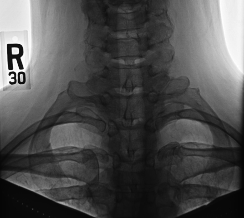
Ossified cervical ribs

X-ray of the cervical ribs

Image: “Cervical Ribs” by Huntsville Hospital Imaging. License: Public Domain

Anatomy and Pathophysiology

Anatomy

Thoracic outlet:

  • Passageway of the neurovasculature between the base of the neck Neck The part of a human or animal body connecting the head to the rest of the body. Peritonsillar Abscess, through the axilla Axilla The axilla is a pyramid-shaped space located between the upper thorax and the arm. The axilla has a base, an apex, and 4 walls (anterior, medial, lateral, posterior). The base of the pyramid is made up of the axillary skin. The apex is the axillary inlet, located between the 1st rib, superior border of the scapula, and clavicle. Axilla and Brachial Plexus: Anatomy, and into the upper limb
  • The thoracic outlet is made up of:
    • Thoracic vertebra (T1) posteriorly
    • 1st pair of ribs Ribs A set of twelve curved bones which connect to the vertebral column posteriorly, and terminate anteriorly as costal cartilage. Together, they form a protective cage around the internal thoracic organs. Chest Wall: Anatomy laterally
    • Clavicle Clavicle A bone on the ventral side of the shoulder girdle, which in humans is commonly called the collar bone. Clavicle Fracture, sternoclavicular joint Sternoclavicular Joint Examination of the Upper Limbs, and manubrium Manubrium The upper or most anterior segment of the sternum which articulates with the clavicle and first two pairs of ribs. Chest Wall: Anatomy of the sternum Sternum A long, narrow, and flat bone commonly known as breastbone occurring in the midsection of the anterior thoracic segment or chest region, which stabilizes the rib cage and serves as the point of origin for several muscles that move the arms, head, and neck. Chest Wall: Anatomy anteriorly

The structures of significance in TOS are:
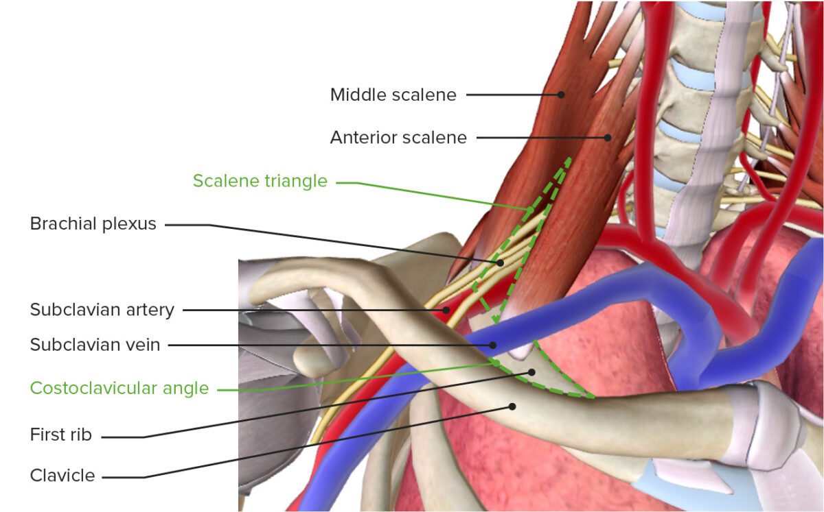
  • Inferior trunk of the brachial plexus Brachial Plexus The large network of nerve fibers which distributes the innervation of the upper extremity. The brachial plexus extends from the neck into the axilla. In humans, the nerves of the plexus usually originate from the lower cervical and the first thoracic spinal cord segments (c5-c8 and T1), but variations are not uncommon. Peripheral Nerve Injuries in the Cervicothoracic Region (C8–T1)
  • Subclavian arteries Arteries Arteries are tubular collections of cells that transport oxygenated blood and nutrients from the heart to the tissues of the body. The blood passes through the arteries in order of decreasing luminal diameter, starting in the largest artery (the aorta) and ending in the small arterioles. Arteries are classified into 3 types: large elastic arteries, medium muscular arteries, and small arteries and arterioles. Arteries: Histology
  • Subclavian veins Veins Veins are tubular collections of cells, which transport deoxygenated blood and waste from the capillary beds back to the heart. Veins are classified into 3 types: small veins/venules, medium veins, and large veins. Each type contains 3 primary layers: tunica intima, tunica media, and tunica adventitia. Veins: Histology
Thoracic outlet

The thoracic outlet featuring the brachial plexus, subclavian vessels, scalene triangle, and costoclavicular angle

Image by BioDigital, edited by Lecturio

Pathophysiology

There are 3 main sites where the compression Compression Blunt Chest Trauma of structures occurs in TOS:
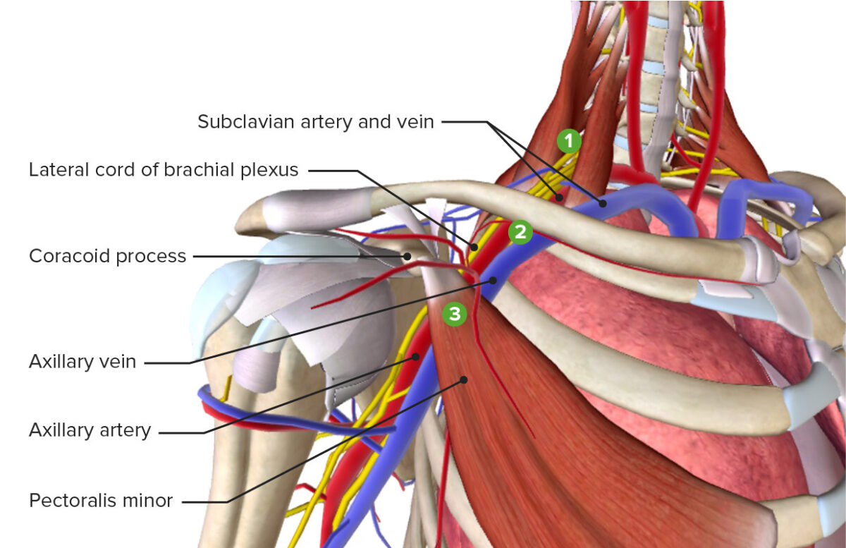
  • Scalene triangle; borders:
    • Anterior scalene muscle anteriorly 
    • Middle scalene muscle posteriorly
    • 1st rib inferiorly
  • Costoclavicular angle or space: the angle made between the clavicle Clavicle A bone on the ventral side of the shoulder girdle, which in humans is commonly called the collar bone. Clavicle Fracture and 1st rib
  • Subcoracoid space: the space behind and below the coracoid process of the scapula
Sites of thoracic outlet syndrome

The 3 most common sites of compression leading to thoracic outlet syndrome: scalene triangle, costoclavicular angle, and subcoracoid space

Image by BioDigital, edited by Lecturio

Clinical Presentation

Neurogenic TOS

  • Paresthesia of the medial side of the arm Arm The arm, or “upper arm” in common usage, is the region of the upper limb that extends from the shoulder to the elbow joint and connects inferiorly to the forearm through the cubital fossa. It is divided into 2 fascial compartments (anterior and posterior). Arm: Anatomy and hand Hand The hand constitutes the distal part of the upper limb and provides the fine, precise movements needed in activities of daily living. It consists of 5 metacarpal bones and 14 phalanges, as well as numerous muscles innervated by the median and ulnar nerves. Hand: Anatomy (can also affect the anterolateral aspect of the chest wall Chest wall The chest wall consists of skin, fat, muscles, bones, and cartilage. The bony structure of the chest wall is composed of the ribs, sternum, and thoracic vertebrae. The chest wall serves as armor for the vital intrathoracic organs and provides the stability necessary for the movement of the shoulders and arms. Chest Wall: Anatomy)
  • Pain Pain An unpleasant sensation induced by noxious stimuli which are detected by nerve endings of nociceptive neurons. Pain: Types and Pathways in the neck Neck The part of a human or animal body connecting the head to the rest of the body. Peritonsillar Abscess, shoulder, arm Arm The arm, or “upper arm” in common usage, is the region of the upper limb that extends from the shoulder to the elbow joint and connects inferiorly to the forearm through the cubital fossa. It is divided into 2 fascial compartments (anterior and posterior). Arm: Anatomy, forearm Forearm The forearm is the region of the upper limb between the elbow and the wrist. The term “forearm” is used in anatomy to distinguish this area from the arm, a term that is commonly used to describe the entire upper limb. The forearm consists of 2 long bones (the radius and the ulna), the interosseous membrane, and multiple arteries, nerves, and muscles. Forearm: Anatomy, and fingers (especially the medial side)
  • Symptoms are aggravated by:
    • Arm Arm The arm, or “upper arm” in common usage, is the region of the upper limb that extends from the shoulder to the elbow joint and connects inferiorly to the forearm through the cubital fossa. It is divided into 2 fascial compartments (anterior and posterior). Arm: Anatomy elevation above shoulder level (reaching, lifting)
    • Sustained use of arms and hands (e.g., typing)
  • Occipital Occipital Part of the back and base of the cranium that encloses the foramen magnum. Skull: Anatomy headache Headache The symptom of pain in the cranial region. It may be an isolated benign occurrence or manifestation of a wide variety of headache disorders. Brain Abscess
  • Progressively weakened grip and difficulty in completing daily activities 
  • Gilliatt-Sumner hand Hand The hand constitutes the distal part of the upper limb and provides the fine, precise movements needed in activities of daily living. It consists of 5 metacarpal bones and 14 phalanges, as well as numerous muscles innervated by the median and ulnar nerves. Hand: Anatomy: wasting ( atrophy Atrophy Decrease in the size of a cell, tissue, organ, or multiple organs, associated with a variety of pathological conditions such as abnormal cellular changes, ischemia, malnutrition, or hormonal changes. Cellular Adaptation) of the muscles of the hand Hand The hand constitutes the distal part of the upper limb and provides the fine, precise movements needed in activities of daily living. It consists of 5 metacarpal bones and 14 phalanges, as well as numerous muscles innervated by the median and ulnar nerves. Hand: Anatomy, especially of the thumb, and thenar eminence (advanced manifestation, rare)

Arterial TOS

  • Fatigue Fatigue The state of weariness following a period of exertion, mental or physical, characterized by a decreased capacity for work and reduced efficiency to respond to stimuli. Fibromyalgia and weakness of the arm Arm The arm, or “upper arm” in common usage, is the region of the upper limb that extends from the shoulder to the elbow joint and connects inferiorly to the forearm through the cubital fossa. It is divided into 2 fascial compartments (anterior and posterior). Arm: Anatomy and forearm Forearm The forearm is the region of the upper limb between the elbow and the wrist. The term “forearm” is used in anatomy to distinguish this area from the arm, a term that is commonly used to describe the entire upper limb. The forearm consists of 2 long bones (the radius and the ulna), the interosseous membrane, and multiple arteries, nerves, and muscles. Forearm: Anatomy
  • Sensation of coldness in the arm Arm The arm, or “upper arm” in common usage, is the region of the upper limb that extends from the shoulder to the elbow joint and connects inferiorly to the forearm through the cubital fossa. It is divided into 2 fascial compartments (anterior and posterior). Arm: Anatomy and forearm Forearm The forearm is the region of the upper limb between the elbow and the wrist. The term “forearm” is used in anatomy to distinguish this area from the arm, a term that is commonly used to describe the entire upper limb. The forearm consists of 2 long bones (the radius and the ulna), the interosseous membrane, and multiple arteries, nerves, and muscles. Forearm: Anatomy, also cold upon palpation Palpation Application of fingers with light pressure to the surface of the body to determine consistency of parts beneath in physical diagnosis; includes palpation for determining the outlines of organs. Dermatologic Examination
  • Upper limb claudication
  • Low BP in the affected arm Arm The arm, or “upper arm” in common usage, is the region of the upper limb that extends from the shoulder to the elbow joint and connects inferiorly to the forearm through the cubital fossa. It is divided into 2 fascial compartments (anterior and posterior). Arm: Anatomy in comparison to the contralateral side
  • Symptoms of hand Hand The hand constitutes the distal part of the upper limb and provides the fine, precise movements needed in activities of daily living. It consists of 5 metacarpal bones and 14 phalanges, as well as numerous muscles innervated by the median and ulnar nerves. Hand: Anatomy ischemia Ischemia A hypoperfusion of the blood through an organ or tissue caused by a pathologic constriction or obstruction of its blood vessels, or an absence of blood circulation. Ischemic Cell Damage secondary to embolization Embolization A method of hemostasis utilizing various agents such as gelfoam, silastic, metal, glass, or plastic pellets, autologous clot, fat, and muscle as emboli. It has been used in the treatment of spinal cord and intracranial arteriovenous malformations, renal arteriovenous fistulas, gastrointestinal bleeding, epistaxis, hypersplenism, certain highly vascular tumors, traumatic rupture of blood vessels, and control of operative hemorrhage. Gastrointestinal Bleeding from subclavian thrombus:
    • Paraesthesia
    • Pallor
    • Pain Pain An unpleasant sensation induced by noxious stimuli which are detected by nerve endings of nociceptive neurons. Pain: Types and Pathways
    • Coldness

Venous TOS

  • Swelling Swelling Inflammation of the arm Arm The arm, or “upper arm” in common usage, is the region of the upper limb that extends from the shoulder to the elbow joint and connects inferiorly to the forearm through the cubital fossa. It is divided into 2 fascial compartments (anterior and posterior). Arm: Anatomy
  • Cyanosis Cyanosis A bluish or purplish discoloration of the skin and mucous membranes due to an increase in the amount of deoxygenated hemoglobin in the blood or a structural defect in the hemoglobin molecule. Pulmonary Examination
  • Pain Pain An unpleasant sensation induced by noxious stimuli which are detected by nerve endings of nociceptive neurons. Pain: Types and Pathways
  • Forearm Forearm The forearm is the region of the upper limb between the elbow and the wrist. The term “forearm” is used in anatomy to distinguish this area from the arm, a term that is commonly used to describe the entire upper limb. The forearm consists of 2 long bones (the radius and the ulna), the interosseous membrane, and multiple arteries, nerves, and muscles. Forearm: Anatomy fatigue Fatigue The state of weariness following a period of exertion, mental or physical, characterized by a decreased capacity for work and reduced efficiency to respond to stimuli. Fibromyalgia
  • Sensation of heaviness or numbness of the arm Arm The arm, or “upper arm” in common usage, is the region of the upper limb that extends from the shoulder to the elbow joint and connects inferiorly to the forearm through the cubital fossa. It is divided into 2 fascial compartments (anterior and posterior). Arm: Anatomy
  • Collateral venous patterning in ipsilateral neck Neck The part of a human or animal body connecting the head to the rest of the body. Peritonsillar Abscess, chest, and shoulder
  • Paget-Schroetter syndrome (see Complications)

Diagnosis

History and physical exam

  • Typical symptoms of pain Pain An unpleasant sensation induced by noxious stimuli which are detected by nerve endings of nociceptive neurons. Pain: Types and Pathways, swelling Swelling Inflammation, and paresthesias Paresthesias Subjective cutaneous sensations (e.g., cold, warmth, tingling, pressure, etc.) that are experienced spontaneously in the absence of stimulation. Posterior Cord Syndrome aggravated by certain activities or positions
  • Unilateral upper-extremity swelling Swelling Inflammation
  • Pulse difference between the upper extremities
  • Weak hand Hand The hand constitutes the distal part of the upper limb and provides the fine, precise movements needed in activities of daily living. It consists of 5 metacarpal bones and 14 phalanges, as well as numerous muscles innervated by the median and ulnar nerves. Hand: Anatomy grip
  • Hand Hand The hand constitutes the distal part of the upper limb and provides the fine, precise movements needed in activities of daily living. It consists of 5 metacarpal bones and 14 phalanges, as well as numerous muscles innervated by the median and ulnar nerves. Hand: Anatomy muscle atrophy Atrophy Decrease in the size of a cell, tissue, organ, or multiple organs, associated with a variety of pathological conditions such as abnormal cellular changes, ischemia, malnutrition, or hormonal changes. Cellular Adaptation

Provocative tests

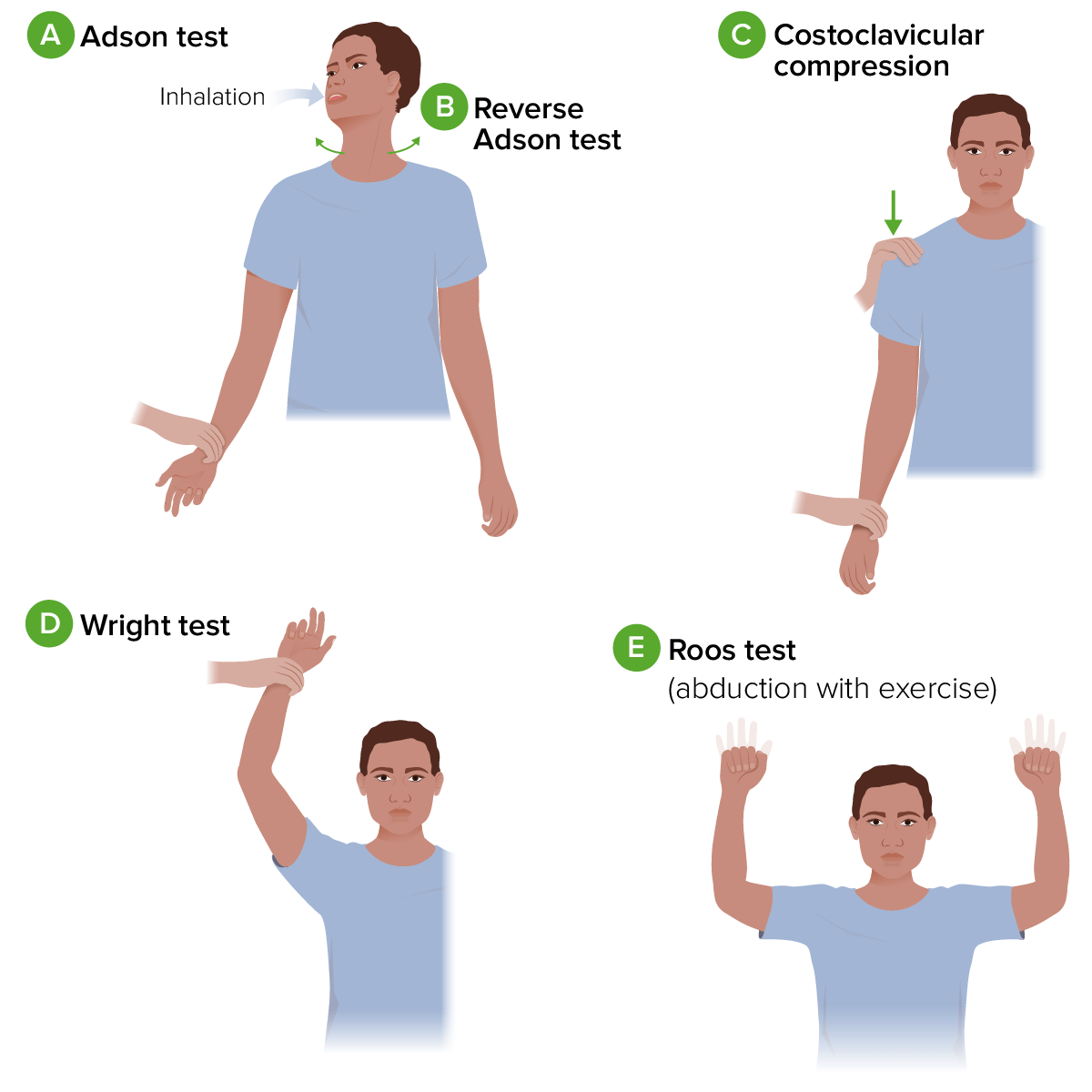
Adson’s test:

  • Procedure:
    1. Fully extend and abduct the patient’s arm Arm The arm, or “upper arm” in common usage, is the region of the upper limb that extends from the shoulder to the elbow joint and connects inferiorly to the forearm through the cubital fossa. It is divided into 2 fascial compartments (anterior and posterior). Arm: Anatomy at approximately 30 degrees.
    2. Palpate the radial pulse during the exam (should be compared to the contralateral side and ipsilateral side in a relaxed position).
    3. Ask the patient to extend their neck Neck The part of a human or animal body connecting the head to the rest of the body. Peritonsillar Abscess, rotate it toward the affected shoulder, take a deep breath Deep breath Respiratory Alkalosis, and hold the position.
  • Positive result: significant reduction or loss of radial pulse, or aggravated paresthesia
  • Prone to false-positive results; limited clinical value

Roos or elevated-arm test:

  • Procedure:
    1. Hold the patient’s arms at 90 degrees of abduction Abduction Examination of the Upper Limbs and external rotation External Rotation Examination of the Upper Limbs, with the elbows flexed and in the frontal Frontal The bone that forms the frontal aspect of the skull. Its flat part forms the forehead, articulating inferiorly with the nasal bone and the cheek bone on each side of the face. Skull: Anatomy plane.
    2. Ask the patient to hold the position and open and close their fists for a few minutes.
  • Positive results:
    • Gradual increase in pain Pain An unpleasant sensation induced by noxious stimuli which are detected by nerve endings of nociceptive neurons. Pain: Types and Pathways or paresthesia at the neck Neck The part of a human or animal body connecting the head to the rest of the body. Peritonsillar Abscess, shoulder, and/or arm Arm The arm, or “upper arm” in common usage, is the region of the upper limb that extends from the shoulder to the elbow joint and connects inferiorly to the forearm through the cubital fossa. It is divided into 2 fascial compartments (anterior and posterior). Arm: Anatomy 
    • Pallor, cyanosis Cyanosis A bluish or purplish discoloration of the skin and mucous membranes due to an increase in the amount of deoxygenated hemoglobin in the blood or a structural defect in the hemoglobin molecule. Pulmonary Examination, and swelling Swelling Inflammation of the arm Arm The arm, or “upper arm” in common usage, is the region of the upper limb that extends from the shoulder to the elbow joint and connects inferiorly to the forearm through the cubital fossa. It is divided into 2 fascial compartments (anterior and posterior). Arm: Anatomy
    • Aggravation of usual symptoms
    • The patient drops their arms in distress before completing the test.

Wright’s hyperabduction test:

  • Procedure:
    1. Passively bring the patient’s arm Arm The arm, or “upper arm” in common usage, is the region of the upper limb that extends from the shoulder to the elbow joint and connects inferiorly to the forearm through the cubital fossa. It is divided into 2 fascial compartments (anterior and posterior). Arm: Anatomy to 90 degrees of abduction Abduction Examination of the Upper Limbs and external rotation External Rotation Examination of the Upper Limbs, with the elbow flexed at 45 degrees.
    2. Palpate the radial pulse while the position is held for 1 minute.
    3. Then, repeat the test with the patient’s arm Arm The arm, or “upper arm” in common usage, is the region of the upper limb that extends from the shoulder to the elbow joint and connects inferiorly to the forearm through the cubital fossa. It is divided into 2 fascial compartments (anterior and posterior). Arm: Anatomy in hyperabduction.
  • Positive result: significant reduction or loss of radial pulse, or aggravated paresthesia

Costoclavicular maneuver:

  • Procedure:
    1. While the patient is standing, passively move their shoulder down and back while they lift their chest. 
    2. Palpate the radial pulse during the motion. 
  • Positive result: significant reduction or loss of the radial pulse, or aggravated paresthesia

Lidocaine Lidocaine A local anesthetic and cardiac depressant used as an antiarrhythmic agent. Its actions are more intense and its effects more prolonged than those of procaine but its duration of action is shorter than that of bupivacaine or prilocaine. Local Anesthetics scalene block test:
  • Procedure: Lidocaine Lidocaine A local anesthetic and cardiac depressant used as an antiarrhythmic agent. Its actions are more intense and its effects more prolonged than those of procaine but its duration of action is shorter than that of bupivacaine or prilocaine. Local Anesthetics is injected into the anterior scalene muscle under imaging guidance.
  • Positive results: significant reduction or complete relief of symptoms
Thoracic outlet syndrome provocative tests

Diagram of provocative tests for thoracic outlet syndrome:
A. Adson test: The individual’s arm is fully extend and abducted. As the examiner palpates the radial pulse, the individual is asked to extend their neck and rotate it towards the affected shoulder. They then take a deep breath and hold this position.
B. Reverse Adson test: similar to the procedure for the Adson test, but the individual is asked to rotate their head to the opposite side.
C. Costoclavicular compression: While palpating the radial pulse, the examiner passively moves the individual’s shoulder down and back while they lift their chest.
D. Wright test: The individual’s arm is passively abducted and externally rotated with a flexed elbow. The position is held for 1 minute while the examiner palpates the radial pulse. The test is then repeated with the patient’s arm in hyperabduction.
E. Roos test: The individual’s arms are held at 90 degrees of abduction and external rotation, with the elbows flexed and in the frontal plane. The position is held while the individual opens and closes their fists for a few minutes.

Image by Lecturio.

Electromyography Electromyography Recording of the changes in electric potential of muscle by means of surface or needle electrodes. Becker Muscular Dystrophy

Imaging

  • Chest X-ray X-ray Penetrating electromagnetic radiation emitted when the inner orbital electrons of an atom are excited and release radiant energy. X-ray wavelengths range from 1 pm to 10 nm. Hard x-rays are the higher energy, shorter wavelength x-rays. Soft x-rays or grenz rays are less energetic and longer in wavelength. The short wavelength end of the x-ray spectrum overlaps the gamma rays wavelength range. The distinction between gamma rays and x-rays is based on their radiation source. Pulmonary Function Tests: can confirm the presence of abnormal structures in the thoracic outlet causing compression Compression Blunt Chest Trauma on the neurovascular bundle
    • Cervical ribs Ribs A set of twelve curved bones which connect to the vertebral column posteriorly, and terminate anteriorly as costal cartilage. Together, they form a protective cage around the internal thoracic organs. Chest Wall: Anatomy
    • Elongated C7 transverse processes
    • Pancoast tumor Tumor Inflammation 
  • Ultrasound:
  • CT angiography Angiography Radiography of blood vessels after injection of a contrast medium. Cardiac Surgery:
    • Yields high-quality images
    • Delineates the anatomical relationship of vessels to the surrounding structures
  • MRI or cervical myelography: can rule out narrowing of the intervertebral foramen Intervertebral Foramen Spinal Stenosis, disk compression Compression Blunt Chest Trauma, and intraspinal space-occupying lesions
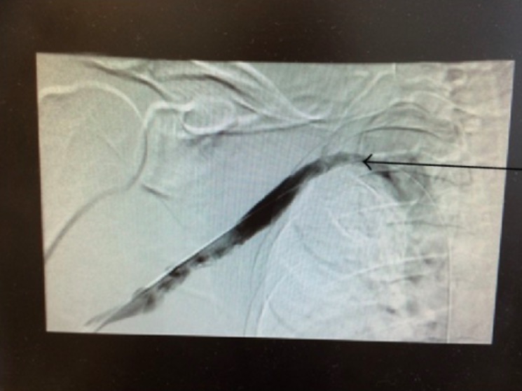
  • Angiogram or venogram: 
    • Can rule out aneurysm Aneurysm An aneurysm is a bulging, weakened area of a blood vessel that causes an abnormal widening of its diameter > 1.5 times the size of the native vessel. Aneurysms occur more often in arteries than in veins and are at risk of dissection and rupture, which can be life-threatening. Thoracic Aortic Aneurysms and thrombosis Thrombosis Formation and development of a thrombus or blood clot in the blood vessel. Epidemic Typhus of the vessels being studied
    • Not always necessary for diagnosis, but can be important for surgical planning

Management and Complications

Management

The management and care of TOS are approached via 2 different methods:

  • Medical management:
    • Posture improvement
    • Stretching
    • Physiotherapy Physiotherapy Spinal Stenosis
    • IM steroid injection
    • Analgesics and/or muscle relaxants
    • Avoiding aggravating activities
    • Use of systemic anticoagulants Anticoagulants Anticoagulants are drugs that retard or interrupt the coagulation cascade. The primary classes of available anticoagulants include heparins, vitamin K-dependent antagonists (e.g., warfarin), direct thrombin inhibitors, and factor Xa inhibitors. Anticoagulants if venous or arterial thrombosis Arterial Thrombosis Hypercoagulable States is present
  • Surgical management indicated for:
    • Vascular symptoms in patients Patients Individuals participating in the health care system for the purpose of receiving therapeutic, diagnostic, or preventive procedures. Clinician–Patient Relationship at low risk for surgery
    • Severe/progressive neurological symptoms that do not improve with medical therapy
    • Surgical approaches:
      • 1st rib resection
      • Cervical rib resection
      • Anterior scalenectomy: The anterior and middle scalene muscles are resected. 
      • Angioplasty Angioplasty Reconstruction or repair of a blood vessel, which includes the widening of a pathological narrowing of an artery or vein by the removal of atheromatous plaque material and/or the endothelial lining as well, or by dilatation (balloon angioplasty) to compress an atheroma. Except for endarterectomy, usually these procedures are performed via catheterization as minimally invasive endovascular procedures. Cardiac Surgery (with resection of the muscle and rib), if an aneurysm Aneurysm An aneurysm is a bulging, weakened area of a blood vessel that causes an abnormal widening of its diameter > 1.5 times the size of the native vessel. Aneurysms occur more often in arteries than in veins and are at risk of dissection and rupture, which can be life-threatening. Thoracic Aortic Aneurysms is present
      • Catheter-directed thrombolysis for venous or arterial thrombi

Complications

  • Frozen shoulder Frozen Shoulder Chronic Shoulder Pain:
    • If left untreated, TOS can cause increased pain Pain An unpleasant sensation induced by noxious stimuli which are detected by nerve endings of nociceptive neurons. Pain: Types and Pathways on the affected side, resulting in the inability to move the upper limb. 
    • Long periods of immobility lead to a frozen shoulder Frozen Shoulder Chronic Shoulder Pain, which is characterized by chronic stiffness and shoulder pain Pain An unpleasant sensation induced by noxious stimuli which are detected by nerve endings of nociceptive neurons. Pain: Types and Pathways.
  • Paget-Schroetter syndrome:
    • Known as effort thrombosis Thrombosis Formation and development of a thrombus or blood clot in the blood vessel. Epidemic Typhus or effort axillary-subclavian vein thrombosis Thrombosis Formation and development of a thrombus or blood clot in the blood vessel. Epidemic Typhus
    • Excess strenuous movement of the upper limb can lead to thrombosis Thrombosis Formation and development of a thrombus or blood clot in the blood vessel. Epidemic Typhus of the subclavian vein (deep vein thrombosis Thrombosis Formation and development of a thrombus or blood clot in the blood vessel. Epidemic Typhus).
  • Cerebrovascular arterial insufficiency:
    • The carotid artery is also present in the thoracic outlet, and distal compression Compression Blunt Chest Trauma of the subclavian artery can lead to carotid artery compression Compression Blunt Chest Trauma and stroke.
    • If the vertebral artery Vertebral artery The first branch of the subclavian artery with distribution to muscles of the neck; vertebrae; spinal cord; cerebellum; and interior of the cerebrum. Lateral Medullary Syndrome (Wallenberg Syndrome) is affected, vision Vision Ophthalmic Exam abnormalities, embolic stroke Embolic stroke An ischemic stroke due to a blood clot, emboli or other types of blockage which forms somewhere other than the brain and subsequently travels near and restricts blood flow to the brain. Most often the origin of the clot is from the heart and is referred to as cardioembolic stroke. Ischemic Stroke, and hypoperfusion of the brainstem and brain Brain The part of central nervous system that is contained within the skull (cranium). Arising from the neural tube, the embryonic brain is comprised of three major parts including prosencephalon (the forebrain); mesencephalon (the midbrain); and rhombencephalon (the hindbrain). The developed brain consists of cerebrum; cerebellum; and other structures in the brain stem. Nervous System: Anatomy, Structure, and Classification are possible.

Differential Diagnosis

  • Carpal tunnel Carpal Tunnel The carpal tunnel is formed by the transverse carpal ligament (flexor retinaculum) superiorly and the carpal bones inferiorly. Carpal Tunnel Syndrome syndrome: a type of peripheral neuropathy Neuropathy Leprosy caused by compression Compression Blunt Chest Trauma of the median nerve Median Nerve A major nerve of the upper extremity. In humans, the fibers of the median nerve originate in the lower cervical and upper thoracic spinal cord (usually C6 to T1), travel via the brachial plexus, and supply sensory and motor innervation to parts of the forearm and hand. Cubital Fossa: Anatomy. Symptoms include numbness, the sensation of heaviness, and decreased grip strength in the hands owing to sensory Sensory Neurons which conduct nerve impulses to the central nervous system. Nervous System: Histology loss in the palmar aspect, the 1st–3rd digits, and the radial half of the 4th digit. Diagnosis is made based on a compression Compression Blunt Chest Trauma test, Tinel’s sign, and Phalen’s sign. Management includes occupational therapy Occupational Therapy Skilled treatment that helps individuals achieve independence in all facets of their lives. It assists in the development of skills needed for independent living. Fetal Alcohol Spectrum Disorder, neutral positioning of a wrist splint, steroid injection, or surgery.
  • De Quervain tenosynovitis: inflammation Inflammation Inflammation is a complex set of responses to infection and injury involving leukocytes as the principal cellular mediators in the body’s defense against pathogenic organisms. Inflammation is also seen as a response to tissue injury in the process of wound healing. The 5 cardinal signs of inflammation are pain, heat, redness, swelling, and loss of function. Inflammation of the 1st extensor compartment of the wrist, which results in radial pain Pain An unpleasant sensation induced by noxious stimuli which are detected by nerve endings of nociceptive neurons. Pain: Types and Pathways and swelling Swelling Inflammation. Diagnosis is made based on positive Finkelstein’s test Finkelstein’S Test Hand and Wrist Pain and an ultrasound, which shows synovial thickening. Management includes physiotherapy Physiotherapy Spinal Stenosis, splinting of the affected joint, steroid injection, or surgery.
  • Horner syndrome Horner syndrome Horner syndrome is a condition resulting from an interruption of the sympathetic innervation of the eyes. The syndrome is usually idiopathic but can be directly caused by head and neck trauma, cerebrovascular disease, or a tumor of the CNS. Horner Syndrome: a neurological disorder resulting from the interruption of the sympathetic innervation of the head, eye, and neck Neck The part of a human or animal body connecting the head to the rest of the body. Peritonsillar Abscess. Horner syndrome Horner syndrome Horner syndrome is a condition resulting from an interruption of the sympathetic innervation of the eyes. The syndrome is usually idiopathic but can be directly caused by head and neck trauma, cerebrovascular disease, or a tumor of the CNS. Horner Syndrome is characterized by a triad of miosis Miosis Pupil: Physiology and Abnormalities, partial ptosis Partial Ptosis Horner Syndrome, and absence of facial sweating, but can also present with pain Pain An unpleasant sensation induced by noxious stimuli which are detected by nerve endings of nociceptive neurons. Pain: Types and Pathways in the face and neck Neck The part of a human or animal body connecting the head to the rest of the body. Peritonsillar Abscess, and atrophy Atrophy Decrease in the size of a cell, tissue, organ, or multiple organs, associated with a variety of pathological conditions such as abnormal cellular changes, ischemia, malnutrition, or hormonal changes. Cellular Adaptation of the hand Hand The hand constitutes the distal part of the upper limb and provides the fine, precise movements needed in activities of daily living. It consists of 5 metacarpal bones and 14 phalanges, as well as numerous muscles innervated by the median and ulnar nerves. Hand: Anatomy and forearm Forearm The forearm is the region of the upper limb between the elbow and the wrist. The term “forearm” is used in anatomy to distinguish this area from the arm, a term that is commonly used to describe the entire upper limb. The forearm consists of 2 long bones (the radius and the ulna), the interosseous membrane, and multiple arteries, nerves, and muscles. Forearm: Anatomy muscles. Diagnosis is clinical and supported by findings from imaging studies. Treatment involves resolving the underlying cause.
  • Raynaud’s disease: an exaggerated vascular response of the digits to cold temperatures or emotional stress, which typically results in sequential Sequential Computed Tomography (CT) digital blanching Blanching Dermatologic Examination, cyanosis Cyanosis A bluish or purplish discoloration of the skin and mucous membranes due to an increase in the amount of deoxygenated hemoglobin in the blood or a structural defect in the hemoglobin molecule. Pulmonary Examination, and rubor Rubor Inflammation. Diagnosis of primary Raynaud’s disease relies on the Allen test Allen test With simultaneous compression of the radial and ulnar arteries, the hand remains pale after alternately releasing. Thromboangiitis Obliterans (Buerger’s Disease) and laboratory workup for secondary causes. Management includes treatment of the underlying cause or therapy with vasodilators Vasodilators Drugs used to cause dilation of the blood vessels. Thromboangiitis Obliterans (Buerger’s Disease).
  • Pectoralis minor syndrome: a rare syndrome that causes pain Pain An unpleasant sensation induced by noxious stimuli which are detected by nerve endings of nociceptive neurons. Pain: Types and Pathways, numbness, and tingling Tingling Posterior Cord Syndrome in the hand Hand The hand constitutes the distal part of the upper limb and provides the fine, precise movements needed in activities of daily living. It consists of 5 metacarpal bones and 14 phalanges, as well as numerous muscles innervated by the median and ulnar nerves. Hand: Anatomy and arm Arm The arm, or “upper arm” in common usage, is the region of the upper limb that extends from the shoulder to the elbow joint and connects inferiorly to the forearm through the cubital fossa. It is divided into 2 fascial compartments (anterior and posterior). Arm: Anatomy, and pain Pain An unpleasant sensation induced by noxious stimuli which are detected by nerve endings of nociceptive neurons. Pain: Types and Pathways or tenderness in the chest wall Chest wall The chest wall consists of skin, fat, muscles, bones, and cartilage. The bony structure of the chest wall is composed of the ribs, sternum, and thoracic vertebrae. The chest wall serves as armor for the vital intrathoracic organs and provides the stability necessary for the movement of the shoulders and arms. Chest Wall: Anatomy below the clavicle Clavicle A bone on the ventral side of the shoulder girdle, which in humans is commonly called the collar bone. Clavicle Fracture and axilla Axilla The axilla is a pyramid-shaped space located between the upper thorax and the arm. The axilla has a base, an apex, and 4 walls (anterior, medial, lateral, posterior). The base of the pyramid is made up of the axillary skin. The apex is the axillary inlet, located between the 1st rib, superior border of the scapula, and clavicle. Axilla and Brachial Plexus: Anatomy. Pectoralis minor syndrome occurs due to compression Compression Blunt Chest Trauma of the neurovascular structures under the pectoralis minor muscle. Diagnosis is clinical and supported by imaging. Management includes physiotherapy Physiotherapy Spinal Stenosis, pectoralis minor muscle block, and surgery.

References

  1. Mohammad Ali Hosseinian, Ali Gharibi Loron, and Yalda Soleimanifard (2017). Evaluation of complications after surgical treatment of thoracic outlet syndrome, NCBI. https://www.ncbi.nlm.nih.gov/pmc/articles/PMC5295481/#:~:text=Complications%20of%20TOS%20surgery%20include,plexus%2C%20severe%20sequelae%20such%20as
  2. Kaoru Goshima (2020). Overview of thoracic outlet syndromes. Retrieved April 8, 2021, from https://www.uptodate.com/contents/overview-of-thoracic-outlet-syndromes
  3. Riki Duncan. Thoracic Outlet Syndrome or Carpal Tunnel Syndrome. KCBJ. Retrieved April 8, 2021, from https://www.kcbj.com/thoracic-outlet-syndrome-or-carpal-tunnel-syndrome
  4. Sian Smale (2013). Thoracic Outlet Syndrome; Rayner and Smale. https://www.raynersmale.com/blog/2013/12/20/thoracic-outlet-syndrome

Create your free account or log in to continue reading!

Sign up now and get free access to Lecturio with concept pages, medical videos, and questions for your medical education.

User Reviews

Unwrap New Skills This Holiday 🎄 Save 30% on all plans now!

Details