The epithelium is a complex of specialized cellular organizations arranged into sheets and lining cavities and covering the surfaces of the body. The cells exhibit polarity, having an apical and a basal pole. Structures important for the epithelial integrity and function involve the basement membrane Basement membrane A darkly stained mat-like extracellular matrix (ecm) that separates cell layers, such as epithelium from endothelium or a layer of connective tissue. The ecm layer that supports an overlying epithelium or endothelium is called basal lamina. Basement membrane (bm) can be formed by the fusion of either two adjacent basal laminae or a basal lamina with an adjacent reticular lamina of connective tissue. Bm, composed mainly of type IV collagen; glycoprotein laminin; and proteoglycan, provides barriers as well as channels between interacting cell layers. Thin Basement Membrane Nephropathy (TBMN), the semipermeable sheet on which the cells rest, and interdigitations, as well as cellular junctions. The epithelium is classified according to the cells (squamous, cuboidal, columnar), the number of layers, and other unique characteristics either due to function (transitional epithelium allowing distention) or appearance (pseudostratified epithelium giving a false impression of multiple layers). The surface epithelium has multiple functions, which include protection, secretion Secretion Coagulation Studies, filtration, and sensory Sensory Neurons which conduct nerve impulses to the central nervous system. Nervous System: Histology reception.
Last updated: Apr 18, 2023
A complex of specialized cellular organizations arranged into sheets and lining cavities and covering the surfaces of the body:
These cells exhibit polarity:
The surface epithelium does not possess blood vessels; therefore, nutrients and oxygen are received from adjacent connective tissue Connective tissue Connective tissues originate from embryonic mesenchyme and are present throughout the body except inside the brain and spinal cord. The main function of connective tissues is to provide structural support to organs. Connective tissues consist of cells and an extracellular matrix. Connective Tissue: Histology.
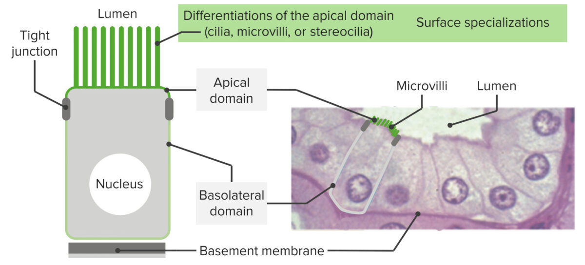
Epithelia have an apex, lateral borders, and a surface sitting on a basement membrane:
As seen in the image, these cells exhibit polarity (have apical domains and basal/basolateral domains). At the apical end, structures encircle the area (like a band around the cell) facilitating cell-to-cell adhesion (tight junctions and adherent junctions). Near the basement membrane, hemidesmosomes anchor the epithelia to the basal lamina. On the right is shown the histology of the intestinal epithelial lining with corresponding structures. On the apical domain, microvilli are seen.
Note that there are tissues that have both:
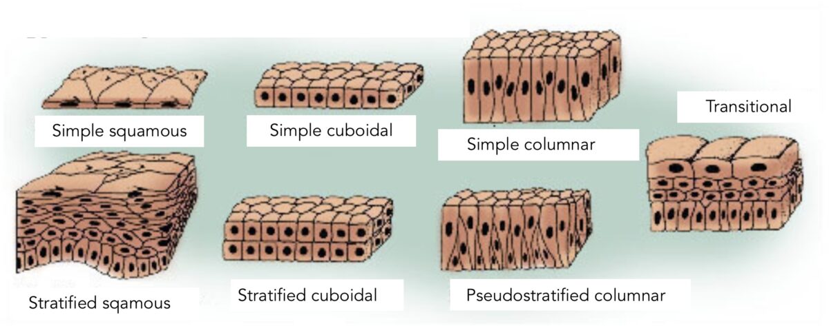
The types of epithelia:
The differentiation and classification of the types are based on the shape of the cell and the layers. Simple epithelium indicates 1 layer of cells. Stratified epithelium indicates multiple layers. Pseudostratified epithelium gives a false impression of > 1 layer owing to different nuclei levels. Transitional epithelium is a type in which the shape of cells “go into transition” depending on the organ function (distention versus relaxation, as seen in the urinary bladder).
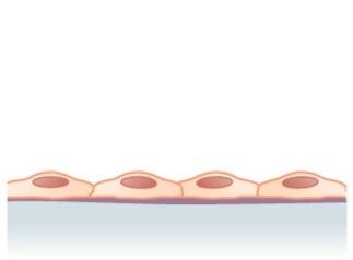
Structure noted in simple squamous epithelium (single layer of flattened cells)
Image: “Simple squamous epithelium” by Phil Schatz. License: CC BY 4.0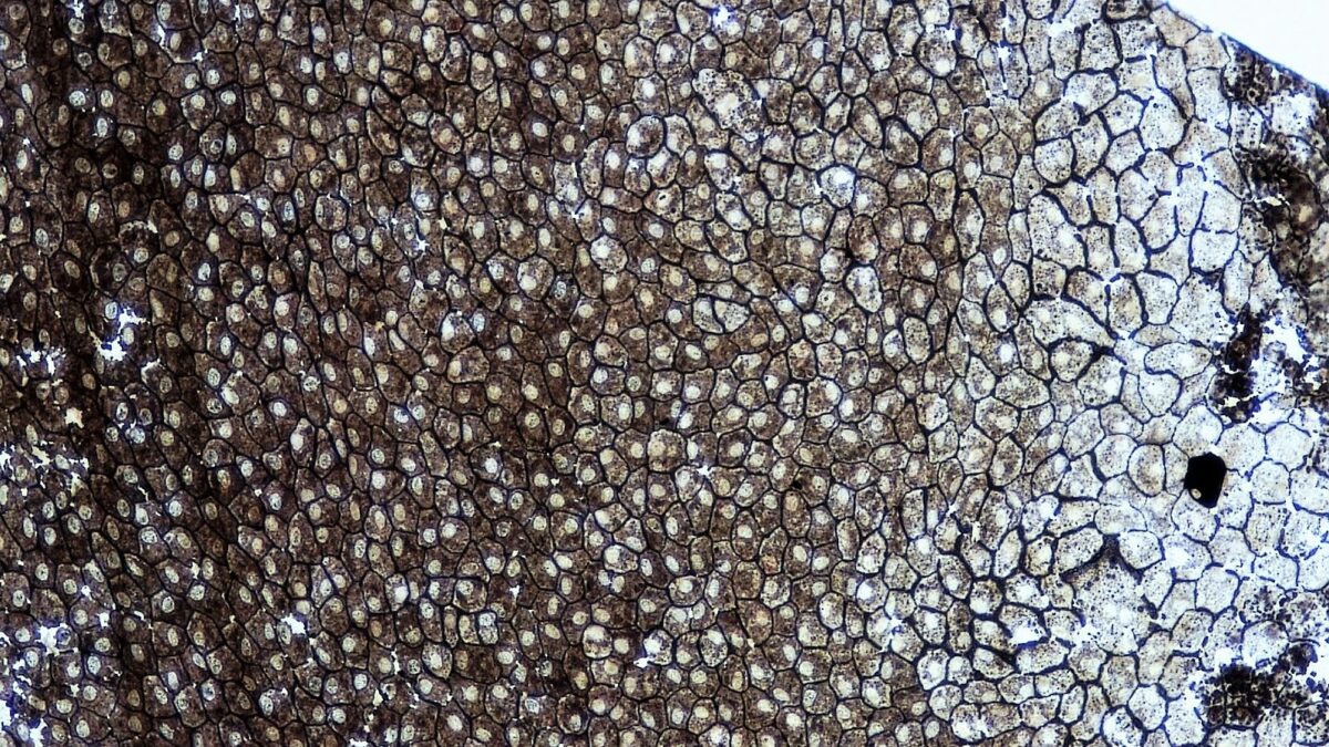
A whole mount of simple squamous epithelium
Image: “Epithelial Tissues Simple Squamous Epithelium” by Berkshire Community College Bioscience Image Library. License: CC0 1.0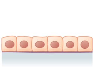
Simple cuboidal epithelium: 1 layer of cuboidal cells
Image: “Simple cuboidal epithelium” by Phil Schatz. License: CC BY 4.0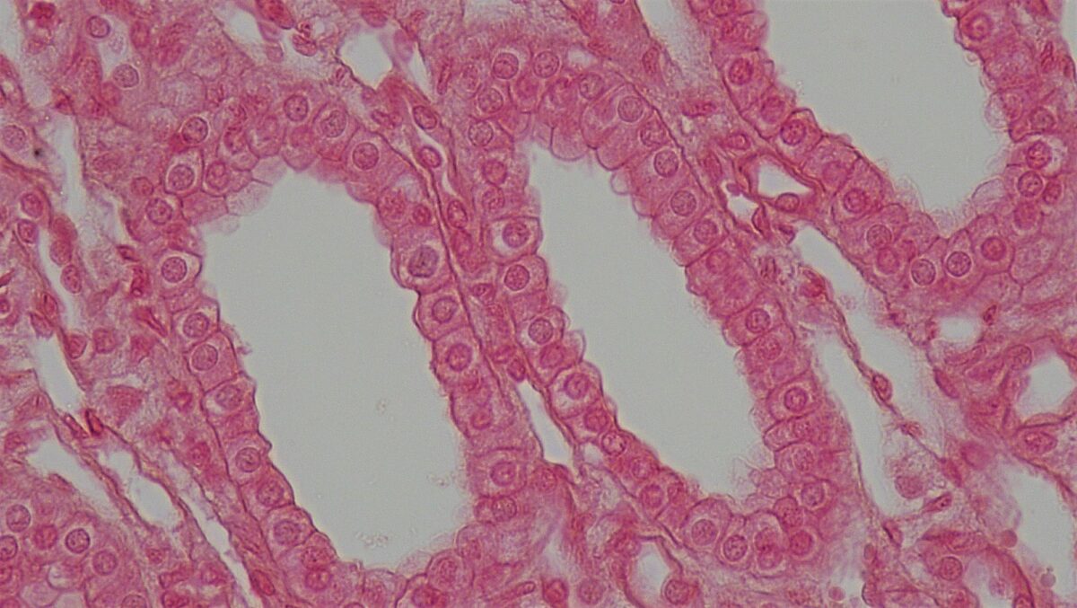
Cross section of a kidney tubule showing a layer of cuboidal cells
Image: “Epithelial Tissues Simple Cuboidal Epithelium” by Berkshire Community College Bioscience Image Library. License: CC0 1.0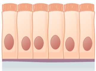
Simple columnar epithelium:
Shown is a layer of columnar cells with cilia
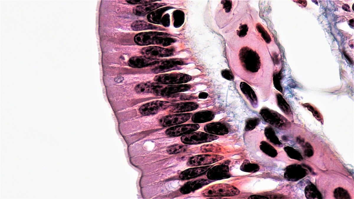
Simple columnar epithelium lining the intestinal tract
Image: “Epithelial Tissues Simple Columnar Epithelium” by Epithelial Tissues: Simple Columnar Epithelium. License: CC0 1.0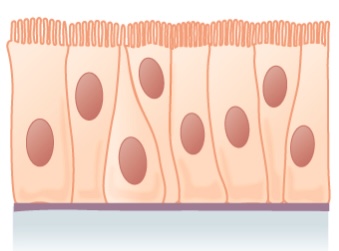
Pseudostratified epithelium:
Shown is a single layer of columnar cells, with varying levels of nuclei
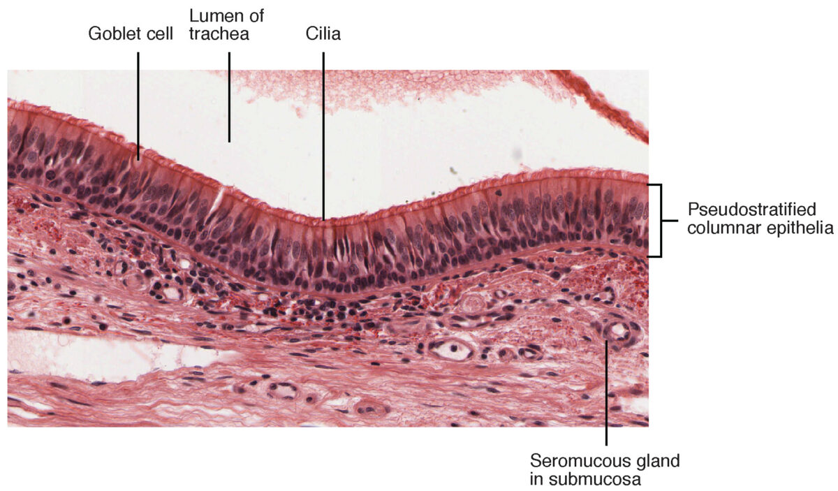
Pseudostratified columnar epithelium found in the trachea
Image: “Cross-section of pseudostratified columnar epithelium” by OpenStax College. License: CC BY 3.0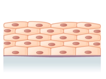
Illustration of stratified squamous epithelium (multiple layers)
Image: “Stratified squamous epithelium” by Phil Schatz. License: CC BY 4.0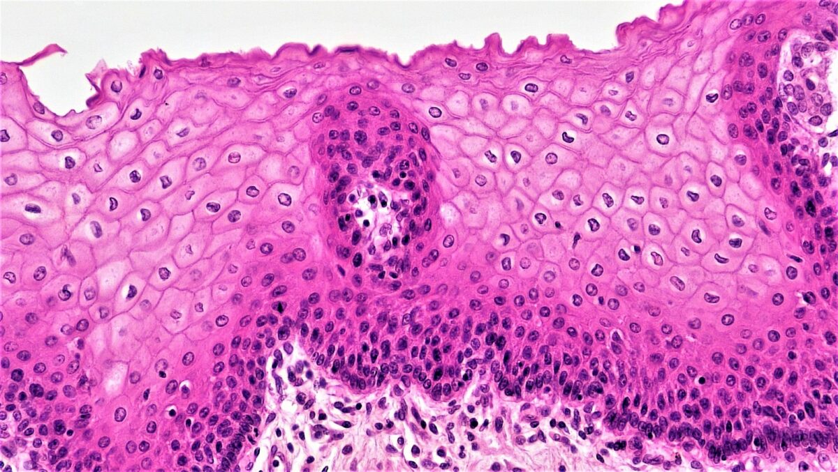
Histologic picture of stratified squamous epithelium
Image: “Epithelial Tissues Stratified Squamous Epithelium” by Berkshire Community College Bioscience Image Library. License: CC0 1.0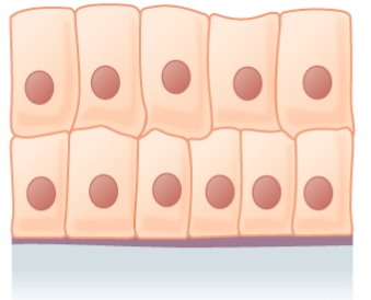
Stratified columnar epithelium
Image: “Stratified columnar epithelium” by Phil Schatz. License: CC BY 4.0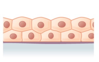
Stratified cuboidal epithelium
Image: “Stratified cuboidal epithelium” by Phil Schatz. License: CC BY 4.0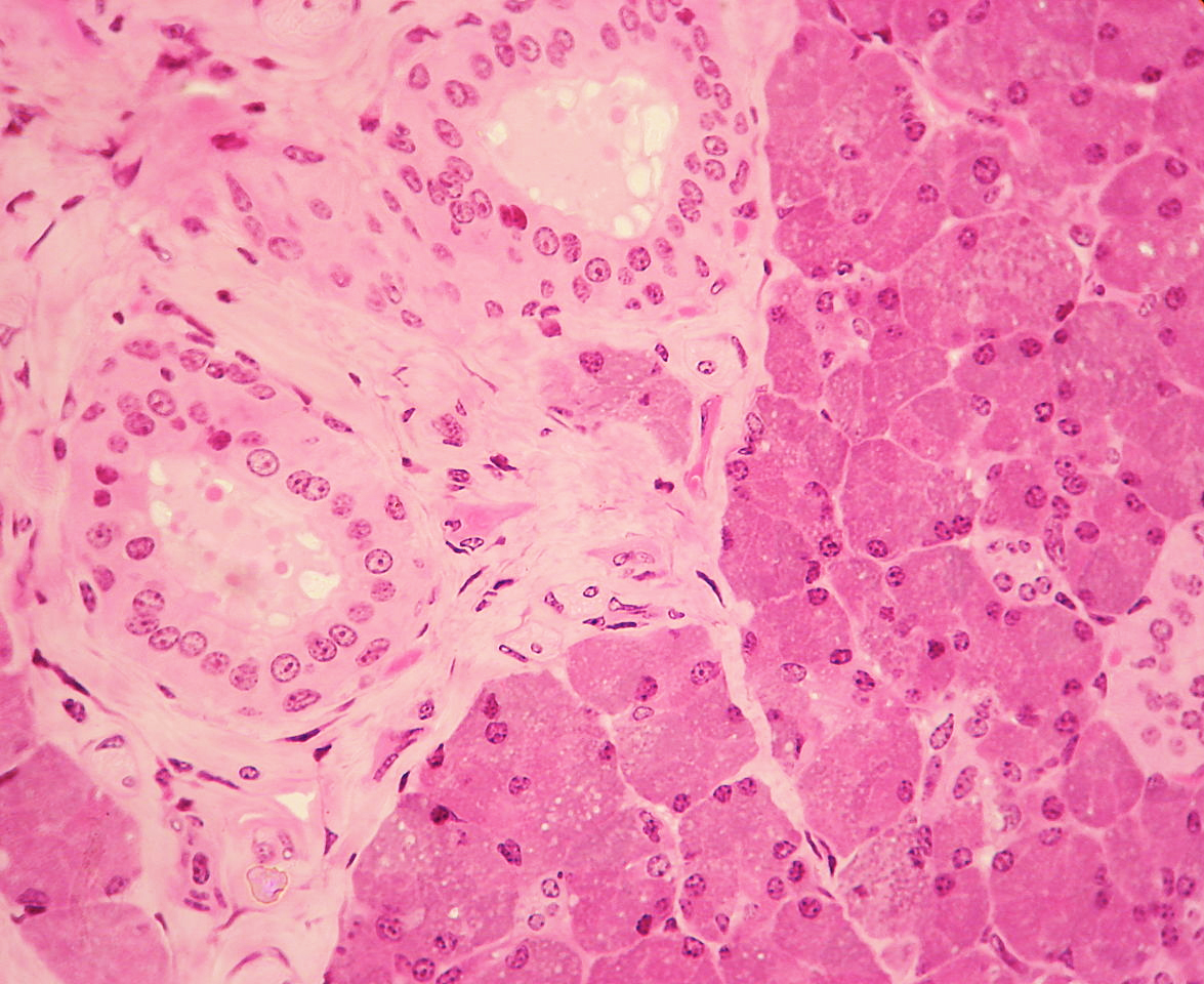
Stratified cuboidal epithelium (seen on the left) is visible in a duct surrounded by connective tissue in the parotid gland.
Image: “WVSOM Parotid Gland1” by Wbensmith. License: CC BY 3.0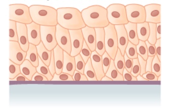
Transitional epithelium
Image: “Transitional epithelium” by Phil Schatz. License: CC BY 4.0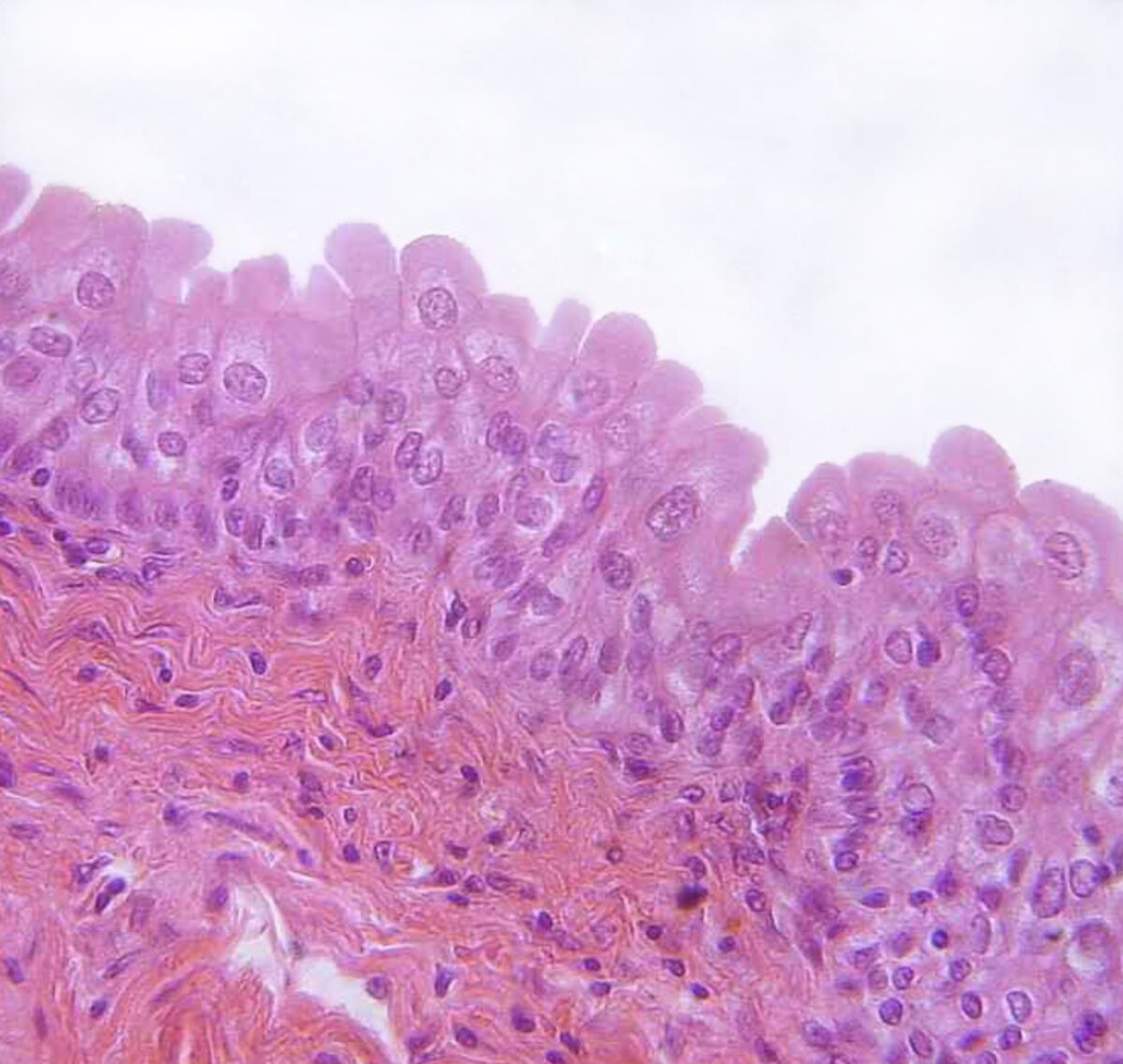
Transitional epithelium found in the urinary bladder
Image: “urinary bladder, urothelium, haemalum-eosin stain” by Polarlys. License: CC BY 2.5