Advertisement

Advertisement

Advertisement

Advertisement

Skin: Structure and Functions

The skin, also referred to as the integumentary system, is the largest organ of the body. The skin is primarily composed of the epidermis (outer layer) and dermis (deep layer). The epidermis is primarily composed of keratinocytes that undergo rapid turnover, while the dermis contains dense layers of connective tissue Connective tissue Connective tissues originate from embryonic mesenchyme and are present throughout the body except inside the brain and spinal cord. The main function of connective tissues is to provide structural support to organs. Connective tissues consist of cells and an extracellular matrix. Connective Tissue: Histology. The skin is composed of surface epithelium Surface epithelium Epithelium is classified according to the cells (squamous, cuboidal, columnar), the number of layers, and other unique characteristics either due to function (transitional epithelium allowing distention) or appearance (pseudostratified epithelium giving a false impression of multiple layers). Surface epithelium has multiple functions, which include protection, secretion, filtration, and sensory reception. Surface Epithelium: Histology, exocrine components, connective tissue Connective tissue Connective tissues originate from embryonic mesenchyme and are present throughout the body except inside the brain and spinal cord. The main function of connective tissues is to provide structural support to organs. Connective tissues consist of cells and an extracellular matrix. Connective Tissue: Histology, muscles, and nerves. The primary role of the skin is to serve as a protective barrier between the internal body and the external environment; it also protects the body from excessive fluid loss.

Last updated: Apr 18, 2023
Editorial responsibility: Stanley Oiseth, Lindsay Jones, Evelin Maza

Advertisement

Advertisement

Advertisement

Advertisement

Advertisement

Advertisement

Structure of the Skin

Cells of the skin

  • Keratinocytes:
    • Primary cell type of the epidermis
    • Undergo continuous mitosis Mitosis A type of cell nucleus division by means of which the two daughter nuclei normally receive identical complements of the number of chromosomes of the somatic cells of the species. Cell Cycle (stimulated by epidermal growth factor)
    • Produce keratin Keratin A class of fibrous proteins or scleroproteins that represents the principal constituent of epidermis; hair; nails; horny tissues, and the organic matrix of tooth enamel. Two major conformational groups have been characterized, alpha-keratin, whose peptide backbone forms a coiled-coil alpha helical structure consisting of type I keratin and a type II keratin, and beta-keratin, whose backbone forms a zigzag or pleated sheet structure. Alpha-keratins have been classified into at least 20 subtypes. In addition multiple isoforms of subtypes have been found which may be due to gene duplication. Seborrheic Keratosis
  • Melanocytes:
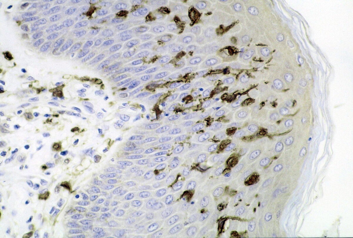
    • Spider-shaped epithelial cells
    • Produce melanin Melanin Insoluble polymers of tyrosine derivatives found in and causing darkness in skin (skin pigmentation), hair, and feathers providing protection against sunburn induced by sunlight. Carotenes contribute yellow and red coloration. Seborrheic Keratosis (UV protection)
    • Melanin Melanin Insoluble polymers of tyrosine derivatives found in and causing darkness in skin (skin pigmentation), hair, and feathers providing protection against sunburn induced by sunlight. Carotenes contribute yellow and red coloration. Seborrheic Keratosis granules accumulate on the apical surface of keratinocytes.
  • Dendritic cells (Langerhans cells):
    • Resident macrophages Macrophages The relatively long-lived phagocytic cell of mammalian tissues that are derived from blood monocytes. Main types are peritoneal macrophages; alveolar macrophages; histiocytes; kupffer cells of the liver; and osteoclasts. They may further differentiate within chronic inflammatory lesions to epithelioid cells or may fuse to form foreign body giant cells or langhans giant cells. Innate Immunity: Phagocytes and Antigen Presentation of the skin
    • Antigen-presenting cells Antigen-presenting cells A heterogeneous group of immunocompetent cells that mediate the cellular immune response by processing and presenting antigens to the T-cells. Traditional antigen-presenting cells include macrophages; dendritic cells; langerhans cells; and B-lymphocytes. Follicular dendritic cells are not traditional antigen-presenting cells, but because they hold antigen on their cell surface in the form of immune complexes for b-cell recognition they are considered so by some authors. Adaptive Immune Response
    • Activate the immune system Immune system The body’s defense mechanism against foreign organisms or substances and deviant native cells. It includes the humoral immune response and the cell-mediated response and consists of a complex of interrelated cellular, molecular, and genetic components. Primary Lymphatic Organs
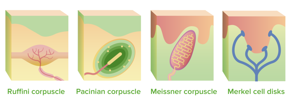
  • Mechanoreceptors:
    • Merkel cells:
    • Ruffini corpuscles:
      • Slowly adapting stretch receptors Receptors Receptors are proteins located either on the surface of or within a cell that can bind to signaling molecules known as ligands (e.g., hormones) and cause some type of response within the cell. Receptors
      • Located in deep dermis Deep Dermis Erysipelas
    • Meissner corpuscles:
      • Rapidly adapting light-touch receptors Receptors Receptors are proteins located either on the surface of or within a cell that can bind to signaling molecules known as ligands (e.g., hormones) and cause some type of response within the cell. Receptors
      • Located in superficial dermis
    • Pacinian corpuscles:

Epidermis

  • Derived from ectoderm Ectoderm The outer of the three germ layers of an embryo. Gastrulation and Neurulation
  • Keratinized stratified squamous epithelium Stratified squamous epithelium Surface Epithelium: Histology
  • 4 or 5 main layers:
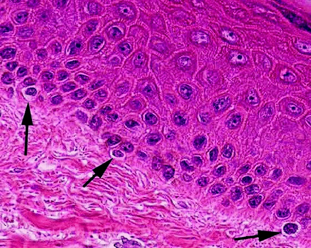
    • Stratum basale (stratum germinativum):
      • Deepest layer
      • Germinating layer
      • Single row of epithelial cells bound to the dermis by hemidesmosomes ( macula Macula An oval area in the retina, 3 to 5 mm in diameter, usually located temporal to the posterior pole of the eye and slightly below the level of the optic disk. It is characterized by the presence of a yellow pigment diffusely permeating the inner layers, contains the fovea centralis in its center, and provides the best phototropic visual acuity. It is devoid of retinal blood vessels, except in its periphery, and receives nourishment from the choriocapillaris of the choroid. Eye: Anatomy adherens Adherens Anchoring points where the cytoskeleton of neighboring cells are connected to each other. They are composed of specialized areas of the plasma membrane where bundles of the actin cytoskeleton attach to the membrane through the transmembrane linkers, cadherins, which in turn attach through their extracellular domains to cadherins in the neighboring cell membranes. In sheets of cells, they form into adhesion belts (zonula adherens) that go all the way around a cell. The Cell: Cell Junctions)
      • Divide constantly while moving toward the surface
      • 10%–15% of the cells are melanocytes.
    • Stratum spinosum (prickly layer):
      • Consists of several layers (thickest layer of epidermis)
      • Web-like system of filaments
      • Cells are linked together by desmosomes Desmosomes A type of junction that attaches one cell to its neighbor. One of a number of differentiated regions which occur, for example, where the cytoplasmic membranes of adjacent epithelial cells are closely apposed. It consists of a circular region of each membrane together with associated intracellular microfilaments and an intercellular material which may include, for example, mucopolysaccharides. Bullous Pemphigoid and Pemphigus Vulgaris.
      • As cells move up through the spinous layer, they accumulate keratohyalin granules.
      • Other elements present are dendritic cells and melanin Melanin Insoluble polymers of tyrosine derivatives found in and causing darkness in skin (skin pigmentation), hair, and feathers providing protection against sunburn induced by sunlight. Carotenes contribute yellow and red coloration. Seborrheic Keratosis granules.
    • Stratum granulosum:
      • Thin layer of 4–6 cells
      • Location of keratinization
      • Cells flatten and nuclei and organelles Organelles A cell is a complex unit that performs several complex functions. An organelle is a specialized subunit within a cell that fulfills a specific role or function. Organelles are enclosed within their own lipid bilayers or are unbound by membranes. The Cell: Organelles begin to disintegrate.
      • Cells acquire 2 types of granules, keratohyalin granules and lamellar granules (protect against water loss).
    • Stratum lucidum (clear layer):
      • Seen ony in thick skin
      • Thin translucent band
      • Made up of 2–3 layers of keratinocytes 
    • Stratum corneum (horny layer):
      • 20–30 layers of flattened anucleate cells
      • Thinner on lens Lens A transparent, biconvex structure of the eye, enclosed in a capsule and situated behind the iris and in front of the vitreous humor (vitreous body). It is slightly overlapped at its margin by the ciliary processes. Adaptation by the ciliary body is crucial for ocular accommodation. Eye: Anatomy of the eye, thicker on calluses 
  • 4 types of cells:
    • Keratinocytes:
      • Deepest, produce keratin Keratin A class of fibrous proteins or scleroproteins that represents the principal constituent of epidermis; hair; nails; horny tissues, and the organic matrix of tooth enamel. Two major conformational groups have been characterized, alpha-keratin, whose peptide backbone forms a coiled-coil alpha helical structure consisting of type I keratin and a type II keratin, and beta-keratin, whose backbone forms a zigzag or pleated sheet structure. Alpha-keratins have been classified into at least 20 subtypes. In addition multiple isoforms of subtypes have been found which may be due to gene duplication. Seborrheic Keratosis (tough fibrous Fibrous Fibrocystic Change protein)
      • Cycle through the epidermis every 30 days
    • Melanocytes:
      • Produce melanin Melanin Insoluble polymers of tyrosine derivatives found in and causing darkness in skin (skin pigmentation), hair, and feathers providing protection against sunburn induced by sunlight. Carotenes contribute yellow and red coloration. Seborrheic Keratosis
      • Live within the stratum basale
    • Merkel cells:
      • Linked with sensory Sensory Neurons which conduct nerve impulses to the central nervous system. Nervous System: Histology nerve endings
      • Only mechanoreceptor with connection to epidermis
    • Langerhans cells:
      • Macrophage-like dendritic cells
      • Activate immune system Immune system The body’s defense mechanism against foreign organisms or substances and deviant native cells. It includes the humoral immune response and the cell-mediated response and consists of a complex of interrelated cellular, molecular, and genetic components. Primary Lymphatic Organs
Layers of the epidermis

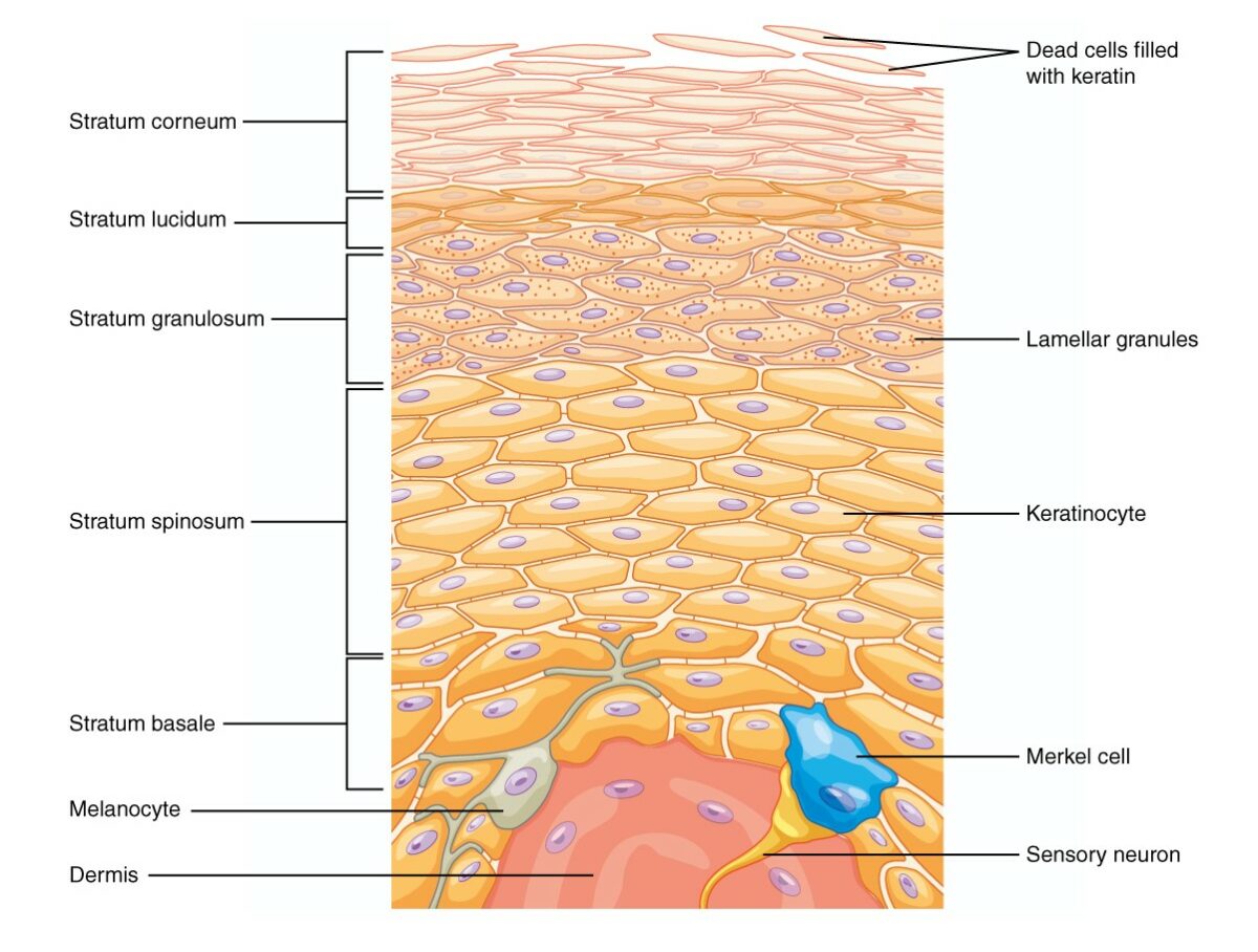
There are 5 layers of epidermis:
Beginning from the cells located in the stratum basale, they differentiate and proliferate toward the skin surface and integrate themselves into the other layers, the last layer being the stratum corneum, which is composed of dead keratinized cells.

Image: “The epidermis of thick skin has five layers: stratum basale, stratum spinosum, stratum granulosum, stratum lucidum, and stratum corneum.” by OpenStax College. License: CC BY 4.0

Dermis

  • Derived from mesoderm Mesoderm The middle germ layer of an embryo derived from three paired mesenchymal aggregates along the neural tube. Gastrulation and Neurulation
  • Components:
    • Strong flexible connective tissue Connective tissue Connective tissues originate from embryonic mesenchyme and are present throughout the body except inside the brain and spinal cord. The main function of connective tissues is to provide structural support to organs. Connective tissues consist of cells and an extracellular matrix. Connective Tissue: Histology (primarily collagen Collagen A polypeptide substance comprising about one third of the total protein in mammalian organisms. It is the main constituent of skin; connective tissue; and the organic substance of bones (bone and bones) and teeth (tooth). Connective Tissue: Histology)
    • Contains fibroblasts Fibroblasts Connective tissue cells which secrete an extracellular matrix rich in collagen and other macromolecules. Sarcoidosis, macrophages Macrophages The relatively long-lived phagocytic cell of mammalian tissues that are derived from blood monocytes. Main types are peritoneal macrophages; alveolar macrophages; histiocytes; kupffer cells of the liver; and osteoclasts. They may further differentiate within chronic inflammatory lesions to epithelioid cells or may fuse to form foreign body giant cells or langhans giant cells. Innate Immunity: Phagocytes and Antigen Presentation, occasional mast cells Mast cells Granulated cells that are found in almost all tissues, most abundantly in the skin and the gastrointestinal tract. Like the basophils, mast cells contain large amounts of histamine and heparin. Unlike basophils, mast cells normally remain in the tissues and do not circulate in the blood. Mast cells, derived from the bone marrow stem cells, are regulated by the stem cell factor. Innate Immunity: Phagocytes and Antigen Presentation, and WBCs
    • Elastic Elastic Connective Tissue: Histology fibers provide flexibility to the dermis
    • Sebaceous glands maintain elasticity Elasticity Resistance and recovery from distortion of shape. Skeletal Muscle Contraction and strength.
  • Supplies the epidermis with nutrients + moisture
  • Contains rich supply of blood vessels, lymphatic vessels Lymphatic Vessels Tubular vessels that are involved in the transport of lymph and lymphocytes. Lymphatic Drainage System: Anatomy, and nerve endings 
  • 2 layers:
    • Papillary layer:
      • Thin superficial layer
      • Made up of elastin and collagen Collagen A polypeptide substance comprising about one third of the total protein in mammalian organisms. It is the main constituent of skin; connective tissue; and the organic substance of bones (bone and bones) and teeth (tooth). Connective Tissue: Histology fibers
      • Contains phagocytes, fibroblasts Fibroblasts Connective tissue cells which secrete an extracellular matrix rich in collagen and other macromolecules. Sarcoidosis, and adipocytes Adipocytes Cells in the body that store fats, usually in the form of triglycerides. White adipocytes are the predominant type and found mostly in the abdominal cavity and subcutaneous tissue. Brown adipocytes are thermogenic cells that can be found in newborns of some species and hibernating mammals. Adipose Tissue: Histology
      • Projections of this layer into the epidermis are called dermal papillae Papillae Lips and Tongue: Anatomy.
      • Dermal papillae Papillae Lips and Tongue: Anatomy form epidermal ridges at the surface of the epidermis (fingerprints).
      • Capillary loops and nerve endings (free nerve endings and Meissner corpuscles) are located in the dermal papillae Papillae Lips and Tongue: Anatomy.
    • Reticular layer:
      • Accounts for 80% of the thickness of the dermis
      • Irregular dense fibrous Fibrous Fibrocystic Change connective tissue Connective tissue Connective tissues originate from embryonic mesenchyme and are present throughout the body except inside the brain and spinal cord. The main function of connective tissues is to provide structural support to organs. Connective tissues consist of cells and an extracellular matrix. Connective Tissue: Histology
      • Well-vascularized cutaneous plexus
      • Gives the skin its elasticity Elasticity Resistance and recovery from distortion of shape. Skeletal Muscle Contraction and helps to toughen the skin
      • Contains sweat glands Sweat glands Sweat-producing structures that are embedded in the dermis. Each gland consists of a single tube, a coiled body, and a superficial duct. Soft Tissue Abscess and hair follicles
Layers of the dermis

The dermis is made up of 2 layers:
The papillary layer is made up of collagen and elastin fibers and contains phagocytes, fibroblasts, and adipocytes. The reticular layer gives the skin its elasticity and helps to toughen the skin. Sweat glands and hair follicles are found at this layer. This layer is also well vascularized.

Image: “This stained slide shows the two components of the dermis—the papillary layer and the reticular layer.” by OpenStax College. License: CC BY 4.0

Function of the Skin

Each layer of the skin has a unique function.

Epidermis and dermis:

  • Mechanical protection
  • Protection against external pathogens
  • Maintenance of cutaneous water loss
  • Temperature regulation:
    • ↑ Temperature → Blood vessels in papillary layer of the dermis dilate.
    • Body also cools by releasing sweat.
  • Protection against UV radiation Radiation Emission or propagation of acoustic waves (sound), electromagnetic energy waves (such as light; radio waves; gamma rays; or x-rays), or a stream of subatomic particles (such as electrons; neutrons; protons; or alpha particles). Osteosarcoma is provided by melanocytes.
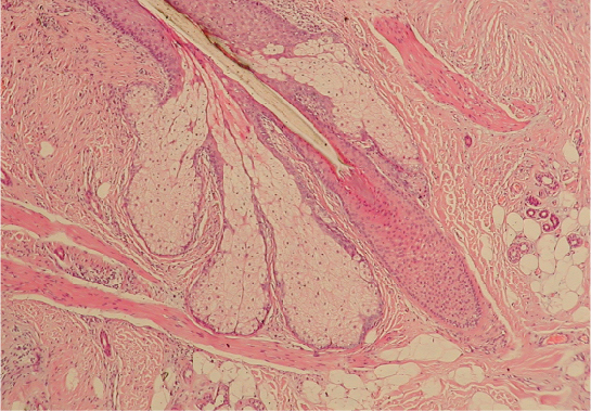
  • Sebaceous glands:
    • Associate with hair follicles
    • Secrete sebum Sebum The oily substance secreted by sebaceous glands. It is composed of keratin, fat, and cellular debris. Infectious Folliculitis to moisturize skin

Hypodermis:

  • Located deep to the dermis
  • Contains adipose tissue Adipose tissue Adipose tissue is a specialized type of connective tissue that has both structural and highly complex metabolic functions, including energy storage, glucose homeostasis, and a multitude of endocrine capabilities. There are three types of adipose tissue, white adipose tissue, brown adipose tissue, and beige or “brite” adipose tissue, which is a transitional form. Adipose Tissue: Histology, which serves as an energy reserve

Skin Appendages

Hair

  • Growth begins in the hair papilla of the dermis.
  • Composed of keratinized cells that emerge and migrate upward inside the hair follicle Hair follicle A tube-like invagination of the epidermis from which the hair shaft develops and into which sebaceous glands open. The hair follicle is lined by a cellular inner and outer root sheath of epidermal origin and is invested with a fibrous sheath derived from the dermis. Follicles of very long hairs extend into the subcutaneous layer of tissue under the skin. Cowden Syndrome
  • Each hair follicle Hair follicle A tube-like invagination of the epidermis from which the hair shaft develops and into which sebaceous glands open. The hair follicle is lined by a cellular inner and outer root sheath of epidermal origin and is invested with a fibrous sheath derived from the dermis. Follicles of very long hairs extend into the subcutaneous layer of tissue under the skin. Cowden Syndrome has an associated sebaceous gland Sebaceous Gland Small, sacculated organs found within the dermis. Each gland has a single duct that emerges from a cluster of oval alveoli. Each alveolus consists of a transparent basement membrane enclosing epithelial cells. The ducts from most sebaceous glands open into a hair follicle, but some open on the general surface of the skin. Sebaceous glands secrete sebum. Hordeolum (Stye).
  • Arrector pili Pili Filamentous or elongated proteinaceous structures which extend from the cell surface in gram-negative bacteria that contain certain types of conjugative plasmid. These pili are the organs associated with genetic transfer and have essential roles in conjugation. Normally, only one or a few pili occur on a given donor cell. This preferred use of ‘pili’ refers to the sexual appendage, to be distinguished from bacterial fimbriae, also known as common pili, which are usually concerned with adhesion. Salmonella muscle causes piloerection of hair shaft (goosebumps).
  • Terminal hairs: coarse hair on scalp, eyebrows, etc ETC The electron transport chain (ETC) sends electrons through a series of proteins, which generate an electrochemical proton gradient that produces energy in the form of adenosine triphosphate (ATP). Electron Transport Chain (ETC).
  • Vellus hair: very fine hair covering a newborn Newborn An infant during the first 28 days after birth. Physical Examination of the Newborn or the cheeks Cheeks The part of the face that is below the eye and to the side of the nose and mouth. Melasma of an adult (peach fuzz)
  • Puberty Puberty Puberty is a complex series of physical, psychosocial, and cognitive transitions usually experienced by adolescents (11-19 years of age). Puberty is marked by a growth in stature and the development of secondary sexual characteristics, achievement of fertility, and changes in most body systems. Puberty:
    • Growth of terminal hairs
    • Accumulates in certain areas (pubic/facial hair)
  • Glabrous skin: Areas of the skin not covered by hair (4%)
  • Growth cycle:

Nails

  • Made up of dense keratinized cells of the epidermis
  • Protect fingertips and toes against injuries
  • Lunula: whitish crescent-shaped area at proximal end of the nail
  • Cuticle:
    • Between lunula and skin
    • Keeps germs from entering below the skin
  • Nail root:
    • Behind the cuticle
    • Forms keratinized cells that push the nail forward
  • Structure of nail:
    • Nail plate: actual fingernail, composed of keratin Keratin A class of fibrous proteins or scleroproteins that represents the principal constituent of epidermis; hair; nails; horny tissues, and the organic matrix of tooth enamel. Two major conformational groups have been characterized, alpha-keratin, whose peptide backbone forms a coiled-coil alpha helical structure consisting of type I keratin and a type II keratin, and beta-keratin, whose backbone forms a zigzag or pleated sheet structure. Alpha-keratins have been classified into at least 20 subtypes. In addition multiple isoforms of subtypes have been found which may be due to gene duplication. Seborrheic Keratosis
    • Nail bed:
      • Below nail plate
      • Continuous with strata basale and spinosum
    • Nail matrix (nail root): 
      • Proximal end of nail, deep to the skin
      • Responsible for new nail formation
    • Eponychium: cuticle
    • Hyponychium: distal extension Extension Examination of the Upper Limbs of nail
Structure of the nail

Structure of the nail:
The whitish crescent-shaped end of the nail (toward the body) is referred to as the lunula (“small moon”). Between the lunula and skin, there is a protective layer, called the cuticle, that keeps pathogens from entering beneath the skin. Behind the cuticle (toward the body), is the nail root, which forms keratinized cells that push the nail forward.
Fingernails grow faster than toenails. The typical growth rate of a fingernail is 1 mm per week; a toenail grows 0.5 mm per week.

Image: “Nails” by OpenStax College. License: CC BY 4.0

Related videos

Clinical Relevance

  • Erysipelas Erysipelas Erysipelas is a bacterial infection of the superficial layer of the skin extending to the skin’s superficial lymphatic vessels. This infection presents as a raised, well-defined, tender, and bright red rash. Typically on the legs or face, but erysipelas can occur anywhere on the skin. Erysipelas: superficial skin infection that affects the dermis in addition to the superficial cutaneous lymphatics. The condition is caused by bacteria Bacteria Bacteria are prokaryotic single-celled microorganisms that are metabolically active and divide by binary fission. Some of these organisms play a significant role in the pathogenesis of diseases. Bacteriology, most commonly Streptococcus Streptococcus Streptococcus is one of the two medically important genera of gram-positive cocci, the other being Staphylococcus. Streptococci are identified as different species on blood agar on the basis of their hemolytic pattern and sensitivity to optochin and bacitracin. There are many pathogenic species of streptococci, including S. pyogenes, S. agalactiae, S. pneumoniae, and the viridans streptococci. Streptococcus pyogenes. The incidence Incidence The number of new cases of a given disease during a given period in a specified population. It also is used for the rate at which new events occur in a defined population. It is differentiated from prevalence, which refers to all cases in the population at a given time. Measures of Disease Frequency of erysipelas Erysipelas Erysipelas is a bacterial infection of the superficial layer of the skin extending to the skin’s superficial lymphatic vessels. This infection presents as a raised, well-defined, tender, and bright red rash. Typically on the legs or face, but erysipelas can occur anywhere on the skin. Erysipelas has been decreasing since the introduction of antibiotics. Possible risk factors include poor hygiene, poor sanitation Sanitation The development and establishment of environmental conditions favorable to the health of the public. Hepatitis E Virus, and a history of lymphedema Lymphedema Edema due to obstruction of lymph vessels or disorders of the lymph nodes. Lymphatic Filariasis (Elephantiasis)
  • Molluscum contagiosum Molluscum contagiosum Molluscum contagiosum is a viral infection limited to the epidermis and is common in children below 5 years of age. Lesions appear as grouped, flesh-colored, dome-shaped papules with central umbilication. Molluscum Contagiosum: viral infection of the skin that only affects humans. The infection is generally mild and should not be a reason for concern. Molluscum contagiosum Molluscum contagiosum Molluscum contagiosum is a viral infection limited to the epidermis and is common in children below 5 years of age. Lesions appear as grouped, flesh-colored, dome-shaped papules with central umbilication. Molluscum Contagiosum causes multiple raised dome-shaped skin-colored papules with central umbilication. The condition is self-limited and thus does not require treatment. The virus Virus Viruses are infectious, obligate intracellular parasites composed of a nucleic acid core surrounded by a protein capsid. Viruses can be either naked (non-enveloped) or enveloped. The classification of viruses is complex and based on many factors, including type and structure of the nucleoid and capsid, the presence of an envelope, the replication cycle, and the host range. Virology belongs to the Poxviridae Poxviridae A family of double-stranded DNA viruses infecting mammals (including humans), birds and insects. There are two subfamilies: chordopoxvirinae, poxviruses of vertebrates, and entomopoxvirinae, poxviruses of insects. Orthopoxvirus family.
  • Basal cell Basal Cell Erythema Multiforme carcinoma: the most common malignant neoplasm of the skin. Basal cell Basal Cell Erythema Multiforme carcinoma is a nonmelanoma skin cancer (NMSC) that forms de novo from cells within the stratum basale. Basal cell Basal Cell Erythema Multiforme carcinoma solely affects the skin with hair and usually appears in areas exposed to UV radiation Radiation Emission or propagation of acoustic waves (sound), electromagnetic energy waves (such as light; radio waves; gamma rays; or x-rays), or a stream of subatomic particles (such as electrons; neutrons; protons; or alpha particles). Osteosarcoma from the sun. The most common locations are the face, head, and neck Neck The part of a human or animal body connecting the head to the rest of the body. Peritonsillar Abscess
  • Melanoma Melanoma Melanoma is a malignant tumor arising from melanocytes, the melanin-producing cells of the epidermis. These tumors are most common in fair-skinned individuals with a history of excessive sun exposure and sunburns. Melanoma: Malignant melanoma Melanoma Melanoma is a malignant tumor arising from melanocytes, the melanin-producing cells of the epidermis. These tumors are most common in fair-skinned individuals with a history of excessive sun exposure and sunburns. Melanoma involves the malignant transformation Transformation Change brought about to an organism’s genetic composition by unidirectional transfer (transfection; transduction, genetic; conjugation, genetic, etc.) and incorporation of foreign DNA into prokaryotic or eukaryotic cells by recombination of part or all of that DNA into the cell’s genome. Bacteriology of melanocytes. Melanoma Melanoma Melanoma is a malignant tumor arising from melanocytes, the melanin-producing cells of the epidermis. These tumors are most common in fair-skinned individuals with a history of excessive sun exposure and sunburns. Melanoma accounts for more than 90% of the deaths caused by skin cancer. Chronic UV light UV light That portion of the electromagnetic spectrum immediately below the visible range and extending into the x-ray frequencies. The longer wavelengths (near-uv or biotic or vital rays) are necessary for the endogenous synthesis of vitamin D and are also called antirachitic rays; the shorter, ionizing wavelengths (far-uv or abiotic or extravital rays) are viricidal, bactericidal, mutagenic, and carcinogenic and are used as disinfectants. Bullous Pemphigoid and Pemphigus Vulgaris exposure, damage caused by sunlight (including sunburns, especially during childhood and adolescence), fair skin type, and the cumulative appearance of melanocytic nevi Melanocytic Nevi Nevus/Nevi (> 100) or the presence of dysplastic nevi Dysplastic Nevi Clinically atypical nevi (usually exceeding 5 mm in diameter and having variable pigmentation and ill defined borders) with an increased risk for development of non-familial cutaneous malignant melanoma. Biopsies show melanocytic dysplasia. Nevi are clinically and histologically identical to the precursor lesions for melanoma in the b-k mole syndrome. Nevus/Nevi represent significant risk factors. The most important risk factor is exposure to sunlight, particularly UVB radiation Radiation Emission or propagation of acoustic waves (sound), electromagnetic energy waves (such as light; radio waves; gamma rays; or x-rays), or a stream of subatomic particles (such as electrons; neutrons; protons; or alpha particles). Osteosarcoma.
  • Squamous cell carcinoma Squamous cell carcinoma Cutaneous squamous cell carcinoma (cSCC) is caused by malignant proliferation of atypical keratinocytes. This condition is the 2nd most common skin malignancy and usually affects sun-exposed areas of fair-skinned patients. The cancer presents as a firm, erythematous, keratotic plaque or papule. Squamous Cell Carcinoma (SCC): nonmelanoma skin cancer arising from the suprabasal epidermal keratinocytes. Squamous cell carcinoma Squamous cell carcinoma Cutaneous squamous cell carcinoma (cSCC) is caused by malignant proliferation of atypical keratinocytes. This condition is the 2nd most common skin malignancy and usually affects sun-exposed areas of fair-skinned patients. The cancer presents as a firm, erythematous, keratotic plaque or papule. Squamous Cell Carcinoma (SCC) is the 2nd most common nonmelanoma skin cancer, after basal cell Basal Cell Erythema Multiforme carcinoma. This carcinoma is seen most frequently in sun-exposed areas in people > 40 years of age. White males are more likely to be affected by this cancer; however, SCC is the most common skin cancer in patients Patients Individuals participating in the health care system for the purpose of receiving therapeutic, diagnostic, or preventive procedures. Clinician–Patient Relationship with darker skin types.

References

  1. Kierszenbaum, A. L., Tres, L. L. (2019). Histology and Cell Biology: An Introduction to Pathology, 5th ed. Elsevier.
  2. Navarette-Dechent, C., et al. (2020). Human surface anatomy terminology for dermatology: a Delphi consensus from the International Skin Imaging Collaboration. Journal of the European Academy of Dermatology and Venereology 34:2659–2663. https://doi.org/10.1111/jdv.16855
  3. Yousef, H., Alhajj, M., Sharma, S. (2020). Anatomy, skin (integument), Epidermis. StatPearls. Retrived October 15, 2021, from https://europepmc.org/article/nbk/nbk470464

Create your free account or log in to continue reading!

Sign up now and get free access to Lecturio with concept pages, medical videos, and questions for your medical education.

User Reviews

Unwrap New Skills This Holiday 🎄 Save 30% on all plans now!

Details