Advertisement

Advertisement

Advertisement
Advertisement

Staphylococcus

Staphylococcus is a medically important genera of Gram-positive Gram-Positive Penicillins, aerobic cocci Cocci Bacteriology. These bacteria Bacteria Bacteria are prokaryotic single-celled microorganisms that are metabolically active and divide by binary fission. Some of these organisms play a significant role in the pathogenesis of diseases. Bacteriology form clusters resembling grapes on culture plates. Staphylococci are ubiquitous for humans, and many strains compose the normal skin Skin The skin, also referred to as the integumentary system, is the largest organ of the body. The skin is primarily composed of the epidermis (outer layer) and dermis (deep layer). The epidermis is primarily composed of keratinocytes that undergo rapid turnover, while the dermis contains dense layers of connective tissue. Skin: Structure and Functions flora. Staphylococcus aureus Staphylococcus aureus Potentially pathogenic bacteria found in nasal membranes, skin, hair follicles, and perineum of warm-blooded animals. They may cause a wide range of infections and intoxications. Brain Abscess is the most virulent species; S. epidermidis and S. saprophyticus are less virulent but are also clinically significant. Infection can cause a wide array of disease, including cellulitis Cellulitis Cellulitis is a common infection caused by bacteria that affects the dermis and subcutaneous tissue of the skin. It is frequently caused by Staphylococcus aureus and Streptococcus pyogenes. The skin infection presents as an erythematous and edematous area with warmth and tenderness. Cellulitis, abscesses, endocarditis Endocarditis Endocarditis is an inflammatory disease involving the inner lining (endometrium) of the heart, most commonly affecting the cardiac valves. Both infectious and noninfectious etiologies lead to vegetations on the valve leaflets. Patients may present with nonspecific symptoms such as fever and fatigue. Endocarditis, osteomyelitis Osteomyelitis Osteomyelitis is an infection of the bone that results from the spread of microorganisms from the blood (hematogenous), nearby infected tissue, or open wounds (non-hematogenous). Infections are most commonly caused by Staphylococcus aureus. Osteomyelitis, and medical device infections Infections Invasion of the host organism by microorganisms or their toxins or by parasites that can cause pathological conditions or diseases. Chronic Granulomatous Disease. Toxins formed by S. aureus can cause gastroenteritis Gastroenteritis Gastroenteritis is inflammation of the stomach and intestines, commonly caused by infections from bacteria, viruses, or parasites. Transmission may be foodborne, fecal-oral, or through animal contact. Common clinical features include abdominal pain, diarrhea, vomiting, fever, and dehydration. Gastroenteritis, SSSS SSSS Staphylococcal scalded skin syndrome (SSSS), also known as Ritter disease and staphylococcal epidermal necrolysis, is a toxin-mediated condition caused by Staphylococcus aureus. The exfoliative toxin produced disseminates and cleaves desmoglein 1 in the epidermis, causing separation and detachment of the skin. Staphylococcal Scalded Skin Syndrome (SSSS), and toxic shock syndrome Toxic Shock Syndrome Toxic shock syndrome (TSS) is an acute, multi-systemic disease caused by the toxin-producing bacteria, Staphylococcus aureus and Streptococcus pyogenes. Staphylococcal TSS is more common and associated with tampons and nasal packing. Toxic Shock Syndrome ( TSS TSS Toxic shock syndrome (TSS) is an acute, multi-systemic disease caused by the toxin-producing bacteria, Staphylococcus aureus and Streptococcus pyogenes. Staphylococcal TSS is more common and associated with tampons and nasal packing. Toxic Shock Syndrome). Antibiotic management varies based on the type of infection, severity, and sensitivity data.

Last updated: Dec 29, 2023

Editorial responsibility: Stanley Oiseth, Lindsay Jones, Evelin Maza

Advertisement

Advertisement

Advertisement

Advertisement

Advertisement

Advertisement

Classification

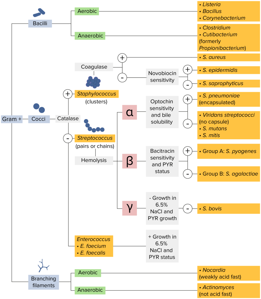
Microbiology flowchart gram-positive bacteria

Gram-positive bacteria:
Most bacteria can be classified according to a lab procedure called Gram staining.
Bacteria with cell walls that have a thick layer of peptidoglycan retain the crystal violet stain utilized in Gram staining but are not affected by the safranin counterstain. These bacteria appear as purple-blue on the stain, indicating that they are gram positive. The bacteria can be further classified according to morphology (branching filaments, bacilli, and cocci in clusters or chains) and their ability to grow in the presence of oxygen (aerobic versus anaerobic). The cocci can also be further identified. Staphylococci can be narrowed down on the basis of the presence of the enzyme coagulase and on their sensitivity to the antibiotic novobiocin. Streptococci are grown on blood agar and classified on the basis of which form of hemolysis they employ (α, β, or γ). Streptococci are further narrowed on the basis of their response to the pyrrolidonyl-β-naphthylamide (PYR) test, their sensitivity to specific antimicrobials (optochin and bacitracin), and their ability to grow on sodium chloride (NaCl) media.
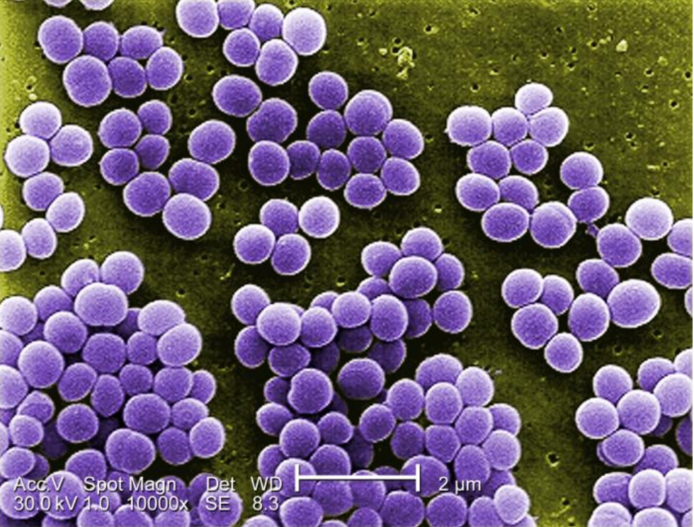
Image by Lecturio.

General Characteristics and Epidemiology

Basic features of Staphylococcus

  • Gram-positive Gram-Positive Penicillins cocci Cocci Bacteriology
    • Grow in clusters
    • Resemble a cluster of grapes
  • Nonmotile
  • Non-spore-forming
  • Aerobic and facultative anaerobic
  • Catalase-positive:
    • An enzyme that cleaves hydrogen peroxide Hydrogen peroxide A strong oxidizing agent used in aqueous solution as a ripening agent, bleach, and topical anti-infective. It is relatively unstable and solutions deteriorate over time unless stabilized by the addition of acetanilide or similar organic materials. Myeloperoxidase Deficiency → water and oxygen 
    • Distinguishing feature from streptococci (which are catalase-negative)
Staphylococcus aureus

Digitally-colorized scanning electron microscopic image under 10,000X magnification of Staphylococcus aureus:
Notice these cocci resemble a “cluster of grapes.”

Image: “10.000x Magnification of Staphylococcus aureus” by CDC/ Matthew J. Arduino, DRPH. License: Public Domain

Clinically relevant species

The clinically relevant species can be classified based on the presence (or absence) of coagulase:

  • Coagulase-positive Coagulase-Positive Staphylococcal Scalded Skin Syndrome (SSSS) staphylococci: S. aureus
  • Coagulase-negative staphylococci (CoNS) species (often grouped together):
    • S. epidermidis 
    • S. saprophyticus
    • Infrequent pathogens (list is not exhaustive):
      • S. lugdunensis
      • S. hominis
      • S. capitis
      • S. haemolyticus

Distinguishing features

Table: Distinguishing features among the common pathogenic Staphylococcus species
Species Coagulase Blood agar Blood agar Nocardia/Nocardiosis Mannitol Mannitol A diuretic and renal diagnostic aid related to sorbitol. It has little significant energy value as it is largely eliminated from the body before any metabolism can take place. It can be used to treat oliguria associated with kidney failure or other manifestations of inadequate renal function and has been used for determination of glomerular filtration rate. Mannitol is also commonly used as a research tool in cell biological studies, usually to control osmolarity. Osmotic Diuretics salt agar Novobiocin*
sensitivity
S. aureus Positive Beta-hemolysis Yellow colonies Sensitive
S. epidermidis Negative Nonhemolytic Pink or red colonies Sensitive
S. saprophyticus Negative Nonhemolytic Yellow or red colonies (depending on the strain) Resistant
*An antibiotic produced by Streptomyces nivens
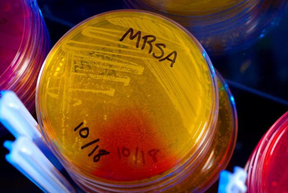
Mrsa

Methicillin-resistant Staphylococcus aureus (MRSA):
Coagulase-positive S. aureus ferments mannitol (red), producing yellow colonies on mannitol salt agar plates.

Image: “Methicillin-resistant Staphylococcus aureus (MRSA) bacteria” by CDC/ Melissa Dankel. License: Public Domain

Epidemiology

  • S. aureus:
    • Colonizes approximately 30% of the general population
    • Major cause of community-acquired and nosocomial infections Infections Invasion of the host organism by microorganisms or their toxins or by parasites that can cause pathological conditions or diseases. Chronic Granulomatous Disease
  • S. epidermidis:
    • Most commonly identified Staphylococcus species from human skin Skin The skin, also referred to as the integumentary system, is the largest organ of the body. The skin is primarily composed of the epidermis (outer layer) and dermis (deep layer). The epidermis is primarily composed of keratinocytes that undergo rapid turnover, while the dermis contains dense layers of connective tissue. Skin: Structure and Functions
    • Common cause of nosocomial bacteremia Bacteremia The presence of viable bacteria circulating in the blood. Fever, chills, tachycardia, and tachypnea are common acute manifestations of bacteremia. The majority of cases are seen in already hospitalized patients, most of whom have underlying diseases or procedures which render their bloodstreams susceptible to invasion. Glycopeptides
  • S. saprophyticus: common cause of urinary tract Urinary tract The urinary tract is located in the abdomen and pelvis and consists of the kidneys, ureters, urinary bladder, and urethra. The structures permit the excretion of urine from the body. Urine flows from the kidneys through the ureters to the urinary bladder and out through the urethra. Urinary Tract: Anatomy infections Infections Invasion of the host organism by microorganisms or their toxins or by parasites that can cause pathological conditions or diseases. Chronic Granulomatous Disease (UTIs) in sexually active young women

Related videos

Pathogenesis

Reservoir Reservoir Animate or inanimate sources which normally harbor disease-causing organisms and thus serve as potential sources of disease outbreaks. Reservoirs are distinguished from vectors (disease vectors) and carriers, which are agents of disease transmission rather than continuing sources of potential disease outbreaks. Humans may serve both as disease reservoirs and carriers. Escherichia coli

Staphylococcus species are part of the normal human flora:

  • Nasal mucosa Nasal mucosa The mucous lining of the nasal cavity, including lining of the nostril (vestibule) and the olfactory mucosa. Nasal mucosa consists of ciliated cells, goblet cells, brush cells, small granule cells, basal cells (stem cells) and glands containing both mucous and serous cells. Nose Anatomy (External & Internal)
  • Respiratory tract
  • Skin Skin The skin, also referred to as the integumentary system, is the largest organ of the body. The skin is primarily composed of the epidermis (outer layer) and dermis (deep layer). The epidermis is primarily composed of keratinocytes that undergo rapid turnover, while the dermis contains dense layers of connective tissue. Skin: Structure and Functions
  • GI tract
  • Genitourinary tract (S. saprophyticus)

Transmission

Staphylococcus aureus Staphylococcus aureus Potentially pathogenic bacteria found in nasal membranes, skin, hair follicles, and perineum of warm-blooded animals. They may cause a wide range of infections and intoxications. Brain Abscess can be transmitted via aerosols Aerosols Colloids with a gaseous dispersing phase and either liquid (fog) or solid (smoke) dispersed phase; used in fumigation or in inhalation therapy; may contain propellant agents. Coxiella/Q Fever and contact with:

  • Infected individuals
  • Bodily fluids
  • Contaminated objects

Risk factors

Risk factors for severe Staphylococcus infection include:

  • Hospitalization Hospitalization The confinement of a patient in a hospital. Delirium
  • Surgery
  • Outpatient procedures (e.g., dialysis Dialysis Renal replacement therapy refers to dialysis and/or kidney transplantation. Dialysis is a procedure by which toxins and excess water are removed from the circulation. Hemodialysis and peritoneal dialysis (PD) are the two types of dialysis, and their primary difference is the location of the filtration process (external to the body in hemodialysis versus inside the body for PD). Peritoneal Dialysis and Hemodialysis)
  • Implanted or prosthetic material:
    • Pacemakers/defibrillators
    • Intravascular catheters
    • Urethral catheters
    • Surgical implants
    • Prosthetic valves
  • Immunocompromised immunocompromised A human or animal whose immunologic mechanism is deficient because of an immunodeficiency disorder or other disease or as the result of the administration of immunosuppressive drugs or radiation. Gastroenteritis individuals
  • Nursing home stays
  • Injection drug use

Virulence factors Virulence factors Those components of an organism that determine its capacity to cause disease but are not required for its viability per se. Two classes have been characterized: toxins, biological and surface adhesion molecules that affect the ability of the microorganism to invade and colonize a host. Haemophilus

Common to most staphylococci:

  • Deoxyribonuclease (DNase): depolymerizes DNA DNA A deoxyribonucleotide polymer that is the primary genetic material of all cells. Eukaryotic and prokaryotic organisms normally contain DNA in a double-stranded state, yet several important biological processes transiently involve single-stranded regions. DNA, which consists of a polysugar-phosphate backbone possessing projections of purines (adenine and guanine) and pyrimidines (thymine and cytosine), forms a double helix that is held together by hydrogen bonds between these purines and pyrimidines (adenine to thymine and guanine to cytosine). DNA Types and Structure, a component of neutrophil extracellular traps → ↓ pus viscosity
  • Fibrinolysin Fibrinolysin A product of the lysis of plasminogen (profibrinolysin) by plasminogen activators. It is composed of two polypeptide chains, light (b) and heavy (a), with a molecular weight of 75, 000. It is the major proteolytic enzyme involved in blood clot retraction or the lysis of fibrin and quickly inactivated by antiplasmins. Hemostasis: lyses fibrin Fibrin A protein derived from fibrinogen in the presence of thrombin, which forms part of the blood clot. Rapidly Progressive Glomerulonephritis walls used to keep an infection contained → aids AIDS Chronic HIV infection and depletion of CD4 cells eventually results in acquired immunodeficiency syndrome (AIDS), which can be diagnosed by the presence of certain opportunistic diseases called AIDS-defining conditions. These conditions include a wide spectrum of bacterial, viral, fungal, and parasitic infections as well as several malignancies and generalized conditions. HIV Infection and AIDS in spread
  • Proteases Proteases Proteins and Peptides: degrade proteins Proteins Linear polypeptides that are synthesized on ribosomes and may be further modified, crosslinked, cleaved, or assembled into complex proteins with several subunits. The specific sequence of amino acids determines the shape the polypeptide will take, during protein folding, and the function of the protein. Energy Homeostasis of target cells, such as components of the:
    • Extracellular matrix Extracellular matrix A meshwork-like substance found within the extracellular space and in association with the basement membrane of the cell surface. It promotes cellular proliferation and provides a supporting structure to which cells or cell lysates in culture dishes adhere. Hypertrophic and Keloid Scars (ECM)
    • Innate immune system Immune system The body’s defense mechanism against foreign organisms or substances and deviant native cells. It includes the humoral immune response and the cell-mediated response and consists of a complex of interrelated cellular, molecular, and genetic components. Primary Lymphatic Organs
  • Biofilm:
    • An extracellular polysaccharide matrix that encases the bacteria Bacteria Bacteria are prokaryotic single-celled microorganisms that are metabolically active and divide by binary fission. Some of these organisms play a significant role in the pathogenesis of diseases. Bacteriology after attaching to foreign material
    • Functions as a protective barrier against antibiotics and the host immune system Immune system The body’s defense mechanism against foreign organisms or substances and deviant native cells. It includes the humoral immune response and the cell-mediated response and consists of a complex of interrelated cellular, molecular, and genetic components. Primary Lymphatic Organs 

Staphylococcus aureus Staphylococcus aureus Potentially pathogenic bacteria found in nasal membranes, skin, hair follicles, and perineum of warm-blooded animals. They may cause a wide range of infections and intoxications. Brain Abscess:

  • Hyaluronidase Hyaluronidase Bacteroides:
    • Splits hyaluronic acid Hyaluronic acid A natural high-viscosity mucopolysaccharide with alternating beta (1-3) glucuronide and beta (1-4) glucosaminidase bonds. It is found in the umbilical cord, in vitreous body and in synovial fluid. A high urinary level is found in progeria. Connective Tissue: Histology 
    • Makes it easier for pathogen invasion into the tissues
  • Lipase Lipase An enzyme of the hydrolase class that catalyzes the reaction of triacylglycerol and water to yield diacylglycerol and a fatty acid anion. It is produced by glands on the tongue and by the pancreas and initiates the digestion of dietary fats. Malabsorption and Maldigestion and hydrolase:
    • Degrade lipids Lipids Lipids are a diverse group of hydrophobic organic molecules, which include fats, oils, sterols, and waxes. Fatty Acids and Lipids
    • Used for invasion
  • Hemolysins Hemolysins Proteins from bacteria and fungi that are soluble enough to be secreted to target erythrocytes and insert into the membrane to form beta-barrel pores. Biosynthesis may be regulated by hemolysin factors. Leptospira/Leptospirosis and leukocidins: causes target cell death Cell death Injurious stimuli trigger the process of cellular adaptation, whereby cells respond to withstand the harmful changes in their environment. Overwhelmed adaptive mechanisms lead to cell injury. Mild stimuli produce reversible injury. If the stimulus is severe or persistent, injury becomes irreversible. Apoptosis is programmed cell death, a mechanism with both physiologic and pathologic effects. Cell Injury and Death, including red blood cells Red blood cells Erythrocytes, or red blood cells (RBCs), are the most abundant cells in the blood. While erythrocytes in the fetus are initially produced in the yolk sac then the liver, the bone marrow eventually becomes the main site of production. Erythrocytes: Histology and immune cells
  • Clumping factor A:
    • Binds fibrinogen Fibrinogen Plasma glycoprotein clotted by thrombin, composed of a dimer of three non-identical pairs of polypeptide chains (alpha, beta, gamma) held together by disulfide bonds. Fibrinogen clotting is a sol-gel change involving complex molecular arrangements: whereas fibrinogen is cleaved by thrombin to form polypeptides a and b, the proteolytic action of other enzymes yields different fibrinogen degradation products. Hemostasis and other proteins Proteins Linear polypeptides that are synthesized on ribosomes and may be further modified, crosslinked, cleaved, or assembled into complex proteins with several subunits. The specific sequence of amino acids determines the shape the polypeptide will take, during protein folding, and the function of the protein. Energy Homeostasis
    • Causes bacteria Bacteria Bacteria are prokaryotic single-celled microorganisms that are metabolically active and divide by binary fission. Some of these organisms play a significant role in the pathogenesis of diseases. Bacteriology clumping in plasma Plasma The residual portion of blood that is left after removal of blood cells by centrifugation without prior blood coagulation. Transfusion Products
    • Particularly important in the pathogenesis of endocarditis Endocarditis Endocarditis is an inflammatory disease involving the inner lining (endometrium) of the heart, most commonly affecting the cardiac valves. Both infectious and noninfectious etiologies lead to vegetations on the valve leaflets. Patients may present with nonspecific symptoms such as fever and fatigue. Endocarditis and endovascular infections Infections Invasion of the host organism by microorganisms or their toxins or by parasites that can cause pathological conditions or diseases. Chronic Granulomatous Disease
  • Coagulase:
    • Converts fibrinogen Fibrinogen Plasma glycoprotein clotted by thrombin, composed of a dimer of three non-identical pairs of polypeptide chains (alpha, beta, gamma) held together by disulfide bonds. Fibrinogen clotting is a sol-gel change involving complex molecular arrangements: whereas fibrinogen is cleaved by thrombin to form polypeptides a and b, the proteolytic action of other enzymes yields different fibrinogen degradation products. Hemostasis to fibrin Fibrin A protein derived from fibrinogen in the presence of thrombin, which forms part of the blood clot. Rapidly Progressive Glomerulonephritis to form a wall around the local area of infection (pseudocapsule)
    • Promotes abscess Abscess Accumulation of purulent material in tissues, organs, or circumscribed spaces, usually associated with signs of infection. Chronic Granulomatous Disease formation
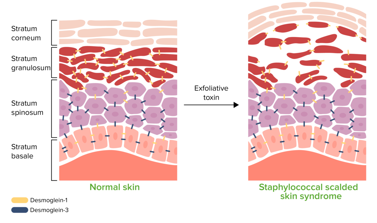
  • Exfoliative toxins A and B:
    • Exotoxins Exotoxins Toxins produced, especially by bacterial or fungal cells, and released into the culture medium or environment. Bacteriology
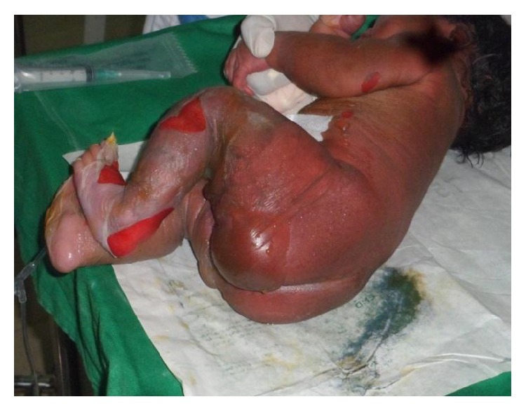
    • Cause SSSS SSSS Staphylococcal scalded skin syndrome (SSSS), also known as Ritter disease and staphylococcal epidermal necrolysis, is a toxin-mediated condition caused by Staphylococcus aureus. The exfoliative toxin produced disseminates and cleaves desmoglein 1 in the epidermis, causing separation and detachment of the skin. Staphylococcal Scalded Skin Syndrome (SSSS)
      • Cleave desmoglein-1 complex in the stratum granulosum Stratum granulosum Skin: Structure and Functions
      • Disrupt keratinocyte-to-keratinocyte adhesion Adhesion The process whereby platelets adhere to something other than platelets, e.g., collagen; basement membrane; microfibrils; or other ‘foreign’ surfaces. Coagulation Studies
      • Cause separation and detachment of the superficial epidermis Epidermis The external, nonvascular layer of the skin. It is made up, from within outward, of five layers of epithelium: (1) basal layer (stratum basale epidermidis); (2) spinous layer (stratum spinosum epidermidis); (3) granular layer (stratum granulosum epidermidis); (4) clear layer (stratum lucidum epidermidis); and (5) horny layer (stratum corneum epidermidis). Skin: Structure and Functions
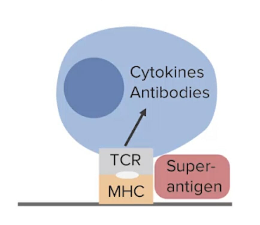
  • Superantigens:
    • Includes the exotoxins Exotoxins Toxins produced, especially by bacterial or fungal cells, and released into the culture medium or environment. Bacteriology:
      • Toxic shock syndrome Toxic Shock Syndrome Toxic shock syndrome (TSS) is an acute, multi-systemic disease caused by the toxin-producing bacteria, Staphylococcus aureus and Streptococcus pyogenes. Staphylococcal TSS is more common and associated with tampons and nasal packing. Toxic Shock Syndrome toxin type-1 ( TSST-1 TSST-1 Toxic Shock Syndrome)
      • Enterotoxin Enterotoxin Substances that are toxic to the intestinal tract causing vomiting, diarrhea, etc. ; most common enterotoxins are produced by bacteria. Diarrhea B
    • Cause toxic shock syndrome Toxic Shock Syndrome Toxic shock syndrome (TSS) is an acute, multi-systemic disease caused by the toxin-producing bacteria, Staphylococcus aureus and Streptococcus pyogenes. Staphylococcal TSS is more common and associated with tampons and nasal packing. Toxic Shock Syndrome ( TSS TSS Toxic shock syndrome (TSS) is an acute, multi-systemic disease caused by the toxin-producing bacteria, Staphylococcus aureus and Streptococcus pyogenes. Staphylococcal TSS is more common and associated with tampons and nasal packing. Toxic Shock Syndrome):
      • Bind BIND Hyperbilirubinemia of the Newborn to MHC class II on antigen-presenting cells Antigen-presenting cells A heterogeneous group of immunocompetent cells that mediate the cellular immune response by processing and presenting antigens to the T-cells. Traditional antigen-presenting cells include macrophages; dendritic cells; langerhans cells; and B-lymphocytes. Follicular dendritic cells are not traditional antigen-presenting cells, but because they hold antigen on their cell surface in the form of immune complexes for b-cell recognition they are considered so by some authors. Adaptive Immune Response → cross-links with the β region of the T cell receptor Receptor Receptors are proteins located either on the surface of or within a cell that can bind to signaling molecules known as ligands (e.g., hormones) and cause some type of response within the cell. Receptors ( TCR TCR Molecules on the surface of T-lymphocytes that recognize and combine with antigens. The receptors are non-covalently associated with a complex of several polypeptides collectively called CD3 antigens. Recognition of foreign antigen and the major histocompatibility complex is accomplished by a single heterodimeric antigen-receptor structure, composed of either alpha-beta or gamma-delta chains. Adaptive Cell-mediated Immunity)
      • Stimulates T cell activation T cell activation Adaptive Cell-mediated Immunity and an exaggerated cytokine release
      • Results in an excessive inflammatory response
  • Other enterotoxins:
    • Heat Heat Inflammation stable exotoxins Exotoxins Toxins produced, especially by bacterial or fungal cells, and released into the culture medium or environment. Bacteriology
    • Mode of action unknown
    • Can cause gastroenteritis Gastroenteritis Gastroenteritis is inflammation of the stomach and intestines, commonly caused by infections from bacteria, viruses, or parasites. Transmission may be foodborne, fecal-oral, or through animal contact. Common clinical features include abdominal pain, diarrhea, vomiting, fever, and dehydration. Gastroenteritis ( food poisoning Food poisoning Acute illnesses, usually affecting the gastrointestinal tract, brought on by consuming contaminated food or beverages. Most of these diseases are infectious, caused by a variety of bacteria, viruses, or parasites that can be foodborne. Sometimes the diseases are caused by harmful toxins from the microbes or other chemicals present in the food. Especially in the latter case, the condition is often called food poisoning. Clostridia)

CoNS:

  • Adhesins Adhesins Cell-surface components or appendages of bacteria that facilitate adhesion (bacterial adhesion) to other cells or to inanimate surfaces. Most fimbriae of gram-negative bacteria function as adhesins, but in many cases it is a minor subunit protein at the tip of the fimbriae that is the actual adhesin. In gram-positive bacteria, a protein or polysaccharide surface layer serves as the specific adhesin. What is sometimes called polymeric adhesin (biofilms) is distinct from protein adhesin. Diarrheagenic E. coli: aid CoNS adhesion Adhesion The process whereby platelets adhere to something other than platelets, e.g., collagen; basement membrane; microfibrils; or other ‘foreign’ surfaces. Coagulation Studies to host cells
  • Urease Urease An enzyme that catalyzes the conversion of urea and water to carbon dioxide and ammonia. Nocardia/Nocardiosis:
    • Produced by S. saprophyticus
    • Hydrolyzes urea Urea A compound formed in the liver from ammonia produced by the deamination of amino acids. It is the principal end product of protein catabolism and constitutes about one half of the total urinary solids. Urea Cycle → C02 and ammonia Ammonia A colorless alkaline gas. It is formed in the body during decomposition of organic materials during a large number of metabolically important reactions. Note that the aqueous form of ammonia is referred to as ammonium hydroxide. Acid-Base Balance → ↑ urine pH pH The quantitative measurement of the acidity or basicity of a solution. Acid-Base Balance
    • Makes it particularly virulent within the urinary tract Urinary tract The urinary tract is located in the abdomen and pelvis and consists of the kidneys, ureters, urinary bladder, and urethra. The structures permit the excretion of urine from the body. Urine flows from the kidneys through the ureters to the urinary bladder and out through the urethra. Urinary Tract: Anatomy

Diseases Caused by S. aureus

Infections Infections Invasion of the host organism by microorganisms or their toxins or by parasites that can cause pathological conditions or diseases. Chronic Granulomatous Disease

Staphylococcus aureus Staphylococcus aureus Potentially pathogenic bacteria found in nasal membranes, skin, hair follicles, and perineum of warm-blooded animals. They may cause a wide range of infections and intoxications. Brain Abscess can cause a wide range of infections Infections Invasion of the host organism by microorganisms or their toxins or by parasites that can cause pathological conditions or diseases. Chronic Granulomatous Disease, including (but not limited to):
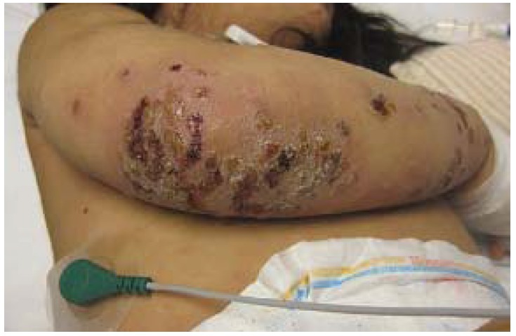
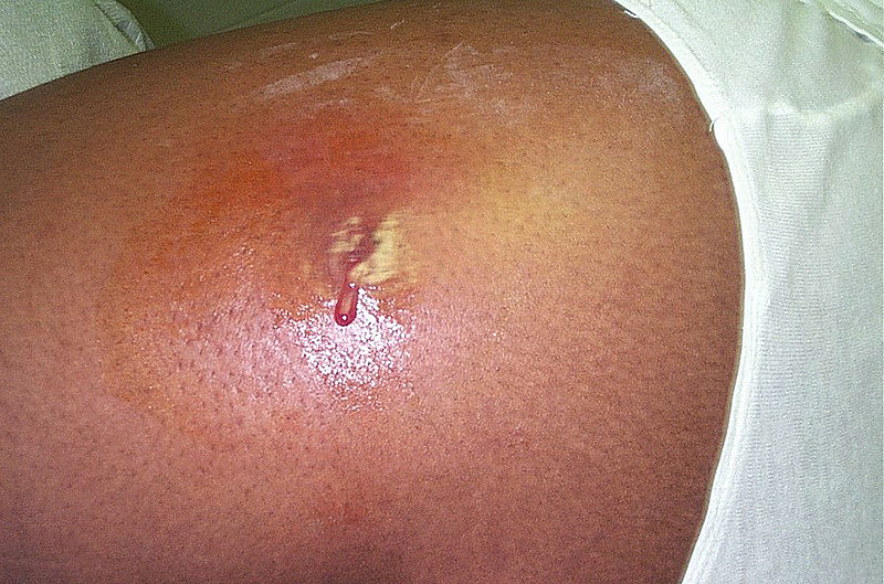
  • Skin Skin The skin, also referred to as the integumentary system, is the largest organ of the body. The skin is primarily composed of the epidermis (outer layer) and dermis (deep layer). The epidermis is primarily composed of keratinocytes that undergo rapid turnover, while the dermis contains dense layers of connective tissue. Skin: Structure and Functions and soft tissue Soft Tissue Soft Tissue Abscess infections Infections Invasion of the host organism by microorganisms or their toxins or by parasites that can cause pathological conditions or diseases. Chronic Granulomatous Disease
    • Folliculitis
    • Impetigo Impetigo Impetigo is a highly contagious superficial bacterial infection typically caused by Staphylococcus aureus (most common) and Streptococcus pyogenes. Impetigo most commonly presents in children aged 2 to 5 years with lesions that evolve from papules to vesicles to pustules, which eventually break down to form characteristic “honey-colored” crusts. Impetigo
    • Cellulitis Cellulitis Cellulitis is a common infection caused by bacteria that affects the dermis and subcutaneous tissue of the skin. It is frequently caused by Staphylococcus aureus and Streptococcus pyogenes. The skin infection presents as an erythematous and edematous area with warmth and tenderness. Cellulitis
    • Abscess Abscess Accumulation of purulent material in tissues, organs, or circumscribed spaces, usually associated with signs of infection. Chronic Granulomatous Disease
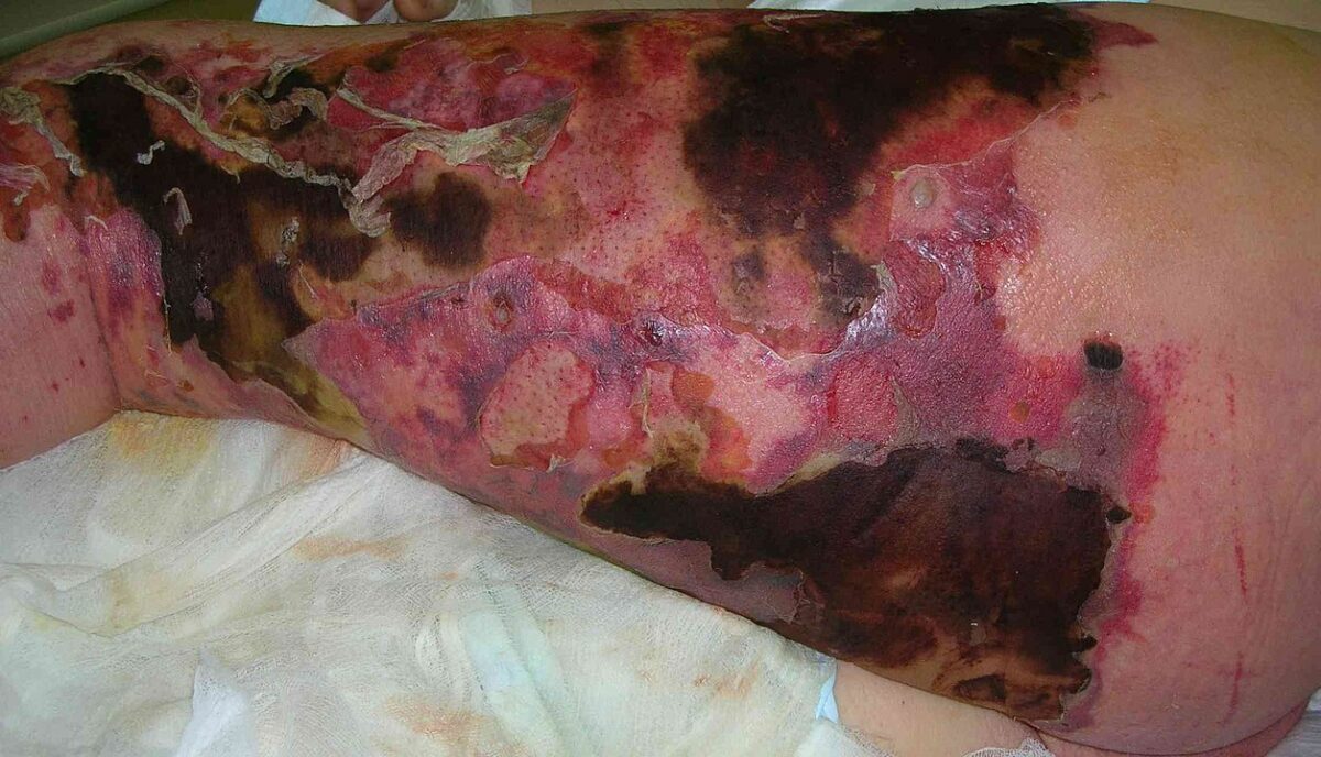
    • Necrotizing fasciitis Necrotizing fasciitis Necrotizing fasciitis is a life-threatening infection that causes rapid destruction and necrosis of the fascia and subcutaneous tissues. Patients may present with significant pain out of proportion to the presenting symptoms and rapidly progressive erythema of the affected area. Necrotizing Fasciitis
    • Surgical wound infection Surgical wound infection Infection occurring at the site of a surgical incision. Surgical Complications
  • Bloodstream infection → can seed infections Infections Invasion of the host organism by microorganisms or their toxins or by parasites that can cause pathological conditions or diseases. Chronic Granulomatous Disease in other tissues
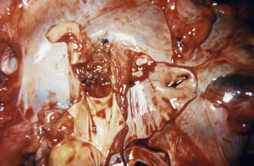
  • Infective endocarditis Infective endocarditis Infective endocarditis (IE) is caused by infection or inflammation of the inner lining of the heart (endocardium), most commonly affecting the heart valves. Endocarditis ( IE IE Infective endocarditis (IE) is caused by infection or inflammation of the inner lining of the heart (endocardium), most commonly affecting the heart valves. Endocarditis)
    • Leads to cardiac valve vegetations
    • Can release septic emboli → embolic complications and/or metastatic infection
    • S. aureus is associated with acute, rapidly progressive infection.
  • Pneumonia Pneumonia Pneumonia or pulmonary inflammation is an acute or chronic inflammation of lung tissue. Causes include infection with bacteria, viruses, or fungi. In more rare cases, pneumonia can also be caused through toxic triggers through inhalation of toxic substances, immunological processes, or in the course of radiotherapy. Pneumonia
    • S. aureus notably causes secondary bacterial pneumonia Secondary bacterial pneumonia Influenza Viruses/Influenza in individuals with influenza Influenza Influenza viruses are members of the Orthomyxoviridae family and the causative organisms of influenza, a highly contagious febrile respiratory disease. There are 3 primary influenza viruses (A, B, and C) and various subtypes, which are classified based on their virulent surface antigens, hemagglutinin (HA) and neuraminidase (NA). Influenza typically presents with a fever, myalgia, headache, and symptoms of an upper respiratory infection. Influenza Viruses/Influenza.
    • Complications:
  • Septic arthritis Arthritis Acute or chronic inflammation of joints. Osteoarthritis
  • Osteomyelitis Osteomyelitis Osteomyelitis is an infection of the bone that results from the spread of microorganisms from the blood (hematogenous), nearby infected tissue, or open wounds (non-hematogenous). Infections are most commonly caused by Staphylococcus aureus. Osteomyelitis
  • Medical device infections Infections Invasion of the host organism by microorganisms or their toxins or by parasites that can cause pathological conditions or diseases. Chronic Granulomatous Disease, such as:

Toxin-mediated diseases

Table: Features of toxin-mediated diseases caused by S. aureus
Disease Exotoxin Clinical presentation
SSSS SSSS Staphylococcal scalded skin syndrome (SSSS), also known as Ritter disease and staphylococcal epidermal necrolysis, is a toxin-mediated condition caused by Staphylococcus aureus. The exfoliative toxin produced disseminates and cleaves desmoglein 1 in the epidermis, causing separation and detachment of the skin. Staphylococcal Scalded Skin Syndrome (SSSS) Exfoliative toxins A and B
Toxic shock syndrome Toxic Shock Syndrome Toxic shock syndrome (TSS) is an acute, multi-systemic disease caused by the toxin-producing bacteria, Staphylococcus aureus and Streptococcus pyogenes. Staphylococcal TSS is more common and associated with tampons and nasal packing. Toxic Shock Syndrome ( TSS TSS Toxic shock syndrome (TSS) is an acute, multi-systemic disease caused by the toxin-producing bacteria, Staphylococcus aureus and Streptococcus pyogenes. Staphylococcal TSS is more common and associated with tampons and nasal packing. Toxic Shock Syndrome) Toxic shock syndrome Toxic Shock Syndrome Toxic shock syndrome (TSS) is an acute, multi-systemic disease caused by the toxin-producing bacteria, Staphylococcus aureus and Streptococcus pyogenes. Staphylococcal TSS is more common and associated with tampons and nasal packing. Toxic Shock Syndrome toxin type-1 ( TSST-1 TSST-1 Toxic Shock Syndrome)
Enterotoxin Enterotoxin Substances that are toxic to the intestinal tract causing vomiting, diarrhea, etc. ; most common enterotoxins are produced by bacteria. Diarrhea B
  • Fever Fever Fever is defined as a measured body temperature of at least 38°C (100.4°F). Fever is caused by circulating endogenous and/or exogenous pyrogens that increase levels of prostaglandin E2 in the hypothalamus. Fever is commonly associated with chills, rigors, sweating, and flushing of the skin. Fever
  • Rash Rash Rocky Mountain Spotted Fever
  • Hypotension Hypotension Hypotension is defined as low blood pressure, specifically < 90/60 mm Hg, and is most commonly a physiologic response. Hypotension may be mild, serious, or life threatening, depending on the cause. Hypotension
  • Multiorgan dysfunction
Gastroenteritis Gastroenteritis Gastroenteritis is inflammation of the stomach and intestines, commonly caused by infections from bacteria, viruses, or parasites. Transmission may be foodborne, fecal-oral, or through animal contact. Common clinical features include abdominal pain, diarrhea, vomiting, fever, and dehydration. Gastroenteritis Enterotoxins
  • Nausea Nausea An unpleasant sensation in the stomach usually accompanied by the urge to vomit. Common causes are early pregnancy, sea and motion sickness, emotional stress, intense pain, food poisoning, and various enteroviruses. Antiemetics and vomiting Vomiting The forcible expulsion of the contents of the stomach through the mouth. Hypokalemia
  • Abdominal cramps Cramps Ion Channel Myopathy
  • Diarrhea Diarrhea Diarrhea is defined as ≥ 3 watery or loose stools in a 24-hour period. There are a multitude of etiologies, which can be classified based on the underlying mechanism of disease. The duration of symptoms (acute or chronic) and characteristics of the stools (e.g., watery, bloody, steatorrheic, mucoid) can help guide further diagnostic evaluation. Diarrhea

Diseases Caused by CoNS

Coagulase-negative staphylococci can cause many of the same conditions as S. aureus. Notable infections Infections Invasion of the host organism by microorganisms or their toxins or by parasites that can cause pathological conditions or diseases. Chronic Granulomatous Disease include:

  • Medical device infections Infections Invasion of the host organism by microorganisms or their toxins or by parasites that can cause pathological conditions or diseases. Chronic Granulomatous Disease (particularly S. epidermidis)
    • IV catheters
    • Prosthetic valves
    • Pacemakers
    • Prosthetic joints Prosthetic joints Prostheses used to partially or totally replace a human or animal joint. Soft Tissue Abscess
    • CNS shunts
    • Breast implants
  • Surgical wound infection Surgical wound infection Infection occurring at the site of a surgical incision. Surgical Complications
  • IE IE Infective endocarditis (IE) is caused by infection or inflammation of the inner lining of the heart (endocardium), most commonly affecting the heart valves. Endocarditis (native and prosthetic valve Prosthetic Valve Soft Tissue Abscess)
  • Osteomyelitis Osteomyelitis Osteomyelitis is an infection of the bone that results from the spread of microorganisms from the blood (hematogenous), nearby infected tissue, or open wounds (non-hematogenous). Infections are most commonly caused by Staphylococcus aureus. Osteomyelitis
  • UTIs (particularly S. saprophyticus)

Antibiotics and Resistance

Antibiotic susceptibility

Staphylococcus species can develop antibiotic resistance Resistance Physiologically, the opposition to flow of air caused by the forces of friction. As a part of pulmonary function testing, it is the ratio of driving pressure to the rate of air flow. Ventilation: Mechanics of Breathing, and identification Identification Defense Mechanisms of susceptibility is necessary for adequate treatment. Species are often designated as:

  • Methicillin-sensitive 
  • Methicillin-resistant 
  • Vancomycin-resistant

Mechanism of resistance Resistance Physiologically, the opposition to flow of air caused by the forces of friction. As a part of pulmonary function testing, it is the ratio of driving pressure to the rate of air flow. Ventilation: Mechanics of Breathing

  • Methicillin Methicillin One of the penicillins which is resistant to penicillinase but susceptible to a penicillin-binding protein. It is inactivated by gastric acid so administered by injection. Penicillins: penicillin-binding protein (PBP)-mediated resistance Resistance Physiologically, the opposition to flow of air caused by the forces of friction. As a part of pulmonary function testing, it is the ratio of driving pressure to the rate of air flow. Ventilation: Mechanics of Breathing
    • Mutations in the PBPs PBPs Bacterial proteins that share the property of binding irreversibly to penicillins and other antibacterial agents derived from lactams. The penicillin-binding proteins are primarily enzymes involved in cell wall biosynthesis including muramoylpentapeptide carboxypeptidase; peptide synthases; transpeptidases; and hexosyltransferases. Penicillins (encoded by mecA gene Gene A category of nucleic acid sequences that function as units of heredity and which code for the basic instructions for the development, reproduction, and maintenance of organisms. Basic Terms of Genetics) → ↓ affinity of penicillins Penicillins Beta-lactam antibiotics contain a beta-lactam ring as a part of their chemical structure. Drugs in this class include penicillin G and V, penicillinase-sensitive and penicillinase-resistant penicillins, cephalosporins, carbapenems, and aztreonam. Penicillins to the PBP 
    • Despite the mutations, these PBPs PBPs Bacterial proteins that share the property of binding irreversibly to penicillins and other antibacterial agents derived from lactams. The penicillin-binding proteins are primarily enzymes involved in cell wall biosynthesis including muramoylpentapeptide carboxypeptidase; peptide synthases; transpeptidases; and hexosyltransferases. Penicillins are still able to produce a cell wall Cell wall The outermost layer of a cell in most plants; bacteria; fungi; and algae. The cell wall is usually a rigid structure that lies external to the cell membrane, and provides a protective barrier against physical or chemical agents. Cell Types: Eukaryotic versus Prokaryotic.
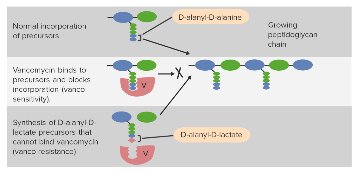
  • Vancomycin Vancomycin Antibacterial obtained from streptomyces orientalis. It is a glycopeptide related to ristocetin that inhibits bacterial cell wall assembly and is toxic to kidneys and the inner ear. Glycopeptides: alteration in peptidoglycan Peptidoglycan Penicillins synthesis Synthesis Polymerase Chain Reaction (PCR)
    • Conferred by vanA gene Gene A category of nucleic acid sequences that function as units of heredity and which code for the basic instructions for the development, reproduction, and maintenance of organisms. Basic Terms of Genetics (on plasmid)
    • Acquired from vancomycin-resistant Enterococcus Enterococcus Enterococcus is a genus of oval-shaped gram-positive cocci that are arranged in pairs or short chains. Distinguishing factors include optochin resistance and the presence of pyrrolidonyl arylamidase (PYR) and Lancefield D antigen. Enterococcus is part of the normal flora of the human GI tract. Enterococcus (VRE)
    • Alters the D-alanyl-D-alanine peptidoglycan Peptidoglycan Penicillins target to D-alanyl-D-lactate or D-alanyl-D-serine → poor glycopeptide binding and no disruption of cell wall Cell wall The outermost layer of a cell in most plants; bacteria; fungi; and algae. The cell wall is usually a rigid structure that lies external to the cell membrane, and provides a protective barrier against physical or chemical agents. Cell Types: Eukaryotic versus Prokaryotic synthesis Synthesis Polymerase Chain Reaction (PCR)

Antibiotic choices

The agent of choice and duration of treatment for Staphylococcus infection depends on the body area affected, the severity of infection, and the antibiotic sensitivity of the strain. 

Table: Antibiotic choices for Staphylococcus based on resistance Resistance Physiologically, the opposition to flow of air caused by the forces of friction. As a part of pulmonary function testing, it is the ratio of driving pressure to the rate of air flow. Ventilation: Mechanics of Breathing
Strain Antibiotic
MSSA and CoNS
  • Penicillinase-resistant penicillins Penicillinase-Resistant Penicillins Penicillins
  • Cephalosporins Cephalosporins Cephalosporins are a group of bactericidal beta-lactam antibiotics (similar to penicillins) that exert their effects by preventing bacteria from producing their cell walls, ultimately leading to cell death. Cephalosporins are categorized by generation and all drug names begin with “cef-” or “ceph-.” Cephalosporins
MRSA
  • Vancomycin Vancomycin Antibacterial obtained from streptomyces orientalis. It is a glycopeptide related to ristocetin that inhibits bacterial cell wall assembly and is toxic to kidneys and the inner ear. Glycopeptides
  • Trimethoprim-sulfamethoxazole
  • Clindamycin Clindamycin An antibacterial agent that is a semisynthetic analog of lincomycin. Lincosamides
  • Linezolid Linezolid An oxazolidinone and acetamide derived anti-bacterial agent and protein synthesis inhibitor that is used in the treatment of gram-positive bacterial infections of the skin and respiratory tract. Oxazolidinones
  • Daptomycin Daptomycin A cyclic lipopeptide antibiotic that inhibits gram-positive bacteria. Lipopeptides and Lipoglycopeptides
  • Tetracyclines Tetracyclines Tetracyclines are a class of broad-spectrum antibiotics indicated for a wide variety of bacterial infections. These medications bind the 30S ribosomal subunit to inhibit protein synthesis of bacteria. Tetracyclines cover gram-positive and gram-negative organisms, as well as atypical bacteria such as chlamydia, mycoplasma, spirochetes, and even protozoa. Tetracyclines
  • Ceftaroline Ceftaroline Cephalosporins
  • Tigecycline Tigecycline A tetracycline derivative that acts as a protein synthesis inhibitor. It is used as an antibacterial agent for the systemic treatment of complicated skin and intra-abdominal infections. It is also used for the treatment of community-acquired pneumonia. Tetracyclines
VRSA

References

  1. Carroll, KC, & Hobden, JA. (2015). The staphylococci. In Brooks, JE, et al. (Eds.), Jawetz, Melnick, & Adelberg’s Medical Microbiology. (27th ed). [VitalSource Bookshelf 9.4.3]. 
  2. Centers for Disease Control and Prevention. (2020). Staphylococcus aureus in Healthcare Settings. https://www.cdc.gov/hai/organisms/staph.html
  3. Fowler, VG, & Holland, TL. Clinical approach to Staphylococcus aureus bacteremia in adults. UpToDate. Retrieved May 13, 2021, from https://www.uptodate.com/contents/clinical-approach-to-staphylococcus-aureus-bacteremia-in-adults
  4. Tufariello, JM, & Lowy, FD. Infection due to coagulase-negative staphylococci: Epidemiology, microbiology, and pathogenesis. UpToDate. Retrieved May 14, 2021, from https://www.uptodate.com/contents/infection-due-to-coagulase-negative-staphylococci-epidemiology-microbiology-and-pathogenesis
  5. Chu, VH. Staphylococcal toxic shock syndrome. UpToDate. Retrieved May 19, 2021, from https://www.uptodate.com/contents/staphylococcal-toxic-shock-syndrome
  6. McMahon, P. Staphylococcal scalded skin syndrome. UpToDate. Retrieved May 19, 2021, from https://www.uptodate.com/contents/staphylococcal-scalded-skin-syndrome
  7. Cong, Y, Yang, S, & Rao, X. (2020). Vancomycin-resistant Staphylococcus aureus infections: A review of case updating and clinical features. Journal of Advanced Research. 21:169-176. https://www.sciencedirect.com/science/article/pii/S2090123219301638
  8. Gardete, S, & Tomasz, A. (2014). Mechanism of vancomycin resistance in Staphylococcus aureus. The Journal of Clinical Investigation. 124(7):2836-40. https://www.ncbi.nlm.nih.gov/pmc/articles/PMC4071404/
  9. Moreillon, P, et al. (1995). Rose of Staphylococcus aureus coagulase and clumping factor in pathogenesis of experimental endocarditis. Infection and Immunity. 63(12):4738-43. https://www.ncbi.nlm.nih.gov/pmc/articles/PMC173679/
  10. Vandenesch, F, Lina, G, & Henry, T. (2012). Staphylococcus aureus hemolysins, bicomponent leukocidins, and cytolytic peptides: A redundant arsenal of membrane-damaging virulence factors? Frontiers in Cellular and Infection Microbiology. 2:12. https://www.ncbi.nlm.nih.gov/pmc/articles/PMC3417661/

Create your free account or log in to continue reading!

Sign up now and get free access to Lecturio with concept pages, medical videos, and questions for your medical education.

User Reviews

Unwrap New Skills This Holiday 🎄 Save 30% on all plans now!

Details