Advertisement

Advertisement

Advertisement

Advertisement

Sjögren’s Syndrome

Sjögren syndrome Sjögren Syndrome Rheumatoid Arthritis ( SS SS Scleroderma (systemic sclerosis) is an autoimmune condition characterized by diffuse collagen deposition and fibrosis. The clinical presentation varies from limited skin involvement to diffuse involvement of internal organs. Scleroderma) is an autoimmune, inflammatory condition where glandular tissues, such as the salivary and lacrimal glands Lacrimal Glands Dacryocystitis, are infiltrated by lymphocytes Lymphocytes Lymphocytes are heterogeneous WBCs involved in immune response. Lymphocytes develop from the bone marrow, starting from hematopoietic stem cells (HSCs) and progressing to common lymphoid progenitors (CLPs). B and T lymphocytes and natural killer (NK) cells arise from the lineage. Lymphocytes: Histology, resulting in decreased tear and saliva Saliva The clear, viscous fluid secreted by the salivary glands and mucous glands of the mouth. It contains mucins, water, organic salts, and ptyalin. Salivary Glands: Anatomy production. The disease mainly affects middle-aged women, and is associated with other autoimmune conditions. Patients Patients Individuals participating in the health care system for the purpose of receiving therapeutic, diagnostic, or preventive procedures. Clinician–Patient Relationship may experience symptoms and complications related to dry eyes and mouth. There is a wide range of extraglandular manifestations, including Raynaud's phenomenon, neuropathy Neuropathy Leprosy, and cutaneous vasculitis Vasculitis Inflammation of any one of the blood vessels, including the arteries; veins; and rest of the vasculature system in the body. Systemic Lupus Erythematosus. Patients Patients Individuals participating in the health care system for the purpose of receiving therapeutic, diagnostic, or preventive procedures. Clinician–Patient Relationship are also at an increased risk of developing non-Hodgkin's lymphoma Lymphoma A general term for various neoplastic diseases of the lymphoid tissue. Imaging of the Mediastinum. Diagnosis is based on the presence of symptoms, and is validated by the clinical examination, serologic studies, or salivary gland Salivary gland Glands that secrete saliva in the mouth. There are three pairs of salivary glands (parotid gland; sublingual gland; submandibular gland). Diseases of the Salivary Glands biopsy Biopsy Removal and pathologic examination of specimens from the living body. Ewing Sarcoma. A multidisciplinary approach is needed to manage patients Patients Individuals participating in the health care system for the purpose of receiving therapeutic, diagnostic, or preventive procedures. Clinician–Patient Relationship and targets symptomatic relief, with immunosuppressive therapy reserved for severe symptoms.

Last updated: May 16, 2024

Editorial responsibility: Stanley Oiseth, Lindsay Jones, Evelin Maza

Advertisement

Advertisement

Advertisement

Advertisement

Advertisement

Advertisement

Overview

Definition

Sjögren’s syndrome ( SS SS Scleroderma (systemic sclerosis) is an autoimmune condition characterized by diffuse collagen deposition and fibrosis. The clinical presentation varies from limited skin involvement to diffuse involvement of internal organs. Scleroderma), also known as sicca syndrome, is a chronic autoimmune and inflammatory condition leading to decreased lacrimal and salivary gland Salivary gland Glands that secrete saliva in the mouth. There are three pairs of salivary glands (parotid gland; sublingual gland; submandibular gland). Diseases of the Salivary Glands function, resulting in dry eyes (xerophthalmia) and mouth (xerostomia).

Epidemiology

  • The 2nd most common rheumatologic disorder
  • Prevalence Prevalence The total number of cases of a given disease in a specified population at a designated time. It is differentiated from incidence, which refers to the number of new cases in the population at a given time. Measures of Disease Frequency: 0.1%–4% of the population 
  • Predominantly seen in women (female : male ratio of 9:1)
  • Age of onset: middle age (40–55 years of age)
  • ½ of patients Patients Individuals participating in the health care system for the purpose of receiving therapeutic, diagnostic, or preventive procedures. Clinician–Patient Relationship have another autoimmune disease (e.g., rheumatoid arthritis Arthritis Acute or chronic inflammation of joints. Osteoarthritis, systemic lupus erythematosus Systemic lupus erythematosus Systemic lupus erythematosus (SLE) is a chronic autoimmune, inflammatory condition that causes immune-complex deposition in organs, resulting in systemic manifestations. Women, particularly those of African American descent, are more commonly affected. Systemic Lupus Erythematosus).

Etiology

Primary SS SS Scleroderma (systemic sclerosis) is an autoimmune condition characterized by diffuse collagen deposition and fibrosis. The clinical presentation varies from limited skin involvement to diffuse involvement of internal organs. Scleroderma:

  • Etiology is unknown.
  • Possible genetic predisposition: 
    • HLA-DR3
    • HLA-DQ2
    • HLA-DR5
  • Possible viral connection: 
    • Epstein-Barr virus Epstein-Barr Virus Epstein-Barr virus (EBV) is a linear, double-stranded DNA virus belonging to the Herpesviridae family. This highly prevalent virus is mostly transmitted through contact with oropharyngeal secretions from an infected individual. The virus can infect epithelial cells and B lymphocytes, where it can undergo lytic replication or latency. Epstein-Barr Virus ( EBV EBV Epstein-barr virus (EBV) is a linear, double-stranded DNA virus belonging to the herpesviridae family. This highly prevalent virus is mostly transmitted through contact with oropharyngeal secretions from an infected individual. The virus can infect epithelial cells and B lymphocytes, where it can undergo lytic replication or latency. Epstein-Barr Virus)
    • Chronic hepatitis C Hepatitis C Hepatitis C is an infection of the liver caused by the hepatitis C virus (HCV). The infection can be transmitted through infectious blood or body fluids and may be transmitted during childbirth or through IV drug use or sexual intercourse. Hepatitis C virus can cause both acute and chronic hepatitis, ranging from a mild to a serious, lifelong illness including liver cirrhosis and hepatocellular carcinoma (HCC). Hepatitis C Virus
    • Human T cell leukemia virus Virus Viruses are infectious, obligate intracellular parasites composed of a nucleic acid core surrounded by a protein capsid. Viruses can be either naked (non-enveloped) or enveloped. The classification of viruses is complex and based on many factors, including type and structure of the nucleoid and capsid, the presence of an envelope, the replication cycle, and the host range. Virology 1
    • Human herpesvirus 6
    • Human immunodeficiency Immunodeficiency Chédiak-Higashi Syndrome virus Virus Viruses are infectious, obligate intracellular parasites composed of a nucleic acid core surrounded by a protein capsid. Viruses can be either naked (non-enveloped) or enveloped. The classification of viruses is complex and based on many factors, including type and structure of the nucleoid and capsid, the presence of an envelope, the replication cycle, and the host range. Virology ( HIV HIV Anti-HIV Drugs)

Secondary SS SS Scleroderma (systemic sclerosis) is an autoimmune condition characterized by diffuse collagen deposition and fibrosis. The clinical presentation varies from limited skin involvement to diffuse involvement of internal organs. Scleroderma:

Presents in association with another autoimmune disease:

  • Rheumatoid arthritis Arthritis Acute or chronic inflammation of joints. Osteoarthritis
  • Systemic lupus erythematosus Systemic lupus erythematosus Systemic lupus erythematosus (SLE) is a chronic autoimmune, inflammatory condition that causes immune-complex deposition in organs, resulting in systemic manifestations. Women, particularly those of African American descent, are more commonly affected. Systemic Lupus Erythematosus
  • Polymyositis Polymyositis Polymyositis (PM) is an autoimmune inflammatory myopathy caused by T cell-mediated muscle injury. The etiology of PM is unclear, but there are several genetic and environmental associations. Polymyositis is most common in middle-aged women and rarely affects children. Polymyositis
  • Scleroderma Scleroderma Scleroderma (systemic sclerosis) is an autoimmune condition characterized by diffuse collagen deposition and fibrosis. The clinical presentation varies from limited skin involvement to diffuse involvement of internal organs. Scleroderma

Pathophysiology

The overall mechanism for SS SS Scleroderma (systemic sclerosis) is an autoimmune condition characterized by diffuse collagen deposition and fibrosis. The clinical presentation varies from limited skin involvement to diffuse involvement of internal organs. Scleroderma is unclear:
  • Process may be set off by an environmental trigger Trigger The type of signal that initiates the inspiratory phase by the ventilator Invasive Mechanical Ventilation in a patient with a genetic predisposition.
  • Dendritic cells Dendritic cells Specialized cells of the hematopoietic system that have branch-like extensions. They are found throughout the lymphatic system, and in non-lymphoid tissues such as skin and the epithelia of the intestinal, respiratory, and reproductive tracts. They trap and process antigens, and present them to T-cells, thereby stimulating cell-mediated immunity. They are different from the non-hematopoietic follicular dendritic cells, which have a similar morphology and immune system function, but with respect to humoral immunity (antibody production). Skin: Structure and Functions migrate into salivary and lacrimal glands Lacrimal Glands Dacryocystitis release Release Release of a virus from the host cell following virus assembly and maturation. Egress can occur by host cell lysis, exocytosis, or budding through the plasma membrane. Virology interferons Interferons Interferon (IFN) is a cytokine with antiviral properties (it interferes with viral infections) and various roles in immunoregulation. The different types are type I IFN (IFN-ɑ and IFN-β), type II IFN (IFN-ɣ), and type III IFN (IFN-ƛ). Interferons
  • Lymphocytes Lymphocytes Lymphocytes are heterogeneous WBCs involved in immune response. Lymphocytes develop from the bone marrow, starting from hematopoietic stem cells (HSCs) and progressing to common lymphoid progenitors (CLPs). B and T lymphocytes and natural killer (NK) cells arise from the lineage. Lymphocytes: Histology (usually CD4 T cells CD4+ T cells A critical subpopulation of T-lymphocytes involved in the induction of most immunological functions. The HIV virus has selective tropism for the T4 cell which expresses the CD4 phenotypic marker, a receptor for HIV. In fact, the key element in the profound immunosuppression seen in HIV infection is the depletion of this subset of T-lymphocytes. T cells: Types and Functions) and B cells B cells Lymphoid cells concerned with humoral immunity. They are short-lived cells resembling bursa-derived lymphocytes of birds in their production of immunoglobulin upon appropriate stimulation. B cells: Types and Functions infiltrate the glands.
  • Interferons Interferons Interferon (IFN) is a cytokine with antiviral properties (it interferes with viral infections) and various roles in immunoregulation. The different types are type I IFN (IFN-ɑ and IFN-β), type II IFN (IFN-ɣ), and type III IFN (IFN-ƛ). Interferons stimulate B cell activating factor (BAFF) production → promotes irregular B cell maturation → B cells B cells Lymphoid cells concerned with humoral immunity. They are short-lived cells resembling bursa-derived lymphocytes of birds in their production of immunoglobulin upon appropriate stimulation. B cells: Types and Functions release Release Release of a virus from the host cell following virus assembly and maturation. Egress can occur by host cell lysis, exocytosis, or budding through the plasma membrane. Virology autoantibodies Autoantibodies Antibodies that react with self-antigens (autoantigens) of the organism that produced them. Blotting Techniques
  • T cells T cells Lymphocytes responsible for cell-mediated immunity. Two types have been identified – cytotoxic (t-lymphocytes, cytotoxic) and helper T-lymphocytes (t-lymphocytes, helper-inducer). They are formed when lymphocytes circulate through the thymus gland and differentiate to thymocytes. When exposed to an antigen, they divide rapidly and produce large numbers of new T cells sensitized to that antigen. T cells: Types and Functions and duct epithelial cells produce more cytokines Cytokines Non-antibody proteins secreted by inflammatory leukocytes and some non-leukocytic cells, that act as intercellular mediators. They differ from classical hormones in that they are produced by a number of tissue or cell types rather than by specialized glands. They generally act locally in a paracrine or autocrine rather than endocrine manner. Adaptive Immune Response.
  • Cytokines Cytokines Non-antibody proteins secreted by inflammatory leukocytes and some non-leukocytic cells, that act as intercellular mediators. They differ from classical hormones in that they are produced by a number of tissue or cell types rather than by specialized glands. They generally act locally in a paracrine or autocrine rather than endocrine manner. Adaptive Immune Response and autoantibodies Autoantibodies Antibodies that react with self-antigens (autoantigens) of the organism that produced them. Blotting Techniques cause damage and atrophy Atrophy Decrease in the size of a cell, tissue, organ, or multiple organs, associated with a variety of pathological conditions such as abnormal cellular changes, ischemia, malnutrition, or hormonal changes. Cellular Adaptation of the glandular and ductal epithelium Epithelium The epithelium is a complex of specialized cellular organizations arranged into sheets and lining cavities and covering the surfaces of the body. The cells exhibit polarity, having an apical and a basal pole. Structures important for the epithelial integrity and function involve the basement membrane, the semipermeable sheet on which the cells rest, and interdigitations, as well as cellular junctions. Surface Epithelium: Histology → ↓ exocrine production (↓ tear and saliva Saliva The clear, viscous fluid secreted by the salivary glands and mucous glands of the mouth. It contains mucins, water, organic salts, and ptyalin. Salivary Glands: Anatomy production).
  • Other exocrine glands Exocrine glands Glands of external secretion that release its secretions to the body’s cavities, organs, or surface, through a duct. Glandular Epithelium: Histology throughout the body can be involved, as well.
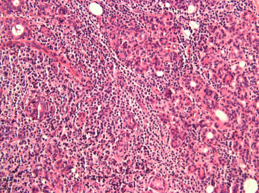
Lymphocytic infiltration of the parotid gland in a patient with sjögren syndrome

Lymphocytic infiltration of the parotid gland in a patient with SS

Image: “Anti-salivary gland protein 1 antibodies in two patients with Sjogren’s syndrome: Two case reports” by Vishwanath S, Shen L, Suresh L, Ambrus JL. License: CC BY 2.0.
Clinical Presentation

Glandular manifestations

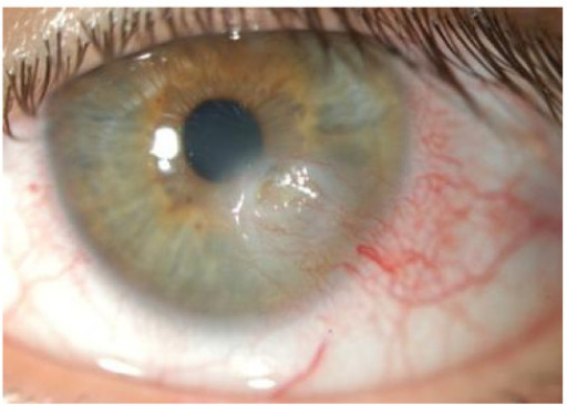
  • Keratoconjunctivitis sicca Keratoconjunctivitis Sicca Rheumatoid Arthritis:
  • Salivary hypofunction:
    • Symptoms:
      • Xerostomia
      • Dysphagia Dysphagia Dysphagia is the subjective sensation of difficulty swallowing. Symptoms can range from a complete inability to swallow, to the sensation of solids or liquids becoming “stuck.” Dysphagia is classified as either oropharyngeal or esophageal, with esophageal dysphagia having 2 sub-types: functional and mechanical. Dysphagia
      • Food sticking in the mouth
      • Change in taste
      • Difficulty speaking for long periods of time
    • Findings:
      • Dry tongue Tongue The tongue, on the other hand, is a complex muscular structure that permits tasting and facilitates the process of mastication and communication. The blood supply of the tongue originates from the external carotid artery, and the innervation is through cranial nerves. Lips and Tongue: Anatomy and lips Lips The lips are the soft and movable most external parts of the oral cavity. The blood supply of the lips originates from the external carotid artery, and the innervation is through cranial nerves. Lips and Tongue: Anatomy
      • Glassy appearance of the palate Palate The palate is the structure that forms the roof of the mouth and floor of the nasal cavity. This structure is divided into soft and hard palates. Palate: Anatomy
      • Absent sublingual salivary pooling
      • Enlarged salivary and parotid glands Parotid glands The largest of the three pairs of salivary glands. They lie on the sides of the face immediately below and in front of the ear. Gastrointestinal Secretions
    • Complications:
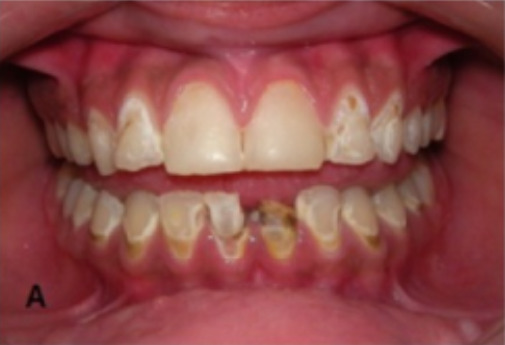
      • Dental caries 
      • Oral candidiasis Candidiasis Candida is a genus of dimorphic, opportunistic fungi. Candida albicans is part of the normal human flora and is the most common cause of candidiasis. The clinical presentation varies and can include localized mucocutaneous infections (e.g., oropharyngeal, esophageal, intertriginous, and vulvovaginal candidiasis) and invasive disease (e.g., candidemia, intraabdominal abscess, pericarditis, and meningitis). Candida/Candidiasis
      • Parotitis Parotitis Inflammation of the parotid gland. Mumps Virus/Mumps
      • Esophagitis Esophagitis Esophagitis is the inflammation or irritation of the esophagus. The major types of esophagitis are medication-induced, infectious, eosinophilic, corrosive, and acid reflux. Patients typically present with odynophagia, dysphagia, and retrosternal chest pain. Esophagitis
      • Weight loss Weight loss Decrease in existing body weight. Bariatric Surgery due to difficulty eating
  • Other glands:

Extraglandular manifestations

  • Cutaneous:
    • Xerosis (dry skin Skin The skin, also referred to as the integumentary system, is the largest organ of the body. The skin is primarily composed of the epidermis (outer layer) and dermis (deep layer). The epidermis is primarily composed of keratinocytes that undergo rapid turnover, while the dermis contains dense layers of connective tissue. Skin: Structure and Functions)
    • Raynaud’s phenomenon
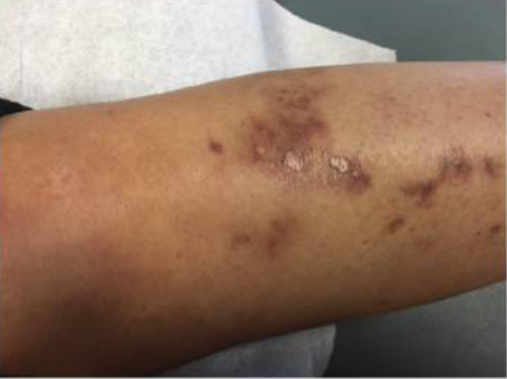
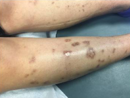
    • Cutaneous vasculitis Vasculitis Inflammation of any one of the blood vessels, including the arteries; veins; and rest of the vasculature system in the body. Systemic Lupus Erythematosus
      • Palpable purpura
      • Urticarial lesions
      • Ulcerations
    • Erythema Erythema Redness of the skin produced by congestion of the capillaries. This condition may result from a variety of disease processes. Chalazion nodosum
    • Livedo reticularis Livedo reticularis A condition characterized by a reticular or fishnet pattern on the skin of lower extremities and other parts of the body. This red and blue pattern is due to deoxygenated blood in unstable dermal blood vessels. The condition is intensified by cold exposure and relieved by rewarming. Chronic Kidney Disease
    • Vitiligo Vitiligo Vitiligo is the most common depigmenting disorder and is caused by the destruction of melanocytes. Patients present with hypo- or depigmented macules or patches which often occur on the face, hands, knees, and/or genitalia. Vitiligo
    • Lichen planus Lichen planus Lichen planus (LP) is an idiopathic, cell-mediated inflammatory skin disease. It is characterized by pruritic, flat-topped, papular, purple skin lesions commonly found on the flexural surfaces of the extremities. Other areas affected include genitalia, nails, scalp, and mucous membranes. Lichen Planus
  • Musculoskeletal:
  • Neurologic: 
    • Cognitive dysfunction
    • Peripheral polyneuropathy Polyneuropathy Polyneuropathy is any disease process affecting the function of or causing damage to multiple nerves of the peripheral nervous system. There are numerous etiologies of polyneuropathy, most of which are systemic and the most common of which is diabetic neuropathy. Polyneuropathy
    • Mononeuritis multiplex Mononeuritis Multiplex Chronic Diabetic Complications
    • Demyelinating disorders Demyelinating disorders Conditions characterized by loss or dysfunction of myelin in the brain, spinal cord, or optic nerves secondary to autoimmune mediated processes. This may take the form of a humoral or cellular immune response directed toward myelin or oligodendroglia associated autoantigens. Posterior Cord Syndrome
  • Psychiatric: depression
  • Respiratory: 
  • Cardiac (rare):
    • Pericarditis Pericarditis Pericarditis is an inflammation of the pericardium, often with fluid accumulation. It can be caused by infection (often viral), myocardial infarction, drugs, malignancies, metabolic disorders, autoimmune disorders, or trauma. Acute, subacute, and chronic forms exist. Pericarditis
    • Myocarditis Myocarditis Myocarditis is an inflammatory disease of the myocardium, which may occur alone or in association with a systemic process. There are numerous etiologies of myocarditis, but all lead to inflammation and myocyte injury, most often leading to signs and symptoms of heart failure. Myocarditis
    • Heart block
  • Gastrointestinal: gastritis Gastritis Gastritis refers to inflammation of the gastric mucosa. Gastritis may occur suddenly (acute gastritis) or slowly over time (chronic gastritis). Gastritis may be asymptomatic or with symptoms, including burning abdominal pain (which either worsens or improves with eating), dyspepsia, nausea, and vomiting. Gastritis
  • Hematologic:
    • Anemia Anemia Anemia is a condition in which individuals have low Hb levels, which can arise from various causes. Anemia is accompanied by a reduced number of RBCs and may manifest with fatigue, shortness of breath, pallor, and weakness. Subtypes are classified by the size of RBCs, chronicity, and etiology. Anemia: Overview and Types
    • Monoclonal gammopathy Monoclonal gammopathy Conditions characterized by the presence of m protein (monoclonal protein) in serum or urine without clinical manifestations of plasma cell dyscrasia. MALT Lymphoma
    • Waldenstrom’s macroglobulinemia
    • Non-Hodgkin’s lymphoma Lymphoma A general term for various neoplastic diseases of the lymphoid tissue. Imaging of the Mediastinum
  • Endocrine:
    • Pancreatitis Pancreatitis Inflammation of the pancreas. Pancreatitis is classified as acute unless there are computed tomographic or endoscopic retrograde cholangiopancreatographic findings of chronic pancreatitis. The two most common forms of acute pancreatitis are alcoholic pancreatitis and gallstone pancreatitis. Acute Pancreatitis
    • Autoimmune thyroid Thyroid The thyroid gland is one of the largest endocrine glands in the human body. The thyroid gland is a highly vascular, brownish-red gland located in the visceral compartment of the anterior region of the neck. Thyroid Gland: Anatomy disease
  • Renal:
    • Interstitial nephritis
    • Glomerulonephritis
    • Renal tubular acidosis Acidosis A pathologic condition of acid accumulation or depletion of base in the body. The two main types are respiratory acidosis and metabolic acidosis, due to metabolic acid build up. Respiratory Acidosis

Diagnosis

Laboratory evaluation

  • Anti-SSA/Ro and anti-SSB/La antibodies Antibodies Immunoglobulins (Igs), also known as antibodies, are glycoprotein molecules produced by plasma cells that act in immune responses by recognizing and binding particular antigens. The various Ig classes are IgG (the most abundant), IgM, IgE, IgD, and IgA, which differ in their biologic features, structure, target specificity, and distribution. Immunoglobulins: Types and Functions (present in 60%–70% of patients Patients Individuals participating in the health care system for the purpose of receiving therapeutic, diagnostic, or preventive procedures. Clinician–Patient Relationship)
  • Antinuclear antibodies Antibodies Immunoglobulins (Igs), also known as antibodies, are glycoprotein molecules produced by plasma cells that act in immune responses by recognizing and binding particular antigens. The various Ig classes are IgG (the most abundant), IgM, IgE, IgD, and IgA, which differ in their biologic features, structure, target specificity, and distribution. Immunoglobulins: Types and Functions
  • Rheumatoid factor Rheumatoid factor Antibodies found in adult rheumatoid arthritis patients that are directed against gamma-chain immunoglobulins. Autoimmune Hepatitis (present in 40%–60% of patients Patients Individuals participating in the health care system for the purpose of receiving therapeutic, diagnostic, or preventive procedures. Clinician–Patient Relationship)
  • Complete blood count:
    • Anemia Anemia Anemia is a condition in which individuals have low Hb levels, which can arise from various causes. Anemia is accompanied by a reduced number of RBCs and may manifest with fatigue, shortness of breath, pallor, and weakness. Subtypes are classified by the size of RBCs, chronicity, and etiology. Anemia: Overview and Types
    • Leukopenia
    • Thrombocytopenia Thrombocytopenia Thrombocytopenia occurs when the platelet count is < 150,000 per microliter. The normal range for platelets is usually 150,000-450,000/µL of whole blood. Thrombocytopenia can be a result of decreased production, increased destruction, or splenic sequestration of platelets. Patients are often asymptomatic until platelet counts are < 50,000/µL. Thrombocytopenia
  • Basic metabolic panel Basic Metabolic Panel Primary vs Secondary Headaches:
    • Creatinine → evaluate for renal involvement
    • Potassium Potassium An element in the alkali group of metals with an atomic symbol k, atomic number 19, and atomic weight 39. 10. It is the chief cation in the intracellular fluid of muscle and other cells. Potassium ion is a strong electrolyte that plays a significant role in the regulation of fluid volume and maintenance of the water-electrolyte balance. Hyperkalemia and bicarbonate Bicarbonate Inorganic salts that contain the -HCO3 radical. They are an important factor in determining the ph of the blood and the concentration of bicarbonate ions is regulated by the kidney. Levels in the blood are an index of the alkali reserve or buffering capacity. Electrolytes → renal tubular acidosis Acidosis A pathologic condition of acid accumulation or depletion of base in the body. The two main types are respiratory acidosis and metabolic acidosis, due to metabolic acid build up. Respiratory Acidosis
  • Erythrocyte sedimentation rate Erythrocyte Sedimentation Rate Soft Tissue Abscess ( ESR ESR Soft Tissue Abscess
  • Urinalysis Urinalysis Examination of urine by chemical, physical, or microscopic means. Routine urinalysis usually includes performing chemical screening tests, determining specific gravity, observing any unusual color or odor, screening for bacteriuria, and examining the sediment microscopically. Urinary Tract Infections (UTIs) in Children → evaluate for renal involvement:

Ophthalmologic evaluation

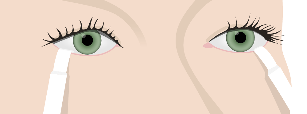
  • Schirmer’s test: 
    • Measures tear production
    • Filter paper is placed in the lower eyelid.
    • Wetness is measured after 5 minutes.
    • < 5 mm MM Multiple myeloma (MM) is a malignant condition of plasma cells (activated B lymphocytes) primarily seen in the elderly. Monoclonal proliferation of plasma cells results in cytokine-driven osteoclastic activity and excessive secretion of IgG antibodies. Multiple Myeloma wetness is considered a positive test.
  • Fluorescein Fluorescein A phthalic indicator dye that appears yellow-green in normal tear film and bright green in a more alkaline medium such as the aqueous humor. Pseudomonas slit-lamp examination Slit-Lamp Examination Blepharitis and red bengal staining: 
    • Evaluates the cornea Cornea The transparent anterior portion of the fibrous coat of the eye consisting of five layers: stratified squamous corneal epithelium; bowman membrane; corneal stroma; descemet membrane; and mesenchymal corneal endothelium. It serves as the first refracting medium of the eye. Eye: Anatomy and conjunctiva Conjunctiva The mucous membrane that covers the posterior surface of the eyelids and the anterior pericorneal surface of the eyeball. Eye: Anatomy for damage
    • Stain highlights areas of devitalized tissue.

Salivary gland Salivary gland Glands that secrete saliva in the mouth. There are three pairs of salivary glands (parotid gland; sublingual gland; submandibular gland). Diseases of the Salivary Glands evaluation

  • Sialometry:
    • Measures the rate of saliva Saliva The clear, viscous fluid secreted by the salivary glands and mucous glands of the mouth. It contains mucins, water, organic salts, and ptyalin. Salivary Glands: Anatomy production
    • Unstimulated saliva Saliva The clear, viscous fluid secreted by the salivary glands and mucous glands of the mouth. It contains mucins, water, organic salts, and ptyalin. Salivary Glands: Anatomy is collected and measured over 15 minutes.
    • Can also be done with stimulation of saliva Saliva The clear, viscous fluid secreted by the salivary glands and mucous glands of the mouth. It contains mucins, water, organic salts, and ptyalin. Salivary Glands: Anatomy (with citric acid or pilocarpine Pilocarpine A slowly hydrolyzed muscarinic agonist with no nicotinic effects. Pilocarpine is used as a miotic and in the treatment of glaucoma. Cholinomimetic Drugs)
  • Scintigraphy:
    • Measures salivary gland Salivary gland Glands that secrete saliva in the mouth. There are three pairs of salivary glands (parotid gland; sublingual gland; submandibular gland). Diseases of the Salivary Glands function through radionuclide uptake
    • Very low uptake is highly specific for SS SS Scleroderma (systemic sclerosis) is an autoimmune condition characterized by diffuse collagen deposition and fibrosis. The clinical presentation varies from limited skin involvement to diffuse involvement of internal organs. Scleroderma.
  • Imaging:
    • Ultrasound:
      • Multiple hypoechoic Hypoechoic A structure that produces a low-amplitude echo (darker grays) Ultrasound (Sonography) areas with convex borders
      • Hyperdense linear bands, cysts Cysts Any fluid-filled closed cavity or sac that is lined by an epithelium. Cysts can be of normal, abnormal, non-neoplastic, or neoplastic tissues. Fibrocystic Change, and calcifications
    • Magnetic resonance imaging (MRI): nodular “honeycomb” appearance of the parotid gland Parotid gland The largest of the three pairs of salivary glands. They lie on the sides of the face immediately below and in front of the ear. Salivary Glands: Anatomy
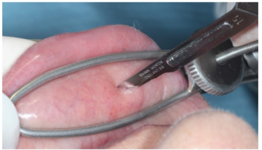
  • Biopsy Biopsy Removal and pathologic examination of specimens from the living body. Ewing Sarcoma
    • Performed on a lower lip salivary gland Salivary gland Glands that secrete saliva in the mouth. There are three pairs of salivary glands (parotid gland; sublingual gland; submandibular gland). Diseases of the Salivary Glands 
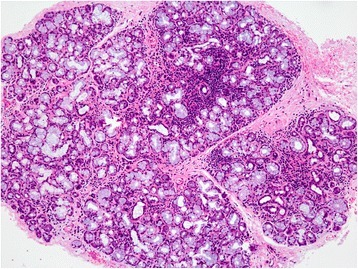
    • Histologic findings:
      • Periductal collections of tightly aggregated lymphocytes Lymphocytes Lymphocytes are heterogeneous WBCs involved in immune response. Lymphocytes develop from the bone marrow, starting from hematopoietic stem cells (HSCs) and progressing to common lymphoid progenitors (CLPs). B and T lymphocytes and natural killer (NK) cells arise from the lineage. Lymphocytes: Histology (focal lymphocytic sialadenitis Sialadenitis Inflammation of salivary tissue (salivary glands), usually due to infection or injuries. Diseases of the Salivary Glands)
      • Parenchymal atrophy Atrophy Decrease in the size of a cell, tissue, organ, or multiple organs, associated with a variety of pathological conditions such as abnormal cellular changes, ischemia, malnutrition, or hormonal changes. Cellular Adaptation
      • Fibrosis Fibrosis Any pathological condition where fibrous connective tissue invades any organ, usually as a consequence of inflammation or other injury. Bronchiolitis Obliterans
      • Scattered lymphoid cells

Diagnostic criteria

Several criteria have been proposed to aid in the diagnosis of SS SS Scleroderma (systemic sclerosis) is an autoimmune condition characterized by diffuse collagen deposition and fibrosis. The clinical presentation varies from limited skin involvement to diffuse involvement of internal organs. Scleroderma. The following is from the American-European Consensus Group (requires 4 of 6 criteria, including a positive biopsy Biopsy Removal and pathologic examination of specimens from the living body. Ewing Sarcoma or autoantibodies Autoantibodies Antibodies that react with self-antigens (autoantigens) of the organism that produced them. Blotting Techniques):

  • Ocular symptoms (at least 1):
  • Oral symptoms (at least 1):
    • Dry mouth for ≥ 3 months
    • Recurrent or persistently swollen salivary glands Salivary glands The salivary glands are exocrine glands positioned in and around the oral cavity. These glands are responsible for secreting saliva into the mouth, which aids in digestion. There are 3 major paired salivary glands: the sublingual, submandibular, and parotid glands. Salivary Glands: Anatomy
    • Need for liquids to swallow dry foods
  • Ocular signs (at least 1):
    • Positive Schirmer’s test
    • Positive dye staining on the surface of the eye
  • Oral signs (at least 1):
    • Positive sialography
    • Positive scintigraphy
  • Positive biopsy Biopsy Removal and pathologic examination of specimens from the living body. Ewing Sarcoma
  • Positive anti-Ro or anti-La (or both)

Management and Complications

Management

  • Multidisciplinary approach (rheumatologist, dentist, and ophthalmologist)
  • Primary SS SS Scleroderma (systemic sclerosis) is an autoimmune condition characterized by diffuse collagen deposition and fibrosis. The clinical presentation varies from limited skin involvement to diffuse involvement of internal organs. Scleroderma with disease limited to ocular and oral symptoms:
    • Oral hygiene
    • Smoking Smoking Willful or deliberate act of inhaling and exhaling smoke from burning substances or agents held by hand. Interstitial Lung Diseases cessation
    • Use of a humidifier 
    • Eye protection
    • Artificial tears and saliva Saliva The clear, viscous fluid secreted by the salivary glands and mucous glands of the mouth. It contains mucins, water, organic salts, and ptyalin. Salivary Glands: Anatomy substitute
    • Muscarinic agonists Muscarinic Agonists Drugs that bind to and activate muscarinic cholinergic receptors (receptors, muscarinic). Muscarinic agonists are most commonly used when it is desirable to increase smooth muscle tone, especially in the GI tract, urinary bladder and the eye. They may also be used to reduce heart rate. Asthma Drugs
      • Pilocarpine Pilocarpine A slowly hydrolyzed muscarinic agonist with no nicotinic effects. Pilocarpine is used as a miotic and in the treatment of glaucoma. Cholinomimetic Drugs
      • Cevimeline
  • Extraglandular involvement:
    • Glucocorticoids Glucocorticoids Glucocorticoids are a class within the corticosteroid family. Glucocorticoids are chemically and functionally similar to endogenous cortisol. There are a wide array of indications, which primarily benefit from the antiinflammatory and immunosuppressive effects of this class of drugs. Glucocorticoids
    • Hydroxychloroquine Hydroxychloroquine A chemotherapeutic agent that acts against erythrocytic forms of malarial parasites. Hydroxychloroquine appears to concentrate in food vacuoles of affected protozoa. It inhibits plasmodial heme polymerase. Immunosuppressants
    • Disease-modifying antirheumatic drugs Disease-modifying antirheumatic drugs Disease-modifying antirheumatic drugs are antiinflammatory medications used to manage rheumatoid arthritis. The medications slow, but do not cure, the progression of the disease. The medications are classified as either synthetic or biologic agents and each has unique mechanisms of action and side effects. Disease-Modifying Antirheumatic Drugs (DMARDs) ( DMARDs DMARDs Disease-modifying antirheumatic drugs are antiinflammatory medications used to manage rheumatoid arthritis. The medications slow, but do not cure, the progression of the disease. The medications are classified as either synthetic or biologic agents and each has unique mechanisms of action and side effects. Disease-Modifying Antirheumatic Drugs (DMARDs)):
      • Methotrexate Methotrexate An antineoplastic antimetabolite with immunosuppressant properties. It is an inhibitor of tetrahydrofolate dehydrogenase and prevents the formation of tetrahydrofolate, necessary for synthesis of thymidylate, an essential component of DNA. Antimetabolite Chemotherapy
      • Leflunomide Leflunomide An isoxazole derivative that inhibits dihydroorotate dehydrogenase, the fourth enzyme in the pyrimidine biosynthetic pathway. It is used an immunosuppressive agent in the treatment of rheumatoid arthritis. Disease-Modifying Antirheumatic Drugs (DMARDs)
      • Azathioprine Azathioprine An immunosuppressive agent used in combination with cyclophosphamide and hydroxychloroquine in the treatment of rheumatoid arthritis. According to the fourth annual report on carcinogens, this substance has been listed as a known carcinogen. Immunosuppressants
      • Sulfasalazine Sulfasalazine A drug that is used in the management of inflammatory bowel diseases. Its activity is generally considered to lie in its metabolic breakdown product, 5-aminosalicylic acid released in the colon. Sulfonamides and Trimethoprim
    • Rituximab Rituximab A murine-derived monoclonal antibody and antineoplastic agent that binds specifically to the cd20 antigen and is used in the treatment of leukemia; lymphoma and rheumatoid arthritis. Immunosuppressants for severe disease that does not respond to the above therapies
  • Secondary SS SS Scleroderma (systemic sclerosis) is an autoimmune condition characterized by diffuse collagen deposition and fibrosis. The clinical presentation varies from limited skin involvement to diffuse involvement of internal organs. Scleroderma can be improved by managing the primary autoimmune disease.

Complications

  • Non-Hodgkin’s lymphoma Lymphoma A general term for various neoplastic diseases of the lymphoid tissue. Imaging of the Mediastinum:
    • Occurs in 5%–10% of patients Patients Individuals participating in the health care system for the purpose of receiving therapeutic, diagnostic, or preventive procedures. Clinician–Patient Relationship
    • Usually presents around 7 years after onset
  • Pregnancy Pregnancy The status during which female mammals carry their developing young (embryos or fetuses) in utero before birth, beginning from fertilization to birth. Pregnancy: Diagnosis, Physiology, and Care complications:
  • Blindness Blindness The inability to see or the loss or absence of perception of visual stimuli. This condition may be the result of eye diseases; optic nerve diseases; optic chiasm diseases; or brain diseases affecting the visual pathways or occipital lobe. Retinopathy of Prematurity due to corneal scarring Scarring Inflammation
  • Renal failure Renal failure Conditions in which the kidneys perform below the normal level in the ability to remove wastes, concentrate urine, and maintain electrolyte balance; blood pressure; and calcium metabolism. Renal insufficiency can be classified by the degree of kidney damage (as measured by the level of proteinuria) and reduction in glomerular filtration rate. Crush Syndrome from progressive renal involvement
  • Pulmonary infections Infections Invasion of the host organism by microorganisms or their toxins or by parasites that can cause pathological conditions or diseases. Chronic Granulomatous Disease
  • Reduced quality Quality Activities and programs intended to assure or improve the quality of care in either a defined medical setting or a program. The concept includes the assessment or evaluation of the quality of care; identification of problems or shortcomings in the delivery of care; designing activities to overcome these deficiencies; and follow-up monitoring to ensure effectiveness of corrective steps. Quality Measurement and Improvement of life

Differential Diagnosis

The differential diagnosis of SS SS Scleroderma (systemic sclerosis) is an autoimmune condition characterized by diffuse collagen deposition and fibrosis. The clinical presentation varies from limited skin involvement to diffuse involvement of internal organs. Scleroderma include differentials based on the presenting symptoms as well as other autoimmune disorders:

  • Medication-induced xerophthalmia: Medications such as anticholinergic Anticholinergic Anticholinergic drugs block the effect of the neurotransmitter acetylcholine at the muscarinic receptors in the central and peripheral nervous systems. Anticholinergic agents inhibit the parasympathetic nervous system, resulting in effects on the smooth muscle in the respiratory tract, vascular system, urinary tract, GI tract, and pupils of the eyes. Anticholinergic Drugs agents, antidepressants, antihistamines Antihistamines Antihistamines are drugs that target histamine receptors, particularly H1 and H2 receptors. H1 antagonists are competitive and reversible inhibitors of H1 receptors. First-generation antihistamines cross the blood-brain barrier and can cause sedation. Antihistamines, and beta blockers may cause eye dryness by inhibiting lacrimal gland function. Xerostomia may also be present. Patients Patients Individuals participating in the health care system for the purpose of receiving therapeutic, diagnostic, or preventive procedures. Clinician–Patient Relationship with medication-induced xerophthalmia will not have the associated systemic symptoms, and a history of medication initiation will provide a clue to the diagnosis. Laboratory abnormalities will not be present. Management includes symptomatic relief or withdrawal of the offending medication.
  • Conjunctivitis Conjunctivitis Conjunctivitis is a common inflammation of the bulbar and/or palpebral conjunctiva. It can be classified into infectious (mostly viral) and noninfectious conjunctivitis, which includes allergic causes. Patients commonly present with red eyes, increased tearing, burning, foreign body sensation, and photophobia. Conjunctivitis: inflammation Inflammation Inflammation is a complex set of responses to infection and injury involving leukocytes as the principal cellular mediators in the body’s defense against pathogenic organisms. Inflammation is also seen as a response to tissue injury in the process of wound healing. The 5 cardinal signs of inflammation are pain, heat, redness, swelling, and loss of function. Inflammation of the conjunctiva Conjunctiva The mucous membrane that covers the posterior surface of the eyelids and the anterior pericorneal surface of the eyeball. Eye: Anatomy, causing redness Redness Inflammation, itching, and decreased visual acuity Visual Acuity Clarity or sharpness of ocular vision or the ability of the eye to see fine details. Visual acuity depends on the functions of retina, neuronal transmission, and the interpretative ability of the brain. Normal visual acuity is expressed as 20/20 indicating that one can see at 20 feet what should normally be seen at that distance. Visual acuity can also be influenced by brightness, color, and contrast. Ophthalmic Exam. Etiologies include viral or bacterial infections Infections Invasion of the host organism by microorganisms or their toxins or by parasites that can cause pathological conditions or diseases. Chronic Granulomatous Disease, and allergies Allergies A medical specialty concerned with the hypersensitivity of the individual to foreign substances and protection from the resultant infection or disorder. Selective IgA Deficiency. Watery or purulent discharge Purulent Discharge Dacryocystitis is usually present, and will differentiate conjunctivitis Conjunctivitis Conjunctivitis is a common inflammation of the bulbar and/or palpebral conjunctiva. It can be classified into infectious (mostly viral) and noninfectious conjunctivitis, which includes allergic causes. Patients commonly present with red eyes, increased tearing, burning, foreign body sensation, and photophobia. Conjunctivitis from SS SS Scleroderma (systemic sclerosis) is an autoimmune condition characterized by diffuse collagen deposition and fibrosis. The clinical presentation varies from limited skin involvement to diffuse involvement of internal organs. Scleroderma. The diagnosis is clinical, and objective tests for SS SS Scleroderma (systemic sclerosis) is an autoimmune condition characterized by diffuse collagen deposition and fibrosis. The clinical presentation varies from limited skin involvement to diffuse involvement of internal organs. Scleroderma will be negative. Management depends on the etiology, but can include topical antihistamines Antihistamines Antihistamines are drugs that target histamine receptors, particularly H1 and H2 receptors. H1 antagonists are competitive and reversible inhibitors of H1 receptors. First-generation antihistamines cross the blood-brain barrier and can cause sedation. Antihistamines, antibiotics, or corticosteroids Corticosteroids Chorioretinitis.
  • Mumps Mumps Mumps is caused by a single-stranded, linear, negative-sense RNA virus of the family Paramyxoviridae. Mumps is typically a disease of childhood, which manifests initially with fever, muscle pain, headache, poor appetite, and a general feeling of malaise, and is classically followed by parotitis. Mumps Virus/Mumps: an acute, contagious, viral disease that presents with painful, bilateral parotid swelling Swelling Inflammation. The parotid swelling Swelling Inflammation in SS SS Scleroderma (systemic sclerosis) is an autoimmune condition characterized by diffuse collagen deposition and fibrosis. The clinical presentation varies from limited skin involvement to diffuse involvement of internal organs. Scleroderma is not painful. Complications include orchitis Orchitis Inflammation of a testis. It has many features of epididymitis, such as swollen scrotum; pain; pyuria; and fever. It is usually related to infections in the urinary tract, which likely spread to the epididymis and then the testis through either the vas deferens or the lymphatics of the spermatic cord. Epididymitis and Orchitis, meningoencephalitis Meningoencephalitis Encephalitis, and pancreatitis Pancreatitis Inflammation of the pancreas. Pancreatitis is classified as acute unless there are computed tomographic or endoscopic retrograde cholangiopancreatographic findings of chronic pancreatitis. The two most common forms of acute pancreatitis are alcoholic pancreatitis and gallstone pancreatitis. Acute Pancreatitis. The diagnosis is clinical, and management is supportive. Mumps Mumps Mumps is caused by a single-stranded, linear, negative-sense RNA virus of the family Paramyxoviridae. Mumps is typically a disease of childhood, which manifests initially with fever, muscle pain, headache, poor appetite, and a general feeling of malaise, and is classically followed by parotitis. Mumps Virus/Mumps can be prevented with a vaccine Vaccine Suspensions of killed or attenuated microorganisms (bacteria, viruses, fungi, protozoa), antigenic proteins, synthetic constructs, or other bio-molecular derivatives, administered for the prevention, amelioration, or treatment of infectious and other diseases. Vaccination.
  • Salivary gland Salivary gland Glands that secrete saliva in the mouth. There are three pairs of salivary glands (parotid gland; sublingual gland; submandibular gland). Diseases of the Salivary Glands tumors: benign Benign Fibroadenoma or malignant tumors of the parotid or submandibular glands Submandibular glands One of two salivary glands in the neck, located in the space bound by the two bellies of the digastric muscle and the angle of the mandible. It discharges through the submandibular duct. The secretory units are predominantly serous although a few mucous alveoli, some with serous demilunes, occur. Gastrointestinal Secretions. Patients Patients Individuals participating in the health care system for the purpose of receiving therapeutic, diagnostic, or preventive procedures. Clinician–Patient Relationship present with a non-tender mass Mass Three-dimensional lesion that occupies a space within the breast Imaging of the Breast, which is usually unilateral. Xerostomia and xerophthalmia are not typically present. The diagnosis is made with a biopsy Biopsy Removal and pathologic examination of specimens from the living body. Ewing Sarcoma. Malignant tumors are managed with excision and radiation Radiation Emission or propagation of acoustic waves (sound), electromagnetic energy waves (such as light; radio waves; gamma rays; or x-rays), or a stream of subatomic particles (such as electrons; neutrons; protons; or alpha particles). Osteosarcoma.
  • Sarcoidosis Sarcoidosis Sarcoidosis is a multisystem inflammatory disease that causes noncaseating granulomas. The exact etiology is unknown. Sarcoidosis usually affects the lungs and thoracic lymph nodes, but it can also affect almost every system in the body, including the skin, heart, and eyes, most commonly. Sarcoidosis: an inflammatory disorder causing non-caseating granulomas Granulomas A relatively small nodular inflammatory lesion containing grouped mononuclear phagocytes, caused by infectious and noninfectious agents. Sarcoidosis in organs and tissues. The etiology is unclear. The parotid and lacrimal glands Lacrimal Glands Dacryocystitis can be affected, causing painless enlargement. Associated xerostomia, xerophthalmia, arthritis Arthritis Acute or chronic inflammation of joints. Osteoarthritis, and interstitial nephritis can also be seen. The diagnosis is made with biopsy Biopsy Removal and pathologic examination of specimens from the living body. Ewing Sarcoma, and corticosteroids Corticosteroids Chorioretinitis are used in the management.

References

  1. Baer, A.N. (2020). Diagnosis and classification of Sjögren’s syndrome. UpToDate. Retrieved January 13, 2021, from https://www.uptodate.com/contents/diagnosis-and-classification-of-sjogrens-syndrome
  2. Baer, A.N. (2020). Overview of the management and prognosis of Sjögren’s syndrome. UpToDate. Retrieved January 13, 2021, from https://www.uptodate.com/contents/overview-of-the-management-and-prognosis-of-sjogrens-syndrome
  3. Baer, A.N. (2020). Clinical manifestations of Sjogren’s syndrome: Exocrine gland disease. In Romain, P.L. (Ed.), Uptodate. Retrieved January 16, 2021, from https://www.uptodate.com/contents/clinical-manifestations-of-sjogrens-syndrome-exocrine-gland-disease
  4. Baer, A.N. (2020). Clinical manifestations of Sjögren’s syndrome: Extraglandular disease. UpToDate. Retrieved January 13, 2021, from https://www.uptodate.com/contents/clinical-manifestations-of-sjogrens-syndrome-extraglandular-disease
  5. Ranatunga, S.K. (2020). Sjogren syndrome. In Diamond, H.S. (Ed.), Medscape. Retrieved January 16, 2021, from https://emedicine.medscape.com/article/332125-overview
  6. Nevares, A.M. (2020). Sjogren syndrome. [online] MSD Manual Professional Version. Retrieved January 16, 2021, from https://www.merckmanuals.com/professional/musculoskeletal-and-connective-tissue-disorders/autoimmune-rheumatic-disorders/sj%C3%B6gren-syndrome
  7. Nair, J.J., & Singh, T.P. (2017). Sjogren’s syndrome: Review of the aetiology, pathophysiology & potential therapeutic interventions. Journal of clinical and experimental dentistry, 9(4): e584‒e589. https://www.ncbi.nlm.nih.gov/pmc/articles/PMC5410683/
  8. Carsons, S.E., & Patel, B.C. (2020). Sjogren syndrome. [online] StatPearls. Retrieved January 16, 2021, from https://www.ncbi.nlm.nih.gov/books/NBK431049/

Create your free account or log in to continue reading!

Sign up now and get free access to Lecturio with concept pages, medical videos, and questions for your medical education.

User Reviews

Unwrap New Skills This Holiday 🎄 Save 30% on all plans now!

Details