Advertisement

Advertisement

Advertisement

Advertisement

Schwannoma

Schwannomas (also known as neurilemmomas) are benign Benign Fibroadenoma nerve sheath tumors in the peripheral nervous system Peripheral nervous system The nervous system outside of the brain and spinal cord. The peripheral nervous system has autonomic and somatic divisions. The autonomic nervous system includes the enteric, parasympathetic, and sympathetic subdivisions. The somatic nervous system includes the cranial and spinal nerves and their ganglia and the peripheral sensory receptors. Nervous System: Anatomy, Structure, and Classification (PNS), arising from Schwann cells that encase the peripheral nerves Peripheral Nerves The nerves outside of the brain and spinal cord, including the autonomic, cranial, and spinal nerves. Peripheral nerves contain non-neuronal cells and connective tissue as well as axons. The connective tissue layers include, from the outside to the inside, the epineurium, the perineurium, and the endoneurium. Nervous System: Histology. Schwannomas are the most common tumors in the PNS. Although schwannomas are typically sporadic Sporadic Selective IgA Deficiency, they can be associated with neurofibromatosis type II and schwannomatosis. The clinical presentation of schwannomas depends on their specific location, but general signs and symptoms include a palpable mass Mass Three-dimensional lesion that occupies a space within the breast Imaging of the Breast on the skin Skin The skin, also referred to as the integumentary system, is the largest organ of the body. The skin is primarily composed of the epidermis (outer layer) and dermis (deep layer). The epidermis is primarily composed of keratinocytes that undergo rapid turnover, while the dermis contains dense layers of connective tissue. Skin: Structure and Functions, pain Pain An unpleasant sensation induced by noxious stimuli which are detected by nerve endings of nociceptive neurons. Pain: Types and Pathways, and/or paresthesias Paresthesias Subjective cutaneous sensations (e.g., cold, warmth, tingling, pressure, etc.) that are experienced spontaneously in the absence of stimulation. Posterior Cord Syndrome due to nerve compression Nerve Compression Brachial Plexus Injuries. The diagnosis is established primarily based on imaging studies (MRI or CT), and the treatment is usually surgical resection.

Last updated: Mar 29, 2023

Editorial responsibility: Stanley Oiseth, Lindsay Jones, Evelin Maza

Advertisement

Advertisement

Advertisement

Advertisement

Advertisement

Advertisement

Overview

Definition

Schwannomas are benign Benign Fibroadenoma, encapsulated Encapsulated Klebsiella nerve sheath tumors in the peripheral nervous system Peripheral nervous system The nervous system outside of the brain and spinal cord. The peripheral nervous system has autonomic and somatic divisions. The autonomic nervous system includes the enteric, parasympathetic, and sympathetic subdivisions. The somatic nervous system includes the cranial and spinal nerves and their ganglia and the peripheral sensory receptors. Nervous System: Anatomy, Structure, and Classification (PNS), arising from Schwann cells.

  • Schwann cells are a type of glial cell found in the PNS:
    • Envelop nerve fibers Nerve Fibers Slender processes of neurons, including the axons and their glial envelopes (myelin sheath). Nerve fibers conduct nerve impulses to and from the central nervous system. Nervous System: Histology 
    • Produce the myelin sheath for peripheral nerves Peripheral Nerves The nerves outside of the brain and spinal cord, including the autonomic, cranial, and spinal nerves. Peripheral nerves contain non-neuronal cells and connective tissue as well as axons. The connective tissue layers include, from the outside to the inside, the epineurium, the perineurium, and the endoneurium. Nervous System: Histology
    • Assist in the regeneration Regeneration The physiological renewal, repair, or replacement of tissue. Wound Healing of damaged nerve fibers Nerve Fibers Slender processes of neurons, including the axons and their glial envelopes (myelin sheath). Nerve fibers conduct nerve impulses to and from the central nervous system. Nervous System: Histology
  • Schwannomas are encapsulated Encapsulated Klebsiella with their cells of origin.
  • Malignant transformation Transformation Change brought about to an organism’s genetic composition by unidirectional transfer (transfection; transduction, genetic; conjugation, genetic, etc.) and incorporation of foreign DNA into prokaryotic or eukaryotic cells by recombination of part or all of that DNA into the cell’s genome. Bacteriology is rare but possible.

Classification of nervous system Nervous system The nervous system is a small and complex system that consists of an intricate network of neural cells (or neurons) and even more glial cells (for support and insulation). It is divided according to its anatomical components as well as its functional characteristics. The brain and spinal cord are referred to as the central nervous system, and the branches of nerves from these structures are referred to as the peripheral nervous system. Nervous System: Anatomy, Structure, and Classification tumors

Table: Classification of nervous system Nervous system The nervous system is a small and complex system that consists of an intricate network of neural cells (or neurons) and even more glial cells (for support and insulation). It is divided according to its anatomical components as well as its functional characteristics. The brain and spinal cord are referred to as the central nervous system, and the branches of nerves from these structures are referred to as the peripheral nervous system. Nervous System: Anatomy, Structure, and Classification tumors
Categories Specific tumors
Neuroepithelial tumors in the CNS
  • Astrocytomas, including glioblastoma multiforme Glioblastoma multiforme Glioblastoma multiforme is a high-grade astrocytoma, an aggressive brain tumor arising from astrocytes, with an unknown cause and a poorly understood link to risk factors. There are two main types: primary, a more aggressive form seen more commonly in older patients, and secondary, developing from lower-grade astrocytomas and seen more commonly in younger patients. Glioblastoma Multiforme
  • Oligodendroglioma Oligodendroglioma Oligodendrogliomas are malignant CNS tumors arising from neural glial cell precursors. Oligodendrogliomas often arise in the frontal lobes of the brain and have a generally favorable prognosis when compared to other gliomas. Oligodendrogliomas are the 3rd most common CNS tumor. The most frequent presenting symptom is a seizure. Oligodendroglioma
  • Ependymoma Ependymoma Ependymomas are glial cell tumors arising from CSF-producing ependymal cells lining the ventricular system. Ependymomas most commonly occur within the posterior fossa in contact with the 4th ventricle, or within the intramedullary spinal cord. Ependymoma and choroid-plexus tumors
  • Medulloblastomas (embryonal tumors)
Meningeal tumors
  • Meningiomas
  • Hemangioblastomas Hemangioblastomas A benign tumor of the nervous system that may occur sporadically or in association with von Hippel-Lindau disease. It accounts for approximately 2% of intracranial tumors, arising most frequently in the cerebellar hemispheres and vermis. Histologically, the tumors are composed of multiple capillary and sinusoidal channels lined with endothelial cells and clusters of lipid-laden pseudoxanthoma cells. Usually solitary, these tumors can be multiple and may also occur in the brain stem, spinal cord, retina, and supratentorial compartment. Cerebellar hemangioblastomas usually present in the third decade with intracranial hypertension, and ataxia. Von Hippel-Lindau Disease
Sellar region tumors
  • Craniopharyngioma Craniopharyngioma Craniopharyngiomas are rare squamous epithelial tumors with a solid and/or cystic structure that arise from the remnants of Rathke’s pouch along the pituitary stalk, in the suprasellar region. Craniopharyngiomas are histologically benign but tend to invade surrounding structures; thus, they should be treated as low-grade malignancies. Craniopharyngioma
  • Pituitary Pituitary A small, unpaired gland situated in the sella turcica. It is connected to the hypothalamus by a short stalk which is called the infundibulum. Hormones: Overview and Types adenoma
  • Pinealoma/pinealoblastoma
Primary CNS lymphoma Lymphoma A general term for various neoplastic diseases of the lymphoid tissue. Imaging of the Mediastinum Primary CNS lymphoma Lymphoma A general term for various neoplastic diseases of the lymphoid tissue. Imaging of the Mediastinum
Metastasis Metastasis The transfer of a neoplasm from one organ or part of the body to another remote from the primary site. Grading, Staging, and Metastasis to the brain Brain The part of central nervous system that is contained within the skull (cranium). Arising from the neural tube, the embryonic brain is comprised of three major parts including prosencephalon (the forebrain); mesencephalon (the midbrain); and rhombencephalon (the hindbrain). The developed brain consists of cerebrum; cerebellum; and other structures in the brain stem. Nervous System: Anatomy, Structure, and Classification (5x more common than primary brain Brain The part of central nervous system that is contained within the skull (cranium). Arising from the neural tube, the embryonic brain is comprised of three major parts including prosencephalon (the forebrain); mesencephalon (the midbrain); and rhombencephalon (the hindbrain). The developed brain consists of cerebrum; cerebellum; and other structures in the brain stem. Nervous System: Anatomy, Structure, and Classification tumors) Most commonly arising from:
  • Lung, breast, and renal cell carcinomas
  • Melanoma Melanoma Melanoma is a malignant tumor arising from melanocytes, the melanin-producing cells of the epidermis. These tumors are most common in fair-skinned individuals with a history of excessive sun exposure and sunburns. Melanoma
Peripheral tumors
  • Schwannomas, including acoustic neuroma Acoustic neuroma Acoustic neuroma, also referred to as vestibular schwannoma, is a benign tumor arising from Schwann cells of the vestibular component of the cranial nerve VIII. Acoustic neuroma forms within the internal auditory meatus and extends into the cerebellopontine angle. Acoustic Neuroma
  • Neuroblastoma Neuroblastoma Neuroblastoma is a malignancy that arises from the neural crest cell derivatives along the sympathetic chain (neuroblasts) and is most commonly located in the adrenal medulla. The tumor often presents in childhood with a flank mass that crosses the midline. Neuroblastoma

Epidemiology

  • Most common tumor Tumor Inflammation of the PNS
  • 60% are vestibular schwannomas Vestibular schwannomas A benign schwannoma of the eighth cranial nerve (vestibulocochlear nerve), mostly arising from the vestibular branch (vestibular nerve) during the fifth or sixth decade of life. Clinical manifestations include hearing loss; headache; vertigo; tinnitus; and facial pain. Bilateral acoustic neuromas are associated with neurofibromatosis 2. Neurofibromatosis Type 2 (also known as acoustic neuromas).
  • Incidence Incidence The number of new cases of a given disease during a given period in a specified population. It also is used for the rate at which new events occur in a defined population. It is differentiated from prevalence, which refers to all cases in the population at a given time. Measures of Disease Frequency: approximately 5 per 100,000 adults per year
  • Age of peak incidence Incidence The number of new cases of a given disease during a given period in a specified population. It also is used for the rate at which new events occur in a defined population. It is differentiated from prevalence, which refers to all cases in the population at a given time. Measures of Disease Frequency: 20 to 60 years
  • No racial/ethnic or sex Sex The totality of characteristics of reproductive structure, functions, phenotype, and genotype, differentiating the male from the female organism. Gender Dysphoria predilections

Etiology

  • Cause is unknown.
  • 90% of cases are sporadic Sporadic Selective IgA Deficiency.
  • Some cases are associated with:
    • Neurofibromatosis type II
    • Schwannomatosis
    • Carney complex Carney complex Autosomal dominant syndrome characterized by cardiac and cutaneous myxomas; lentiginosis (spotty pigmentation of the skin), and endocrinopathy and its associated endocrine tumors. The cardiac myxomas may lead to sudden cardiac death and other complications in carney complex patients. The gene coding for the prkar1a protein is one of the causative genetic loci (type 1). A second locus is at chromosome 2p16 (type 2). Cardiac Myxoma (disorder characterized by multiple tumors)

Pathophysiology

  • NF2 NF2 Neurofibromatosis type 2 is a neurocutaneous disorder that can arise from mutations in the NF2 gene located in chromosome 22 and may be inherited in an autosomal dominant fashion or occur from de novo mutations. The main clinical features are bilateral vestibular schwannomas, intracranial/spinal meningioma, and intramedullary and extramedullary spinal tumors. Neurofibromatosis Type 2 gene Gene A category of nucleic acid sequences that function as units of heredity and which code for the basic instructions for the development, reproduction, and maintenance of organisms. Basic Terms of Genetics on chromosome Chromosome In a prokaryotic cell or in the nucleus of a eukaryotic cell, a structure consisting of or containing DNA which carries the genetic information essential to the cell. Basic Terms of Genetics 22:
    • Appears to play an essential role in sporadic Sporadic Selective IgA Deficiency and syndromic schwannoma development
    • Encodes for the merlin Merlin A membrane protein homologous to the erm (ezrin-radixin-moesin) family of cytoskeleton-associated proteins which regulate physical properties of membranes. Alterations in neurofibromin 2 are the cause of neurofibromatosis 2. Neurofibromatosis Type 2 protein (also known as schwannomin Schwannomin A membrane protein homologous to the erm (ezrin-radixin-moesin) family of cytoskeleton-associated proteins which regulate physical properties of membranes. Alterations in neurofibromin 2 are the cause of neurofibromatosis 2. Neurofibromatosis Type 2)
    • Merlin Merlin A membrane protein homologous to the erm (ezrin-radixin-moesin) family of cytoskeleton-associated proteins which regulate physical properties of membranes. Alterations in neurofibromin 2 are the cause of neurofibromatosis 2. Neurofibromatosis Type 2 protein: a tumor Tumor Inflammation suppressor protein involved in cytoskeletal organization
    • NF2 NF2 Neurofibromatosis type 2 is a neurocutaneous disorder that can arise from mutations in the NF2 gene located in chromosome 22 and may be inherited in an autosomal dominant fashion or occur from de novo mutations. The main clinical features are bilateral vestibular schwannomas, intracranial/spinal meningioma, and intramedullary and extramedullary spinal tumors. Neurofibromatosis Type 2 gene Gene A category of nucleic acid sequences that function as units of heredity and which code for the basic instructions for the development, reproduction, and maintenance of organisms. Basic Terms of Genetics mutations → ↓ merlin Merlin A membrane protein homologous to the erm (ezrin-radixin-moesin) family of cytoskeleton-associated proteins which regulate physical properties of membranes. Alterations in neurofibromin 2 are the cause of neurofibromatosis 2. Neurofibromatosis Type 2 formation 
    • Inactivation of both alleles of NF2 NF2 Neurofibromatosis type 2 is a neurocutaneous disorder that can arise from mutations in the NF2 gene located in chromosome 22 and may be inherited in an autosomal dominant fashion or occur from de novo mutations. The main clinical features are bilateral vestibular schwannomas, intracranial/spinal meningioma, and intramedullary and extramedullary spinal tumors. Neurofibromatosis Type 2 is observed in most schwannoma cases.
  • In Carney complex Carney complex Autosomal dominant syndrome characterized by cardiac and cutaneous myxomas; lentiginosis (spotty pigmentation of the skin), and endocrinopathy and its associated endocrine tumors. The cardiac myxomas may lead to sudden cardiac death and other complications in carney complex patients. The gene coding for the prkar1a protein is one of the causative genetic loci (type 1). A second locus is at chromosome 2p16 (type 2). Cardiac Myxoma: loss of expression of PRKAR1A (which codes for a regulatory subunit of protein kinase Protein kinase A family of enzymes that catalyze the conversion of ATP and a protein to adp and a phosphoprotein. Interferons A)
  • Most cases arise sporadically and with only a single lesion: Multiple lesions are more common with an associated genetic condition.

Classification

Schwannomas can be classified into 1 of 4 primary types based on histology.

Table: Classification of schwannomas
Type of schwannoma Key points
Classic schwannoma
Cellular schwannoma
  • ↑ Cellularity and mitotic activity
  • No malignant potential
  • Recurrence is higher than in classic schwannoma.
  • Primarily found in the paravertebral region
Plexiform schwannoma
  • Aggregation Aggregation The attachment of platelets to one another. This clumping together can be induced by a number of agents (e.g., thrombin; collagen) and is part of the mechanism leading to the formation of a thrombus. Coagulation Studies of multiple schwannomas
  • Forms along a nerve plexus (e.g., brachial plexus Brachial Plexus The large network of nerve fibers which distributes the innervation of the upper extremity. The brachial plexus extends from the neck into the axilla. In humans, the nerves of the plexus usually originate from the lower cervical and the first thoracic spinal cord segments (c5-c8 and T1), but variations are not uncommon. Peripheral Nerve Injuries in the Cervicothoracic Region, lumbosacral plexus)
  • Can be associated with neurofibromatosis type II and schwannomatosis
  • High local recurrence rate
  • No malignant potential
Melanotic schwannoma
  • Malignant potential
  • Rare, arising around spinal nerve roots
  • Can produce melanin Melanin Insoluble polymers of tyrosine derivatives found in and causing darkness in skin (skin pigmentation), hair, and feathers providing protection against sunburn induced by sunlight. Carotenes contribute yellow and red coloration. Seborrheic Keratosis A pigmentation

Clinical Presentation

Locations

Aside from acoustic neuromas (which arise from the 8th cranial nerve), schwannomas may form in a variety of locations throughout the body:

  • Most commonly in the upper limbs
  • Other common locations:
    • Head
    • Trunk
    • Flexor surfaces of the lower extremities
  • Less common/rare locations:
    • Spinal roots
    • Posterior mediastinum Posterior mediastinum Mediastinum and Great Vessels: Anatomy
    • Retroperitoneum
    • Bone Bone Bone is a compact type of hardened connective tissue composed of bone cells, membranes, an extracellular mineralized matrix, and central bone marrow. The 2 primary types of bone are compact and spongy. Bones: Structure and Types
    • GI tract
    • Liver Liver The liver is the largest gland in the human body. The liver is found in the superior right quadrant of the abdomen and weighs approximately 1.5 kilograms. Its main functions are detoxification, metabolism, nutrient storage (e.g., iron and vitamins), synthesis of coagulation factors, formation of bile, filtration, and storage of blood. Liver: Anatomy and pancreas Pancreas The pancreas lies mostly posterior to the stomach and extends across the posterior abdominal wall from the duodenum on the right to the spleen on the left. This organ has both exocrine and endocrine tissue. Pancreas: Anatomy
    • Thyroid Thyroid The thyroid gland is one of the largest endocrine glands in the human body. The thyroid gland is a highly vascular, brownish-red gland located in the visceral compartment of the anterior region of the neck. Thyroid Gland: Anatomy and adrenal glands Adrenal Glands The adrenal glands are a pair of retroperitoneal endocrine glands located above the kidneys. The outer parenchyma is called the adrenal cortex and has 3 distinct zones, each with its own secretory products. Beneath the cortex lies the adrenal medulla, which secretes catecholamines involved in the fight-or-flight response. Adrenal Glands: Anatomy

Symptom development

  • Signs and symptoms of schwannoma depend on the specific location of the tumor Tumor Inflammation
  • Symptoms develop due to local compression Compression Blunt Chest Trauma of the adjacent nerve.
  • Schwannomas typically grow slowly and may exist for years without symptoms; most are asymptomatic.
  • Average time from onset of symptoms to diagnosis is 5.5 years.

Signs and symptoms

When symptoms do occur, they may include:

  • Circular mass Mass Three-dimensional lesion that occupies a space within the breast Imaging of the Breast palpable on the skin Skin The skin, also referred to as the integumentary system, is the largest organ of the body. The skin is primarily composed of the epidermis (outer layer) and dermis (deep layer). The epidermis is primarily composed of keratinocytes that undergo rapid turnover, while the dermis contains dense layers of connective tissue. Skin: Structure and Functions
  • Tenderness to palpation Palpation Application of fingers with light pressure to the surface of the body to determine consistency of parts beneath in physical diagnosis; includes palpation for determining the outlines of organs. Dermatologic Examination
  • Paresthesias Paresthesias Subjective cutaneous sensations (e.g., cold, warmth, tingling, pressure, etc.) that are experienced spontaneously in the absence of stimulation. Posterior Cord Syndrome due to nerve compression Nerve Compression Brachial Plexus Injuries
  • Weakness and/or sensory Sensory Neurons which conduct nerve impulses to the central nervous system. Nervous System: Histology loss along the distribution of the affected peripheral nerve:
    • Radicular pain Radicular Pain Spinal Disk Herniation from spinal nerve or plexus impingement
    • Sciatic nerve Sciatic Nerve A nerve which originates in the lumbar and sacral spinal cord (l4 to s3) and supplies motor and sensory innervation to the lower extremity. The sciatic nerve, which is the main continuation of the sacral plexus, is the largest nerve in the body. It has two major branches, the tibial nerve and the peroneal nerve. Gluteal Region: Anatomy schwannomas: can present similarly to disk herniation Herniation Omphalocele with low back and lower extremity radicular symptoms
    • C7 nerve root involvement: may produce thoracic outlet Thoracic Outlet Thoracic Outlet Syndrome syndrome
    • Ankle: tarsal tunnel symptoms
    • Wrist: carpal tunnel Carpal Tunnel The carpal tunnel is formed by the transverse carpal ligament (flexor retinaculum) superiorly and the carpal bones inferiorly. Carpal Tunnel Syndrome symptoms
    • Trigeminal involvement: causes numbness or pain Pain An unpleasant sensation induced by noxious stimuli which are detected by nerve endings of nociceptive neurons. Pain: Types and Pathways in the trigeminal nerve Trigeminal nerve The 5th and largest cranial nerve. The trigeminal nerve is a mixed motor and sensory nerve. The larger sensory part forms the ophthalmic, mandibular, and maxillary nerves which carry afferents sensitive to external or internal stimuli from the skin, muscles, and joints of the face and mouth and from the teeth. Most of these fibers originate from cells of the trigeminal ganglion and project to the trigeminal nucleus of the brain stem. The smaller motor part arises from the brain stem trigeminal motor nucleus and innervates the muscles of mastication. The 12 Cranial Nerves: Overview and Functions distribution
  • Acoustic neuromas present with:
    • Decreased hearing
    • Tinnitus Tinnitus A nonspecific symptom of hearing disorder characterized by the sensation of buzzing, ringing, clicking, pulsations, and other noises in the ear. Objective tinnitus refers to noises generated from within the ear or adjacent structures that can be heard by other individuals. The term subjective tinnitus is used when the sound is audible only to the affected individual. Tinnitus may occur as a manifestation of cochlear diseases; vestibulocochlear nerve diseases; intracranial hypertension; craniocerebral trauma; and other conditions. Cranial Nerve Palsies
    • Imbalance

Diagnosis

Following a suggestive history and exam, the diagnosis is primarily established via imaging and can be confirmed on biopsy Biopsy Removal and pathologic examination of specimens from the living body. Ewing Sarcoma.
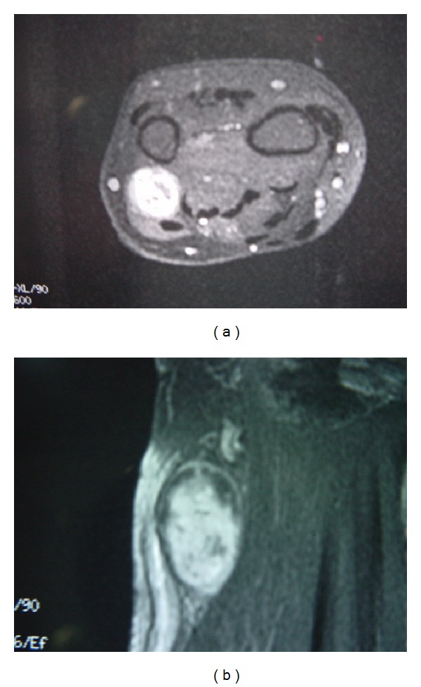
Imaging

  • MRI scan:
    • Imaging modality of choice
    • Oval or round mass Mass Three-dimensional lesion that occupies a space within the breast Imaging of the Breast that enhances uniformly with gadolinium Gadolinium An element of the rare earth family of metals. It has the atomic symbol gd, atomic number 64, and atomic weight 157. 25. Its oxide is used in the control rods of some nuclear reactors. Magnetic Resonance Imaging (MRI) contrast
    • T1-weighted MRI: isodense (or hypodense) to muscle
    • T2-weighted MRI: hyperdense to muscle
    • Target sign Target sign Pseudomembranous Colitis may be present:
      • Central area of hypodensity with a peripheral T2 signal hyperintensity
      • Most specific sign of a peripheral nerve sheath tumor Tumor Inflammation
      • Not specific for schwannoma
  • CT scan:
    • Hypodense to muscle
    • Enhances with contrast
  • Ultrasound:
    • Can detect schwannomas under the skin Skin The skin, also referred to as the integumentary system, is the largest organ of the body. The skin is primarily composed of the epidermis (outer layer) and dermis (deep layer). The epidermis is primarily composed of keratinocytes that undergo rapid turnover, while the dermis contains dense layers of connective tissue. Skin: Structure and Functions
    • Useful during surgery
Mri showing schwannoma

MRI showing schwannoma involving dorsal cutaneous branch of ulnar nerve

Image: “Schwannoma in the upper limbs” by Tang CY, Fung B, Fok M, Zhu J. License: CC BY 3.0

Biopsy Biopsy Removal and pathologic examination of specimens from the living body. Ewing Sarcoma and histopathology

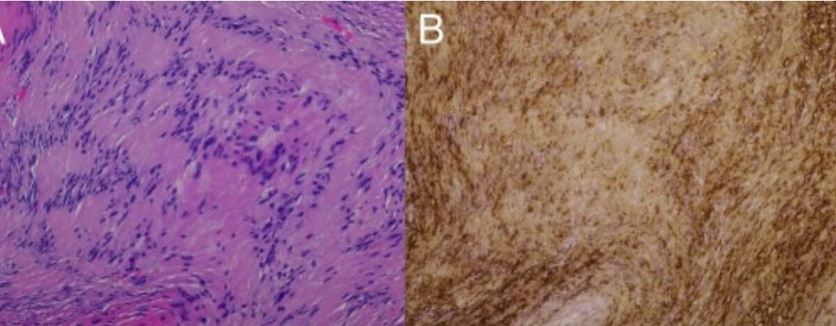
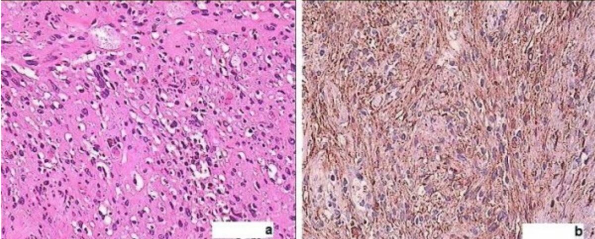
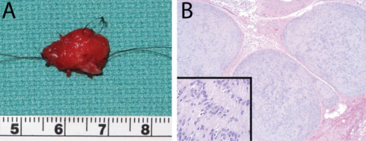
Classic schwannomas:

  • Surrounded by a well-defined fibrous capsule Fibrous capsule Hip Joint: Anatomy
  • Contains 2 distinct histologic regions:
    • Antoni A tissue: 
      • Dense hypercellular area
      • Predominantly benign Benign Fibroadenoma spindle cells Spindle Cells Acoustic Neuroma in intersecting bundles
      • May palisade around eosinophilic areas (Verocay bodies)
      • Positive for S100 S100 A family of highly acidic calcium-binding proteins found in large concentration in the brain and believed to be glial in origin. They are also found in other organs in the body. They have in common the ef-hand motif (ef hand motifs) found on a number of calcium binding proteins. The name of this family derives from the property of being soluble in a 100% saturated ammonium sulfate solution. Acoustic Neuroma protein staining
    • Antoni B tissue:
  • Degenerative changes Degenerative Changes Spinal Stenosis in longstanding (sometimes referred to as “ancient”) schwannomas:
    • Marked nuclear pleomorphism
    • Degenerative cysts Cysts Any fluid-filled closed cavity or sac that is lined by an epithelium. Cysts can be of normal, abnormal, non-neoplastic, or neoplastic tissues. Fibrocystic Change
    • Signs of remote hemorrhage and calcifications
    • Blood vessel hyalinization

Distinctive features of other types:

  • Cellular schwannomas:
    • Predominantly Antoni A tissue without Verocay bodies
    • ↑ Cellularity and mitotic activity
    • May be locally destructive
    • Will stain positive for S100 S100 A family of highly acidic calcium-binding proteins found in large concentration in the brain and believed to be glial in origin. They are also found in other organs in the body. They have in common the ef-hand motif (ef hand motifs) found on a number of calcium binding proteins. The name of this family derives from the property of being soluble in a 100% saturated ammonium sulfate solution. Acoustic Neuroma protein
  • Plexiform schwannomas:
    • Aggregations of multiple schwannomas: plexiform pattern of growth involving multiple fascicles
    • Less well circumscribed and may even lack a capsule Capsule An envelope of loose gel surrounding a bacterial cell which is associated with the virulence of pathogenic bacteria. Some capsules have a well-defined border, whereas others form a slime layer that trails off into the medium. Most capsules consist of relatively simple polysaccharides but there are some bacteria whose capsules are made of polypeptides. Bacteroides
    • Predominantly Antoni A tissue
    • ↑ Cellularity and mitotic activity
  • Melanotic schwannomas: 
    • Show dense melanin Melanin Insoluble polymers of tyrosine derivatives found in and causing darkness in skin (skin pigmentation), hair, and feathers providing protection against sunburn induced by sunlight. Carotenes contribute yellow and red coloration. Seborrheic Keratosis pigmentation
    • May stain positive for:
      • MelanA
      • Glial fibrillary acidic protein (GFAP, more commonly seen in astrocytomas)
      • Neurofilament protein

Grading Grading Methods which attempt to express in replicable terms the level of cell differentiation in neoplasms as increasing anaplasia correlates with the aggressiveness of the neoplasm. Grading, Staging, and Metastasis

Schwannomas are commonly classified according to the Enneking system of benign Benign Fibroadenoma lesions. There are 3 grades, and most schwannomas are grade II or III lesions.

  • Grade I: inactive lesions
  • Grade II: not destructive or locally aggressive but may cause deformation of surrounding tissues
  • Grade III: locally aggressive and may invade local tissues but are not metastatic

Management

Asymptomatic cases

  • Observation and follow-up
  • Consider a biopsy Biopsy Removal and pathologic examination of specimens from the living body. Ewing Sarcoma.

Symptomatic cases

For patients Patients Individuals participating in the health care system for the purpose of receiving therapeutic, diagnostic, or preventive procedures. Clinician–Patient Relationship with functional deficits and/or pain Pain An unpleasant sensation induced by noxious stimuli which are detected by nerve endings of nociceptive neurons. Pain: Types and Pathways due to nerve compression Nerve Compression Brachial Plexus Injuries, treatment is recommended.

  • Surgical excision (mainstay of treatment):
  • Radiation Radiation Emission or propagation of acoustic waves (sound), electromagnetic energy waves (such as light; radio waves; gamma rays; or x-rays), or a stream of subatomic particles (such as electrons; neutrons; protons; or alpha particles). Osteosarcoma therapy:
    • May be considered, especially in patients Patients Individuals participating in the health care system for the purpose of receiving therapeutic, diagnostic, or preventive procedures. Clinician–Patient Relationship with acoustic neuromas or tumors near vital blood vessels or nerves
    • Typically not needed due to low risk of metastasis Metastasis The transfer of a neoplasm from one organ or part of the body to another remote from the primary site. Grading, Staging, and Metastasis and recurrence

Differential Diagnosis

  • Neurofibroma Neurofibroma A moderately firm, benign, encapsulated tumor resulting from proliferation of schwann cells and fibroblasts that includes portions of nerve fibers. The tumors usually develop along peripheral or cranial nerves and are a central feature of neurofibromatosis 1, where they may occur intracranially or involve spinal roots. Pathologic features include fusiform enlargement of the involved nerve. Microscopic examination reveals a disorganized and loose cellular pattern with elongated nuclei intermixed with fibrous strands. Neurofibromatosis Type 1: another benign Benign Fibroadenoma tumor Tumor Inflammation of the peripheral nervous system Peripheral nervous system The nervous system outside of the brain and spinal cord. The peripheral nervous system has autonomic and somatic divisions. The autonomic nervous system includes the enteric, parasympathetic, and sympathetic subdivisions. The somatic nervous system includes the cranial and spinal nerves and their ganglia and the peripheral sensory receptors. Nervous System: Anatomy, Structure, and Classification, arising from a mix of Schwann cells, perineural-like cells, nerve fibers Nerve Fibers Slender processes of neurons, including the axons and their glial envelopes (myelin sheath). Nerve fibers conduct nerve impulses to and from the central nervous system. Nervous System: Histology, myxoid stroma, and fibroblasts Fibroblasts Connective tissue cells which secrete an extracellular matrix rich in collagen and other macromolecules. Sarcoidosis. Neurofibromas can arise sporadically or in association with neurofibromatosis type I. Neurofibromas present as a palpable circular mass Mass Three-dimensional lesion that occupies a space within the breast Imaging of the Breast on the skin Skin The skin, also referred to as the integumentary system, is the largest organ of the body. The skin is primarily composed of the epidermis (outer layer) and dermis (deep layer). The epidermis is primarily composed of keratinocytes that undergo rapid turnover, while the dermis contains dense layers of connective tissue. Skin: Structure and Functions accompanied by pain Pain An unpleasant sensation induced by noxious stimuli which are detected by nerve endings of nociceptive neurons. Pain: Types and Pathways, paresthesia, and bleeding. Diagnosis is made by imaging studies and biopsy Biopsy Removal and pathologic examination of specimens from the living body. Ewing Sarcoma, and treatment is either observation or, in symptomatic patients Patients Individuals participating in the health care system for the purpose of receiving therapeutic, diagnostic, or preventive procedures. Clinician–Patient Relationship, surgical resection. 
  • Malignant peripheral nerve sheath tumor Tumor Inflammation (MPNST): also referred to as malignant schwannomas, these are highly malignant, locally invasive sarcomas. These tumors do not arise from benign Benign Fibroadenoma schwannomas; rather, they arise from de novo mutations or degeneration of plexiform neurofibromas. Malignant peripheral nerve sheath tumors are strongly associated with neurofibromatosis type I and mutations in the NF1 NF1 Neurofibromatosis type 1 (NF1), also known as phakomatosis, is a neurocutaneous disorder that is most commonly of autosomal dominant inheritance due to mutations in the NF1 gene. Neurofibromatosis type 1 presents a range of clinical manifestations with the most prominent features being various pigmented skin lesions called café au lait macules (CALMs), neurofibromas, freckling of the inguinal and axillary regions, and iris hamartomas. Neurofibromatosis Type 1 gene Gene A category of nucleic acid sequences that function as units of heredity and which code for the basic instructions for the development, reproduction, and maintenance of organisms. Basic Terms of Genetics. These tumors may appear similar to cellular or plexiform schwannomas in particular, but MPNSTs generally do not express S100 S100 A family of highly acidic calcium-binding proteins found in large concentration in the brain and believed to be glial in origin. They are also found in other organs in the body. They have in common the ef-hand motif (ef hand motifs) found on a number of calcium binding proteins. The name of this family derives from the property of being soluble in a 100% saturated ammonium sulfate solution. Acoustic Neuroma protein.
  • Perineurioma: benign Benign Fibroadenoma PNS tumors arising from the perineural cells. Perineuriomas are most commonly sporadic Sporadic Selective IgA Deficiency and present with palpable skin Skin The skin, also referred to as the integumentary system, is the largest organ of the body. The skin is primarily composed of the epidermis (outer layer) and dermis (deep layer). The epidermis is primarily composed of keratinocytes that undergo rapid turnover, while the dermis contains dense layers of connective tissue. Skin: Structure and Functions masses along the large nerves, pain Pain An unpleasant sensation induced by noxious stimuli which are detected by nerve endings of nociceptive neurons. Pain: Types and Pathways, and paresthesias Paresthesias Subjective cutaneous sensations (e.g., cold, warmth, tingling, pressure, etc.) that are experienced spontaneously in the absence of stimulation. Posterior Cord Syndrome. As with schwannomas, diagnosis is made via imaging and biopsy Biopsy Removal and pathologic examination of specimens from the living body. Ewing Sarcoma, and the treatment is surgical resection in symptomatic patients Patients Individuals participating in the health care system for the purpose of receiving therapeutic, diagnostic, or preventive procedures. Clinician–Patient Relationship
  • Ganglioneuroma: slow-growing, large, encapsulated Encapsulated Klebsiella tumors arising from the sympathetic ganglion cells Ganglion Cells The Visual Pathway and Related Disorders. Ganglioneuromas can occur anywhere along the sympathetic chain. Clinical presentation depends on their specific location, with common locations including the mediastinum Mediastinum The mediastinum is the thoracic area between the 2 pleural cavities. The mediastinum contains vital structures of the circulatory, respiratory, digestive, and nervous systems including the heart and esophagus, and major thoracic vessels. Mediastinum and Great Vessels: Anatomy, retroperitoneum, and adrenal glands Adrenal Glands The adrenal glands are a pair of retroperitoneal endocrine glands located above the kidneys. The outer parenchyma is called the adrenal cortex and has 3 distinct zones, each with its own secretory products. Beneath the cortex lies the adrenal medulla, which secretes catecholamines involved in the fight-or-flight response. Adrenal Glands: Anatomy, and the symptoms are due to mass Mass Three-dimensional lesion that occupies a space within the breast Imaging of the Breast effect (e.g., constipation Constipation Constipation is common and may be due to a variety of causes. Constipation is generally defined as bowel movement frequency < 3 times per week. Patients who are constipated often strain to pass hard stools. The condition is classified as primary (also known as idiopathic or functional constipation) or secondary, and as acute or chronic. Constipation, radicular pain Radicular Pain Spinal Disk Herniation). Diagnosis is made by imaging studies and biopsy Biopsy Removal and pathologic examination of specimens from the living body. Ewing Sarcoma, and treatment is surgical resection. 
  • Ganglion cyst Ganglion cyst Nodular tumor-like lesions or mucoid flesh, arising from tendon sheaths, ligaments, or joint capsule, especially of the hands, wrists, or feet. They are not true cysts as they lack epithelial wall. They are distinguished from synovial cysts by the lack of communication with a joint cavity or the synovial membrane. Examination of the Upper Limbs: benign Benign Fibroadenoma cystic Cystic Fibrocystic Change tumors filled with a mucinous, gelatinous fluid that arise from a herniation Herniation Omphalocele of connective tissue Connective tissue Connective tissues originate from embryonic mesenchyme and are present throughout the body except inside the brain and spinal cord. The main function of connective tissues is to provide structural support to organs. Connective tissues consist of cells and an extracellular matrix. Connective Tissue: Histology associated with nearby joints or tendons. Ganglion cysts Cysts Any fluid-filled closed cavity or sac that is lined by an epithelium. Cysts can be of normal, abnormal, non-neoplastic, or neoplastic tissues. Fibrocystic Change form primarily on the dorsal wrist, and presentation is related to pressure on surrounding nerves with resultant pain Pain An unpleasant sensation induced by noxious stimuli which are detected by nerve endings of nociceptive neurons. Pain: Types and Pathways, numbness, and weakness. Diagnosis is made by history and physical exam findings, as well as plain X-rays X-rays X-rays are high-energy particles of electromagnetic radiation used in the medical field for the generation of anatomical images. X-rays are projected through the body of a patient and onto a film, and this technique is called conventional or projectional radiography. X-rays, MRI, or ultrasound. Symptomatic management is with aspiration or surgery (open or arthroscopic).
  • Lipoma Lipoma A lipoma is a benign neoplasm of fat cells (adipocytes) and the most common soft tissue tumor in adults. The etiology is unknown, but obesity is a predisposing factor; genetics also play a role, with multiple lipomas occurring in various inherited disorders. Lipoma: benign Benign Fibroadenoma tumors of the subcutaneous fat Subcutaneous fat Fatty tissue under the skin throughout the body. Erythema Nodosum that can occur on any part of the body. Lipomas are typically soft, painless nodules. Diagnosis is typically clinical, though ultrasound can help distinguish a lipoma Lipoma A lipoma is a benign neoplasm of fat cells (adipocytes) and the most common soft tissue tumor in adults. The etiology is unknown, but obesity is a predisposing factor; genetics also play a role, with multiple lipomas occurring in various inherited disorders. Lipoma from cystic Cystic Fibrocystic Change structures. Treatment is observation or surgical excision.

References

  1. Gilchrist, JM, Donahue, JE. (2021). Peripheral nerve tumors. UpToDate. Retrieved May 16, 2021, from https://www.uptodate.com/contents/peripheral-nerve-tumors 
  2. Laufer, I, Bilsky, M. (2021). Intradural nerve sheath tumors. UpToDate. Retrieved May 16, 2021, from https://www.uptodate.com/contents/intradural-nerve-sheath-tumors 
  3. Dickey, ID. (2020). Neurilemmoma (Schwannoma): Practice essentials, etiology, epidemiology. Medscape. Retrieved May 17, 2021, from https://emedicine.medscape.com/article/1256405-overview
  4. Davis, DD, Kane, SM. (2020). Neurilemmoma. StatPearls. Retrieved May 15, 2021, from https://pubmed.ncbi.nlm.nih.gov/32809662/
  5. Sheikh, MM, De Jesus, O. (2021). Schwannoma. StatPearls. Retrieved May 16, 2021, from https://www.statpearls.com/articlelibrary/viewarticle/28766/ 

Create your free account or log in to continue reading!

Sign up now and get free access to Lecturio with concept pages, medical videos, and questions for your medical education.

User Reviews

Unwrap New Skills This Holiday 🎄 Save 30% on all plans now!

Details