Advertisement

Advertisement

Advertisement

Advertisement

Refractive Errors

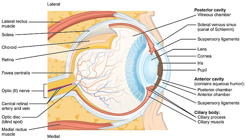
By refraction, the light that enters the eye is focused onto a particular point of the retina Retina The ten-layered nervous tissue membrane of the eye. It is continuous with the optic nerve and receives images of external objects and transmits visual impulses to the brain. Its outer surface is in contact with the choroid and the inner surface with the vitreous body. The outermost layer is pigmented, whereas the inner nine layers are transparent. Eye: Anatomy. The main refractive components of the eye are the cornea Cornea The transparent anterior portion of the fibrous coat of the eye consisting of five layers: stratified squamous corneal epithelium; bowman membrane; corneal stroma; descemet membrane; and mesenchymal corneal endothelium. It serves as the first refracting medium of the eye. Eye: Anatomy and the lens Lens A transparent, biconvex structure of the eye, enclosed in a capsule and situated behind the iris and in front of the vitreous humor (vitreous body). It is slightly overlapped at its margin by the ciliary processes. Adaptation by the ciliary body is crucial for ocular accommodation. Eye: Anatomy. When the corneal curvature, the refractive power of the lens Lens A transparent, biconvex structure of the eye, enclosed in a capsule and situated behind the iris and in front of the vitreous humor (vitreous body). It is slightly overlapped at its margin by the ciliary processes. Adaptation by the ciliary body is crucial for ocular accommodation. Eye: Anatomy, does not match the size of the eye, ametropia or a refractive error Error Refers to any act of commission (doing something wrong) or omission (failing to do something right) that exposes patients to potentially hazardous situations. Disclosure of Information occurs. The types of refractive errors include myopia (nearsightedness), hyperopia (farsightedness), and astigmatism (which can occur in both myopia and hyperopia). The use of a proper refractive device helps correct the visual impairment. Laser in situ keratomileusis (LASIK) is the most common corrective surgical procedure.

Last updated: May 17, 2024
Editorial responsibility: Stanley Oiseth, Lindsay Jones, Evelin Maza

Advertisement

Advertisement

Advertisement

Advertisement

Advertisement

Advertisement

Definitions and Epidemiology

Refraction

  • Light entering the eye is bent and focused onto the retina Retina The ten-layered nervous tissue membrane of the eye. It is continuous with the optic nerve and receives images of external objects and transmits visual impulses to the brain. Its outer surface is in contact with the choroid and the inner surface with the vitreous body. The outermost layer is pigmented, whereas the inner nine layers are transparent. Eye: Anatomy to create an image. 
  • Affects visual acuity Visual Acuity Clarity or sharpness of ocular vision or the ability of the eye to see fine details. Visual acuity depends on the functions of retina, neuronal transmission, and the interpretative ability of the brain. Normal visual acuity is expressed as 20/20 indicating that one can see at 20 feet what should normally be seen at that distance. Visual acuity can also be influenced by brightness, color, and contrast. Ophthalmic Exam
  • Light is refracted as it moves through structures of the eye (listed in order of encounter by light rays):
    1. Cornea Cornea The transparent anterior portion of the fibrous coat of the eye consisting of five layers: stratified squamous corneal epithelium; bowman membrane; corneal stroma; descemet membrane; and mesenchymal corneal endothelium. It serves as the first refracting medium of the eye. Eye: Anatomy
      • Avascular Avascular Corneal Abrasions, Erosion, and Ulcers, transparent structure in the front of the eye
      • Primary site of refraction (⅔ of the eye’s refractive power)
      • Curvature of the cornea Cornea The transparent anterior portion of the fibrous coat of the eye consisting of five layers: stratified squamous corneal epithelium; bowman membrane; corneal stroma; descemet membrane; and mesenchymal corneal endothelium. It serves as the first refracting medium of the eye. Eye: Anatomy must match eye size; if it does not → refractive error Error Refers to any act of commission (doing something wrong) or omission (failing to do something right) that exposes patients to potentially hazardous situations. Disclosure of Information
    2. Aqueous humor Humor Defense Mechanisms
    3. Through the pupil Pupil The pupil is the space within the eye that permits light to project onto the retina. Anatomically located in front of the lens, the pupil’s size is controlled by the surrounding iris. The pupil provides insight into the function of the central and autonomic nervous systems. Pupil: Physiology and Abnormalities (dilation adjusted by iris)
    4. Lens Lens A transparent, biconvex structure of the eye, enclosed in a capsule and situated behind the iris and in front of the vitreous humor (vitreous body). It is slightly overlapped at its margin by the ciliary processes. Adaptation by the ciliary body is crucial for ocular accommodation. Eye: Anatomy
      • Second greatest site of refraction
      • Able to adjust refraction
      • Accommodation: adjustment in lens Lens A transparent, biconvex structure of the eye, enclosed in a capsule and situated behind the iris and in front of the vitreous humor (vitreous body). It is slightly overlapped at its margin by the ciliary processes. Adaptation by the ciliary body is crucial for ocular accommodation. Eye: Anatomy shape to change focus from distant to near images ( lens Lens A transparent, biconvex structure of the eye, enclosed in a capsule and situated behind the iris and in front of the vitreous humor (vitreous body). It is slightly overlapped at its margin by the ciliary processes. Adaptation by the ciliary body is crucial for ocular accommodation. Eye: Anatomy shape changed by action of ciliary muscle)
    5. Vitreous body Vitreous body The transparent, semigelatinous substance that fills the cavity behind the crystalline lens of the eye and in front of the retina. It is contained in a thin hyaloid membrane and forms about four fifths of the optic globe. Eye: Anatomy
    6. Retina Retina The ten-layered nervous tissue membrane of the eye. It is continuous with the optic nerve and receives images of external objects and transmits visual impulses to the brain. Its outer surface is in contact with the choroid and the inner surface with the vitreous body. The outermost layer is pigmented, whereas the inner nine layers are transparent. Eye: Anatomy (inner surface of the eye containing photoreceptors)

Ametropia or refractive error Error Refers to any act of commission (doing something wrong) or omission (failing to do something right) that exposes patients to potentially hazardous situations. Disclosure of Information

  • When light entering the eye does not focus on the retina Retina The ten-layered nervous tissue membrane of the eye. It is continuous with the optic nerve and receives images of external objects and transmits visual impulses to the brain. Its outer surface is in contact with the choroid and the inner surface with the vitreous body. The outermost layer is pigmented, whereas the inner nine layers are transparent. Eye: Anatomy
  • The focusing power of the cornea Cornea The transparent anterior portion of the fibrous coat of the eye consisting of five layers: stratified squamous corneal epithelium; bowman membrane; corneal stroma; descemet membrane; and mesenchymal corneal endothelium. It serves as the first refracting medium of the eye. Eye: Anatomy and lens Lens A transparent, biconvex structure of the eye, enclosed in a capsule and situated behind the iris and in front of the vitreous humor (vitreous body). It is slightly overlapped at its margin by the ciliary processes. Adaptation by the ciliary body is crucial for ocular accommodation. Eye: Anatomy (corneal curvature, refractive power of lens Lens A transparent, biconvex structure of the eye, enclosed in a capsule and situated behind the iris and in front of the vitreous humor (vitreous body). It is slightly overlapped at its margin by the ciliary processes. Adaptation by the ciliary body is crucial for ocular accommodation. Eye: Anatomy) does not match eye size ( axial Axial Computed Tomography (CT) length of globe).
  • The condition requires a refractive device.
  • Unit of measurement of refractive error Error Refers to any act of commission (doing something wrong) or omission (failing to do something right) that exposes patients to potentially hazardous situations. Disclosure of Information: diopter
  • Types:
    • Myopia: nearsightedness
    • Hyperopia: farsightedness
    • Astigmatism: uneven focus

Epidemiology

  • Refractive error Error Refers to any act of commission (doing something wrong) or omission (failing to do something right) that exposes patients to potentially hazardous situations. Disclosure of Information affects approximately ⅓ of the population > 40 years of age in the United States and Western Europe.
  • Myopia, the most common refractive error Error Refers to any act of commission (doing something wrong) or omission (failing to do something right) that exposes patients to potentially hazardous situations. Disclosure of Information, decreases with increasing age; high prevalence Prevalence The total number of cases of a given disease in a specified population at a designated time. It is differentiated from incidence, which refers to the number of new cases in the population at a given time. Measures of Disease Frequency among Asians
  • Hyperopia and astigmatism increase with age.

Further definitions

  • Emmetropia
    • Normal vision Vision Ophthalmic Exam
    • Light is focused on the retina Retina The ten-layered nervous tissue membrane of the eye. It is continuous with the optic nerve and receives images of external objects and transmits visual impulses to the brain. Its outer surface is in contact with the choroid and the inner surface with the vitreous body. The outermost layer is pigmented, whereas the inner nine layers are transparent. Eye: Anatomy
  • Presbyopia
    • Not a refractive error Error Refers to any act of commission (doing something wrong) or omission (failing to do something right) that exposes patients to potentially hazardous situations. Disclosure of Information but affects visual acuity Visual Acuity Clarity or sharpness of ocular vision or the ability of the eye to see fine details. Visual acuity depends on the functions of retina, neuronal transmission, and the interpretative ability of the brain. Normal visual acuity is expressed as 20/20 indicating that one can see at 20 feet what should normally be seen at that distance. Visual acuity can also be influenced by brightness, color, and contrast. Ophthalmic Exam
    • Affected by physiologic loss of accommodation
    • In aging, the lens Lens A transparent, biconvex structure of the eye, enclosed in a capsule and situated behind the iris and in front of the vitreous humor (vitreous body). It is slightly overlapped at its margin by the ciliary processes. Adaptation by the ciliary body is crucial for ocular accommodation. Eye: Anatomy loses elasticity Elasticity Resistance and recovery from distortion of shape. Skeletal Muscle Contraction, thus has less ability to become more rounded → inability to focus at reading distance
  • Anisometropia: each eye with different refractive power

Clinical Presentation

Myopia Hyperopia Astigmatism
Focal point In front of retina Retina The ten-layered nervous tissue membrane of the eye. It is continuous with the optic nerve and receives images of external objects and transmits visual impulses to the brain. Its outer surface is in contact with the choroid and the inner surface with the vitreous body. The outermost layer is pigmented, whereas the inner nine layers are transparent. Eye: Anatomy Behind retina Retina The ten-layered nervous tissue membrane of the eye. It is continuous with the optic nerve and receives images of external objects and transmits visual impulses to the brain. Its outer surface is in contact with the choroid and the inner surface with the vitreous body. The outermost layer is pigmented, whereas the inner nine layers are transparent. Eye: Anatomy Multiple sites
Causes Eye too long, cornea Cornea The transparent anterior portion of the fibrous coat of the eye consisting of five layers: stratified squamous corneal epithelium; bowman membrane; corneal stroma; descemet membrane; and mesenchymal corneal endothelium. It serves as the first refracting medium of the eye. Eye: Anatomy with too much curvature Eye too short, cornea Cornea The transparent anterior portion of the fibrous coat of the eye consisting of five layers: stratified squamous corneal epithelium; bowman membrane; corneal stroma; descemet membrane; and mesenchymal corneal endothelium. It serves as the first refracting medium of the eye. Eye: Anatomy with too little curvature Uneven curvature of the cornea Cornea The transparent anterior portion of the fibrous coat of the eye consisting of five layers: stratified squamous corneal epithelium; bowman membrane; corneal stroma; descemet membrane; and mesenchymal corneal endothelium. It serves as the first refracting medium of the eye. Eye: Anatomy
Result Cannot focus distant objects Cannot focus near objects Blurry vision Vision Ophthalmic Exam

Myopia

  • Axial Axial Computed Tomography (CT) length of the eye is too long or the cornea Cornea The transparent anterior portion of the fibrous coat of the eye consisting of five layers: stratified squamous corneal epithelium; bowman membrane; corneal stroma; descemet membrane; and mesenchymal corneal endothelium. It serves as the first refracting medium of the eye. Eye: Anatomy is too curved.
  • Light is focused in front of the retina Retina The ten-layered nervous tissue membrane of the eye. It is continuous with the optic nerve and receives images of external objects and transmits visual impulses to the brain. Its outer surface is in contact with the choroid and the inner surface with the vitreous body. The outermost layer is pigmented, whereas the inner nine layers are transparent. Eye: Anatomy
  • Ability to focus on near objects only; faraway objects will be out of focus
  • Risk factors:
    • Genetics Genetics Genetics is the study of genes and their functions and behaviors. Basic Terms of Genetics
    • Reading (↑ axial Axial Computed Tomography (CT) length during development)
    • Medications (sulfa, diuretics Diuretics Agents that promote the excretion of urine through their effects on kidney function. Heart Failure and Angina Medication, cholinergics): transient effect
    • Diabetes Diabetes Diabetes mellitus (DM) is a metabolic disease characterized by hyperglycemia and dysfunction of the regulation of glucose metabolism by insulin. Type 1 DM is diagnosed mostly in children and young adults as the result of autoimmune destruction of β cells in the pancreas and the resulting lack of insulin. Type 2 DM has a significant association with obesity and is characterized by insulin resistance. Diabetes Mellitus mellitus: transient; change in serum osmolarity Osmolarity The concentration of osmotically active particles in solution expressed in terms of osmoles of solute per liter of solution. Osmolality is expressed in terms of osmoles of solute per kilogram of solvent. Hypernatremia increases osmotic fluid into the lens Lens A transparent, biconvex structure of the eye, enclosed in a capsule and situated behind the iris and in front of the vitreous humor (vitreous body). It is slightly overlapped at its margin by the ciliary processes. Adaptation by the ciliary body is crucial for ocular accommodation. Eye: Anatomy lens Lens A transparent, biconvex structure of the eye, enclosed in a capsule and situated behind the iris and in front of the vitreous humor (vitreous body). It is slightly overlapped at its margin by the ciliary processes. Adaptation by the ciliary body is crucial for ocular accommodation. Eye: Anatomy becomes swollen → increased refractive power
    • Trauma
    • Excessive accommodation
    • Elevated intraocular pressure Intraocular Pressure The pressure of the fluids in the eye. Ophthalmic Exam
    • ↑ maternal age and maternal smoking Smoking Willful or deliberate act of inhaling and exhaling smoke from burning substances or agents held by hand. Interstitial Lung Diseases during pregnancy Pregnancy The status during which female mammals carry their developing young (embryos or fetuses) in utero before birth, beginning from fertilization to birth. Pregnancy: Diagnosis, Physiology, and Care
Myopia

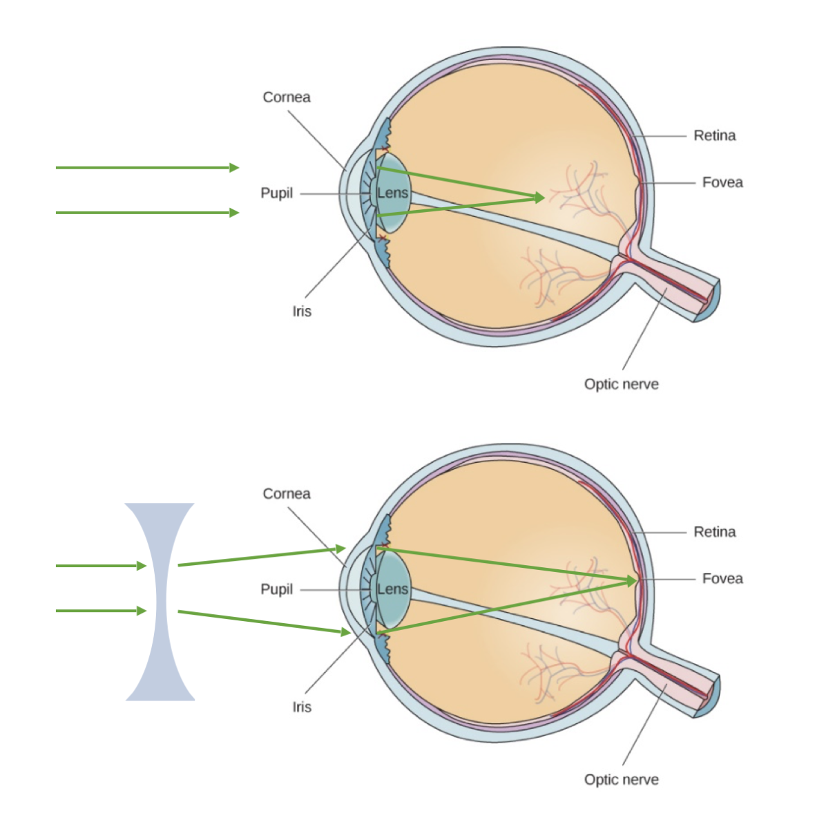
This figure illustrates myopia (nearsightedness). The top image shows light as it is refracted too sharply by the cornea, resulting in a focal point in front of the retina.
With corrective negative lenses (bottom image), the light can be dispersed through the concave lens and focus appropriately onto the retina for clear vision.
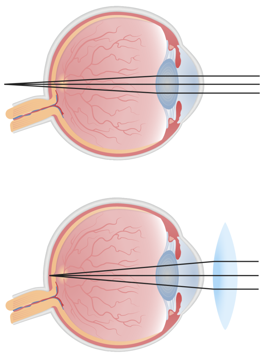
Image: “myopia” by OpenStax-CNX. License: CC BY 4.0

Hyperopia

  • Axial Axial Computed Tomography (CT) length of the eye is too short or the cornea Cornea The transparent anterior portion of the fibrous coat of the eye consisting of five layers: stratified squamous corneal epithelium; bowman membrane; corneal stroma; descemet membrane; and mesenchymal corneal endothelium. It serves as the first refracting medium of the eye. Eye: Anatomy is flat.
  • Light is focused behind the retina Retina The ten-layered nervous tissue membrane of the eye. It is continuous with the optic nerve and receives images of external objects and transmits visual impulses to the brain. Its outer surface is in contact with the choroid and the inner surface with the vitreous body. The outermost layer is pigmented, whereas the inner nine layers are transparent. Eye: Anatomy.
  • Ability to focus on faraway objects only; near objects will be out of focus.
  • Risk factors:
    • Trauma
    • A mass Mass Three-dimensional lesion that occupies a space within the breast Imaging of the Breast or lesion behind the eye
    • Surgery ( lens Lens A transparent, biconvex structure of the eye, enclosed in a capsule and situated behind the iris and in front of the vitreous humor (vitreous body). It is slightly overlapped at its margin by the ciliary processes. Adaptation by the ciliary body is crucial for ocular accommodation. Eye: Anatomy removal)
    • Medications: anticholinergics Anticholinergics Anticholinergic drugs block the effect of the neurotransmitter acetylcholine at the muscarinic receptors in the central and peripheral nervous systems. Anticholinergic agents inhibit the parasympathetic nervous system, resulting in effects on the smooth muscle in the respiratory tract, vascular system, urinary tract, GI tract, and pupils of the eyes. Anticholinergic Drugs
    • Rapid decline in glucose Glucose A primary source of energy for living organisms. It is naturally occurring and is found in fruits and other parts of plants in its free state. It is used therapeutically in fluid and nutrient replacement. Lactose Intolerance (transient)
Hyperopia

This figure illustrates hyperopia (farsightedness). The top image illustrates uncorrected refraction, where the light is not refracted sharply enough, resulting in a focal point behind the retina.
With corrective positive lenses (bottom image), the light can be dispersed through the convex lens and focus appropriately onto the retina for clear vision.

Image by Lecturio.

Astigmatism

  • Uneven curvature of the cornea Cornea The transparent anterior portion of the fibrous coat of the eye consisting of five layers: stratified squamous corneal epithelium; bowman membrane; corneal stroma; descemet membrane; and mesenchymal corneal endothelium. It serves as the first refracting medium of the eye. Eye: Anatomy ( cornea Cornea The transparent anterior portion of the fibrous coat of the eye consisting of five layers: stratified squamous corneal epithelium; bowman membrane; corneal stroma; descemet membrane; and mesenchymal corneal endothelium. It serves as the first refracting medium of the eye. Eye: Anatomy shape is “more like a football than a basketball”)
  • Multiple focal points = different refractive powers along different meridians
  • Results in blurry vision Vision Ophthalmic Exam at all viewing distances
Astigmatism

This figure contrasts the normal corneal curvature with that of an astigmatic cornea. The cornea with astigmatism distorts the focus point of light in front of and/or behind the retina.

Image by Lecturio.

Diagnosis

Adults

  • No consensus on routine vision Vision Ophthalmic Exam screening Screening Preoperative Care for adults > 65 years of age without vision Vision Ophthalmic Exam problems
  • Vision Vision Ophthalmic Exam is integral to maintaining independence in activities of daily living ( ADL ADL Lymphatic Filariasis (Elephantiasis)), so the need for optical correction has to be determined.
  • An eye specialist referral is recommended for unexplained vision Vision Ophthalmic Exam change/loss. 
  • Snellen chart
    • High-contrast symbols (black letters/numbers on white background) with varying sizes read at a standard distance (20 feet (6 meters))
    • Often used to evaluate visual acuity Visual Acuity Clarity or sharpness of ocular vision or the ability of the eye to see fine details. Visual acuity depends on the functions of retina, neuronal transmission, and the interpretative ability of the brain. Normal visual acuity is expressed as 20/20 indicating that one can see at 20 feet what should normally be seen at that distance. Visual acuity can also be influenced by brightness, color, and contrast. Ophthalmic Exam

Children

Management

Non-surgical treatment

Myopia

  1. Corrected with concave (diverging) lens Lens A transparent, biconvex structure of the eye, enclosed in a capsule and situated behind the iris and in front of the vitreous humor (vitreous body). It is slightly overlapped at its margin by the ciliary processes. Adaptation by the ciliary body is crucial for ocular accommodation. Eye: Anatomy, which spreads out the light
    • Functions to decrease the focusing power (“minus” power lenses)
    • Eyeglass prescriptions for myopia are in negative diopters (less focusing power), i.e., –3.00. 
  2. In general, school-aged children are treated for myopia of > 1.52 diopters (interferes with education and/or social function).

Hyperopia

  • Corrected with convex (converging) lens Lens A transparent, biconvex structure of the eye, enclosed in a capsule and situated behind the iris and in front of the vitreous humor (vitreous body). It is slightly overlapped at its margin by the ciliary processes. Adaptation by the ciliary body is crucial for ocular accommodation. Eye: Anatomy, which brings the focus forward onto the retina Retina The ten-layered nervous tissue membrane of the eye. It is continuous with the optic nerve and receives images of external objects and transmits visual impulses to the brain. Its outer surface is in contact with the choroid and the inner surface with the vitreous body. The outermost layer is pigmented, whereas the inner nine layers are transparent. Eye: Anatomy
    • Functions to increase the focusing power (“plus” lenses)
    • Eyeglass prescriptions for hyperopia are in positive diopters (more focusing power), i.e., +2.00.
  • In children, mild hyperopia is normal. Correction recommended in high hyperopia, i.e. > 4 diopters (interferes with education and at risk for refractive amblyopia Amblyopia A nonspecific term referring to impaired vision. Major subcategories include stimulus deprivation-induced amblyopia and toxic amblyopia. Stimulus deprivation-induced amblyopia is a developmental disorder of the visual cortex. A discrepancy between visual information received by the visual cortex from each eye results in abnormal cortical development. Strabismus and refractive errors may cause this condition. Toxic amblyopia is a disorder of the optic nerve which is associated with alcoholism, tobacco smoking, and other toxins and as an adverse effect of the use of some medications. Strabismus).

Astigmatism

  • Corrected with cylindrical lenses (to even out focusing power)
  • Eyeglass prescription for astigmatism indicates the cylinder and axis ( orientation Orientation Awareness of oneself in relation to time, place and person. Psychiatric Assessment of astigmatism)

Presbyopia

  • Use of reading glasses (convex lens Lens A transparent, biconvex structure of the eye, enclosed in a capsule and situated behind the iris and in front of the vitreous humor (vitreous body). It is slightly overlapped at its margin by the ciliary processes. Adaptation by the ciliary body is crucial for ocular accommodation. Eye: Anatomy)

Eyeglass prescription

  • OS (oculus sinister): left eye
  • OD (oculus dexter): right eye
  • OU (oculus uterque): both eyes
  • SPH: sphere/degree of near- or farsightedness)
    • Numbers represent diopters: the further away from zero, the stronger the prescription is
    • “Minus” sign: myopia, i.e. -3.00 = 3 diopters of nearsightedness
    • “Plus” sign: hyperopia, i.e. +2.25 = 2 and ¼ diopters of farsightedness
  • CYL/cylinder: indicating astigmatic component x AXIS (radial degrees of the cylinder)
    • CYL can be “plus” (farsighted astigmatism) or “minus” (nearsighted astigmatism)
    • AXIS is 0180 degrees; indicates where the difference in curvature is
  • Add: additional lens Lens A transparent, biconvex structure of the eye, enclosed in a capsule and situated behind the iris and in front of the vitreous humor (vitreous body). It is slightly overlapped at its margin by the ciliary processes. Adaptation by the ciliary body is crucial for ocular accommodation. Eye: Anatomy power, i.e., bifocals (instead of having separate reading glasses)

Example:

A patient with hyperopia, more on the left, also with astigmatism in both eyes, would be given the following prescription:

  • OS +3.25 + 2.50 x 090
  • OD +3.00 + 2.50 x 090

Surgical treatment

  • Typically performed in adults
  • LASIK: laser-assisted in situ keratomileusis
    • Most widely used procedure
    • Uses excimer laser to reshape the surface of the cornea Cornea The transparent anterior portion of the fibrous coat of the eye consisting of five layers: stratified squamous corneal epithelium; bowman membrane; corneal stroma; descemet membrane; and mesenchymal corneal endothelium. It serves as the first refracting medium of the eye. Eye: Anatomy ( epithelium Epithelium The epithelium is a complex of specialized cellular organizations arranged into sheets and lining cavities and covering the surfaces of the body. The cells exhibit polarity, having an apical and a basal pole. Structures important for the epithelial integrity and function involve the basement membrane, the semipermeable sheet on which the cells rest, and interdigitations, as well as cellular junctions. Surface Epithelium: Histology and stroma)
    • Minimal pain Pain An unpleasant sensation induced by noxious stimuli which are detected by nerve endings of nociceptive neurons. Pain: Types and Pathways
    • For myopia: flattens the cornea Cornea The transparent anterior portion of the fibrous coat of the eye consisting of five layers: stratified squamous corneal epithelium; bowman membrane; corneal stroma; descemet membrane; and mesenchymal corneal endothelium. It serves as the first refracting medium of the eye. Eye: Anatomy
    • For hyperopia: Laser is applied at the periphery to steepen the central cornea Cornea The transparent anterior portion of the fibrous coat of the eye consisting of five layers: stratified squamous corneal epithelium; bowman membrane; corneal stroma; descemet membrane; and mesenchymal corneal endothelium. It serves as the first refracting medium of the eye. Eye: Anatomy.
    • For astigmatism: combines peripheral and central laser treatments
  • LASEK: laser epithelial keratomileusis
    • Corneal epithelium Epithelium The epithelium is a complex of specialized cellular organizations arranged into sheets and lining cavities and covering the surfaces of the body. The cells exhibit polarity, having an apical and a basal pole. Structures important for the epithelial integrity and function involve the basement membrane, the semipermeable sheet on which the cells rest, and interdigitations, as well as cellular junctions. Surface Epithelium: Histology flap is formed with use of alcohol, then laser is applied
    • Mild-to-moderate postoperative pain Pain An unpleasant sensation induced by noxious stimuli which are detected by nerve endings of nociceptive neurons. Pain: Types and Pathways
    • Slower optical recovery than LASIK 
    • May need postoperative medications for up to 3 months
  • PRK: photorefractive keratectomy
    • The corneal epithelium Epithelium The epithelium is a complex of specialized cellular organizations arranged into sheets and lining cavities and covering the surfaces of the body. The cells exhibit polarity, having an apical and a basal pole. Structures important for the epithelial integrity and function involve the basement membrane, the semipermeable sheet on which the cells rest, and interdigitations, as well as cellular junctions. Surface Epithelium: Histology is removed and then laser treatment is applied.
    • Mild-to-moderate postoperative pain Pain An unpleasant sensation induced by noxious stimuli which are detected by nerve endings of nociceptive neurons. Pain: Types and Pathways, may require systemic analgesic
    • May take up to 3 months for visual acuity Visual Acuity Clarity or sharpness of ocular vision or the ability of the eye to see fine details. Visual acuity depends on the functions of retina, neuronal transmission, and the interpretative ability of the brain. Normal visual acuity is expressed as 20/20 indicating that one can see at 20 feet what should normally be seen at that distance. Visual acuity can also be influenced by brightness, color, and contrast. Ophthalmic Exam to stabilize

Differential Diagnosis

The differential diagnosis of refractive errors includes the following conditions:

  • Cataracts: A leading cause of blindness Blindness The inability to see or the loss or absence of perception of visual stimuli. This condition may be the result of eye diseases; optic nerve diseases; optic chiasm diseases; or brain diseases affecting the visual pathways or occipital lobe. Retinopathy of Prematurity worldwide. It is characterized by a decrease in vision Vision Ophthalmic Exam due to cloudiness or loss of transparency of the lens Lens A transparent, biconvex structure of the eye, enclosed in a capsule and situated behind the iris and in front of the vitreous humor (vitreous body). It is slightly overlapped at its margin by the ciliary processes. Adaptation by the ciliary body is crucial for ocular accommodation. Eye: Anatomy, which develops areas of opaqueness that require surgical extraction.
  • Glaucoma Glaucoma Glaucoma is an optic neuropathy characterized by typical visual field defects and optic nerve atrophy seen as optic disc cupping on examination. The acute form of glaucoma is a medical emergency. Glaucoma is often, but not always, caused by increased intraocular pressure (IOP). Glaucoma: An optic neuropathy Neuropathy Leprosy with distinctive changes in the optic cup and visual field Visual Field The Visual Pathway and Related Disorders defect. It is often associated with increased pressure within the eyeball (commonly within the anterior and posterior chambers), which results in gradual vision Vision Ophthalmic Exam loss. This can occur acutely due to a blockage of the aqueous humor Humor Defense Mechanisms drainage ( acute angle-closure glaucoma Acute Angle-Closure Glaucoma Glaucoma) which is an emergency. The majority of cases are due to open-angle glaucoma Glaucoma Glaucoma is an optic neuropathy characterized by typical visual field defects and optic nerve atrophy seen as optic disc cupping on examination. The acute form of glaucoma is a medical emergency. Glaucoma is often, but not always, caused by increased intraocular pressure (IOP). Glaucoma.
  • Uveitis Uveitis Uveitis is the inflammation of the uvea, the pigmented middle layer of the eye, which comprises the iris, ciliary body, and choroid. The condition is categorized based on the site of disease; anterior uveitis is the most common. Diseases of the Uvea: acute inflammation Acute Inflammation Inflammation of the uvea Uvea The pigmented vascular coat of the eyeball, consisting of the choroid; ciliary body; and iris, which are continuous with each other. Eye: Anatomy (vascular layer of the eyeball) and of the ciliary body Ciliary body A ring of tissue extending from the scleral spur to the ora serrata of the retina. It consists of the uveal portion and the epithelial portion. The ciliary muscle is in the uveal portion and the ciliary processes are in the epithelial portion. Eye: Anatomy. Affected patients Patients Individuals participating in the health care system for the purpose of receiving therapeutic, diagnostic, or preventive procedures. Clinician–Patient Relationship often suffer from blurred vision Blurred Vision Retinal Detachment, painful (anterior uveitis Uveitis Uveitis is the inflammation of the uvea, the pigmented middle layer of the eye, which comprises the iris, ciliary body, and choroid. The condition is categorized based on the site of disease; anterior uveitis is the most common. Diseases of the Uvea) or painless ( posterior uveitis Posterior Uveitis Diseases of the Uvea) redness Redness Inflammation of the eye.

References

  1. Shahzad, M., Gardiner, M. & Givens, J. (2019). Visual impairment in adults: Refractive disorders and presbyopia. UpToDate. Retrieved September 1, 2020, from https://www.uptodate.com/contents/visual-impairment-in-adults-refractive-disorders-and-presbyopia
  2. Bower, K.; Jacobs, D. & Givens, J. (2020). Laser refractive surgery. UpToDate. Retrieved September 1, 2020, from https://www.uptodate.com/contents/laser-refractive-surgery
  3. Bope, Edward T.; Kellerman, Rick D. (2015). Conn’s Current Therapy 2016. Elsevier Health Sciences. p. 354. ISBN 9780323355353

Create your free account or log in to continue reading!

Sign up now and get free access to Lecturio with concept pages, medical videos, and questions for your medical education.

User Reviews

Unwrap New Skills This Holiday 🎄 Save 30% on all plans now!

Details