Advertisement

Advertisement

Advertisement

Advertisement

Posterior Cord Syndrome

Posterior cord syndrome (PCS) refers to a group of symptoms caused by an incomplete spinal cord injury that affects the dorsal columns, the corticospinal tracts Corticospinal Tracts Central Cord Syndrome ( CSTs CSTs Central Cord Syndrome), and descending autonomic tracts to the bladder Bladder A musculomembranous sac along the urinary tract. Urine flows from the kidneys into the bladder via the ureters, and is held there until urination. Pyelonephritis and Perinephric Abscess. Posterior cord syndrome is rare but has a diverse range of etiologies, including demyelinating disorders, degenerative spinal conditions, neoplasms Neoplasms New abnormal growth of tissue. Malignant neoplasms show a greater degree of anaplasia and have the properties of invasion and metastasis, compared to benign neoplasms. Benign Bone Tumors, vascular abnormalities, and hereditary neurodegenerative disorders. Clinical symptoms include gait ataxia Gait ataxia Impairment of the ability to coordinate the movements required for normal ambulation (walking) which may result from impairments of motor function or sensory feedback. This condition may be associated with brain diseases (including cerebellar diseases and basal ganglia diseases); spinal cord diseases; or peripheral nervous system diseases. Friedreich Ataxia, paresthesias with loss of position and vibration Vibration A continuing periodic change in displacement with respect to a fixed reference. Neurological Examination sense, and urinary incontinence Urinary incontinence Urinary incontinence (UI) is involuntary loss of bladder control or unintentional voiding, which represents a hygienic or social problem to the patient. Urinary incontinence is a symptom, a sign, and a disorder. The 5 types of UI include stress, urge, mixed, overflow, and functional. Urinary Incontinence. The diagnosis is made clinically and with neuroimaging Neuroimaging Non-invasive methods of visualizing the central nervous system, especially the brain, by various imaging modalities. Febrile Infant. Management addresses treatment of the underlying cause.

Last updated: Mar 29, 2023

Editorial responsibility: Stanley Oiseth, Lindsay Jones, Evelin Maza

Advertisement

Advertisement

Advertisement

Advertisement

Advertisement

Advertisement

Overview

Definition

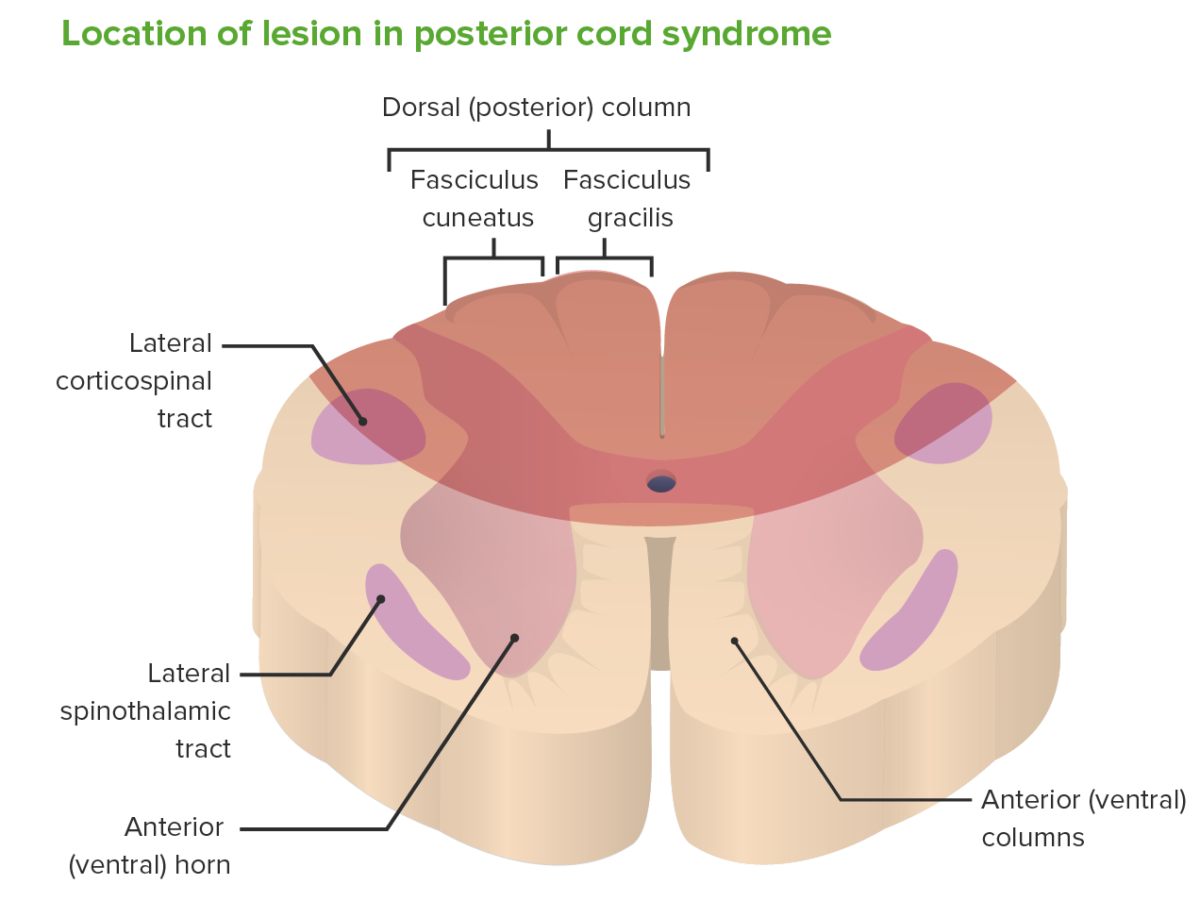
PosteriorPosterior cord syndrome refers to a group of symptoms caused by an incomplete spinal cord Spinal cord The spinal cord is the major conduction pathway connecting the brain to the body; it is part of the CNS. In cross section, the spinal cord is divided into an H-shaped area of gray matter (consisting of synapsing neuronal cell bodies) and a surrounding area of white matter (consisting of ascending and descending tracts of myelinated axons). Spinal Cord: Anatomy injury that affects the dorsal columns, the corticospinal tracts Corticospinal Tracts Central Cord Syndrome ( CSTs CSTs Central Cord Syndrome), and descending autonomic tracts to the bladder Bladder A musculomembranous sac along the urinary tract. Urine flows from the kidneys into the bladder via the ureters, and is held there until urination. Pyelonephritis and Perinephric Abscess.
Epidemiology

  • Overall incidence Incidence The number of new cases of a given disease during a given period in a specified population. It also is used for the rate at which new events occur in a defined population. It is differentiated from prevalence, which refers to all cases in the population at a given time. Measures of Disease Frequency: rare (reported as low as 2% in some studies)
  • More common in older individuals
  • Least common of the spinal cord Spinal cord The spinal cord is the major conduction pathway connecting the brain to the body; it is part of the CNS. In cross section, the spinal cord is divided into an H-shaped area of gray matter (consisting of synapsing neuronal cell bodies) and a surrounding area of white matter (consisting of ascending and descending tracts of myelinated axons). Spinal Cord: Anatomy (SC) injury syndromes 

Etiology

  • Demyelinating causes:
    • Multiple sclerosis Sclerosis A pathological process consisting of hardening or fibrosis of an anatomical structure, often a vessel or a nerve. Wilms Tumor (primary progressive form)
    • Tabes dorsalis Tabes dorsalis Parenchymatous neurosyphilis marked by slowly progressive degeneration of the posterior columns, posterior roots, and ganglia of the spinal cord. The condition tends to present 15 to 20 years after the initial infection and is characterized by lightning-like pains in the lower extremities, urinary incontinence; ataxia; severely impaired position and vibratory sense, abnormal gait, optic atrophy; Argyll-Robertson pupils, hypotonia, hyperreflexia, and trophic joint degeneration. Syphilis ( neurosyphilis Neurosyphilis Infections of the central nervous system caused by treponema pallidum which present with a variety of clinical syndromes. The initial phase of infection usually causes a mild or asymptomatic meningeal reaction. The meningovascular form may present acutely as brain infarction. The infection may also remain subclinical for several years. Late syndromes include general paresis; tabes dorsalis; meningeal syphilis; syphilitic optic atrophy; and spinal syphilis. General paresis is characterized by progressive dementia; dysarthria; tremor; myoclonus; seizures; and argyll-robertson pupils. Syphilis, a long term complication of untreated syphilis Syphilis Syphilis is a bacterial infection caused by the spirochete Treponema pallidum pallidum (T. p. pallidum), which is usually spread through sexual contact. Syphilis has 4 clinical stages: primary, secondary, latent, and tertiary. Syphilis
  • Infectious: AIDS-related myelopathy
  • Mechanical compression Compression Blunt Chest Trauma/disruption:
  • Vascular/ischemic: posterior spinal artery occlusion
  • Metabolic: subacute combined degeneration
    • Due to vitamin B12 deficiency
    • Has also been seen with copper Copper A heavy metal trace element with the atomic symbol cu, atomic number 29, and atomic weight 63. 55. Trace Elements deficiency myeloneuropathy
  • Hereditary neurodegenerative disorders:
    • Friedreich ataxia Ataxia Impairment of the ability to perform smoothly coordinated voluntary movements. This condition may affect the limbs, trunk, eyes, pharynx, larynx, and other structures. Ataxia may result from impaired sensory or motor function. Sensory ataxia may result from posterior column injury or peripheral nerve diseases. Motor ataxia may be associated with cerebellar diseases; cerebral cortex diseases; thalamic diseases; basal ganglia diseases; injury to the red nucleus; and other conditions. Ataxia-telangiectasia
    • Leukoencephalopathy with brain Brain The part of central nervous system that is contained within the skull (cranium). Arising from the neural tube, the embryonic brain is comprised of three major parts including prosencephalon (the forebrain); mesencephalon (the midbrain); and rhombencephalon (the hindbrain). The developed brain consists of cerebrum; cerebellum; and other structures in the brain stem. Nervous System: Anatomy, Structure, and Classification stem and SC involvement and lactate elevation (LBSL)
  • Trauma

Pathophysiology

  • Degeneration, mechanical compression Compression Blunt Chest Trauma, or ischemia Ischemia A hypoperfusion of the blood through an organ or tissue caused by a pathologic constriction or obstruction of its blood vessels, or an absence of blood circulation. Ischemic Cell Damage → neurologic sequelae 
  • Dorsal columns affected → bilateral loss of joint position and vibration Vibration A continuing periodic change in displacement with respect to a fixed reference. Neurological Examination sense:
  • Descending autonomic tracts affecting the parasympathetic system Parasympathetic system The craniosacral division of the autonomic nervous system. The cell bodies of the parasympathetic preganglionic fibers are in brain stem nuclei and in the sacral spinal cord. They synapse in cranial autonomic ganglia or in terminal ganglia near target organs. The parasympathetic nervous system generally acts to conserve resources and restore homeostasis, often with effects reciprocal to the sympathetic nervous system. Autonomic Nervous System: Anatomy urinary incontinence Urinary incontinence Urinary incontinence (UI) is involuntary loss of bladder control or unintentional voiding, which represents a hygienic or social problem to the patient. Urinary incontinence is a symptom, a sign, and a disorder. The 5 types of UI include stress, urge, mixed, overflow, and functional. Urinary Incontinence
  • Variable Variable Variables represent information about something that can change. The design of the measurement scales, or of the methods for obtaining information, will determine the data gathered and the characteristics of that data. As a result, a variable can be qualitative or quantitative, and may be further classified into subgroups. Types of Variables degrees of corticospinal tract involvement → motor Motor Neurons which send impulses peripherally to activate muscles or secretory cells. Nervous System: Histology weakness, reflex dysfunction
  • Spinothalamic tract preserved: intact pain Pain An unpleasant sensation induced by noxious stimuli which are detected by nerve endings of nociceptive neurons. Pain: Types and Pathways and temperature sensation
Area affected by posterior cord syndrome

Area affected by posterior cord syndrome

Image by Lecturio.

Clinical Presentation, Diagnosis, and Management

Understanding the structures affected by a posterior cord lesion is essential to their correlation Correlation Determination of whether or not two variables are correlated. This means to study whether an increase or decrease in one variable corresponds to an increase or decrease in the other variable. Causality, Validity, and Reliability with clinical signs and symptoms. Diagnosis of PCS is by clinical exam and diagnostic imaging. 
Clinical Presentation

  • History:
    • Unsteady walking
    • Poor balance; falls
    • Paresthesias (tingling and “pins-and-needles” sensations)
    • Possible weakness or urinary incontinence Urinary incontinence Urinary incontinence (UI) is involuntary loss of bladder control or unintentional voiding, which represents a hygienic or social problem to the patient. Urinary incontinence is a symptom, a sign, and a disorder. The 5 types of UI include stress, urge, mixed, overflow, and functional. Urinary Incontinence
  • Physical examination:
    • Findings are almost always bilateral.
    • Sensory Sensory Neurons which conduct nerve impulses to the central nervous system. Nervous System: Histology deficits:
      • Loss of vibration Vibration A continuing periodic change in displacement with respect to a fixed reference. Neurological Examination sense
      • Loss of proprioception Proprioception Sensory functions that transduce stimuli received by proprioceptive receptors in joints, tendons, muscles, and the inner ear into neural impulses to be transmitted to the central nervous system. Proprioception provides sense of stationary positions and movements of one’s body parts, and is important in maintaining kinesthesia and postural balance. Neurological Examination below the site of injury
    • If corticospinal tract fibers are affected:
    • Preserved:

Diagnosis

  • Physical exam
  • Lab testing:
    • Vitamin B12 and copper Copper A heavy metal trace element with the atomic symbol cu, atomic number 29, and atomic weight 63. 55. Trace Elements levels
    • Rapid plasma reagin test Rapid plasma reagin test Treponema for syphilis Syphilis Syphilis is a bacterial infection caused by the spirochete Treponema pallidum pallidum (T. p. pallidum), which is usually spread through sexual contact. Syphilis has 4 clinical stages: primary, secondary, latent, and tertiary. Syphilis
  • Imaging:
    • X-ray X-ray Penetrating electromagnetic radiation emitted when the inner orbital electrons of an atom are excited and release radiant energy. X-ray wavelengths range from 1 pm to 10 nm. Hard x-rays are the higher energy, shorter wavelength x-rays. Soft x-rays or grenz rays are less energetic and longer in wavelength. The short wavelength end of the x-ray spectrum overlaps the gamma rays wavelength range. The distinction between gamma rays and x-rays is based on their radiation source. Pulmonary Function Tests of the cervical spine Spine The human spine, or vertebral column, is the most important anatomical and functional axis of the human body. It consists of 7 cervical vertebrae, 12 thoracic vertebrae, and 5 lumbar vertebrae and is limited cranially by the skull and caudally by the sacrum. Vertebral Column: Anatomy may show:
      • Degenerative spine Spine The human spine, or vertebral column, is the most important anatomical and functional axis of the human body. It consists of 7 cervical vertebrae, 12 thoracic vertebrae, and 5 lumbar vertebrae and is limited cranially by the skull and caudally by the sacrum. Vertebral Column: Anatomy disorders
      • Vertebral fractures
      • Atlantoaxial subluxation Atlantoaxial Subluxation Rheumatoid Arthritis
    • CT/MRI to look for:
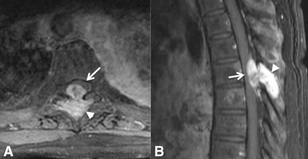
Mri of a patient with pcs

Axial (A) and sagittal (B) MRIs of a patient with posterior cord syndrome due to breast cancer metastasis to the spine:
The arrows and arrowheads show a bony lesion emerging from the spine and compressing the posterior aspect of the spinal cord.

Image: “A 61-year-old woman with breast cancer metastatic to the spine leading to spinal cord compression syndrome” by Marcos D Guimaraes, et al. License: CC BY 4.0

Management

  • Depends on the underlying cause
  • Medical management:
    • Vitamin B12 replacement for subacute combined degeneration
    • Penicillin Penicillin Rheumatic Fever for neurosyphilis Neurosyphilis Infections of the central nervous system caused by treponema pallidum which present with a variety of clinical syndromes. The initial phase of infection usually causes a mild or asymptomatic meningeal reaction. The meningovascular form may present acutely as brain infarction. The infection may also remain subclinical for several years. Late syndromes include general paresis; tabes dorsalis; meningeal syphilis; syphilitic optic atrophy; and spinal syphilis. General paresis is characterized by progressive dementia; dysarthria; tremor; myoclonus; seizures; and argyll-robertson pupils. Syphilis
    • Treatment for multiple sclerosis Sclerosis A pathological process consisting of hardening or fibrosis of an anatomical structure, often a vessel or a nerve. Wilms Tumor if present
    • Treatment for cancer if present → inpatient SC rehabilitation → functional improvement and improved bowel/ bladder Bladder A musculomembranous sac along the urinary tract. Urine flows from the kidneys into the bladder via the ureters, and is held there until urination. Pyelonephritis and Perinephric Abscess continence
  • Surgical laminectomy Laminectomy A surgical procedure that entails removing all (laminectomy) or part (laminotomy) of selected vertebral lamina to relieve pressure on the spinal cord and/or spinal nerve roots. Vertebral lamina is the thin flattened posterior wall of vertebral arch that forms the vertebral foramen through which pass the spinal cord and nerve roots. Neurosurgery for spinal decompression if PSC PSC Primary sclerosing cholangitis (PSC) is an inflammatory disease that causes fibrosis and strictures of the bile ducts. The exact etiology is unknown, but there is a strong association with IBD. Patients typically present with an insidious onset of fatigue, pruritus, and jaundice, which can progress to cirrhosis and complications related to biliary obstruction. Primary Sclerosing Cholangitis is trauma-related.

Differential Diagnoses

  • Anterior cord syndrome Anterior cord syndrome Anterior cord syndrome (ACS) is an incomplete cord syndrome predominantly affecting the anterior (ventral) …” of the spinal cord while sparing the dorsal columns. Anterior cord syndrome can be caused by occlusion of the anterior spinal artery or by trauma, which results in disc herniation and bone fragments disrupting the spinal cord. Anterior Cord Syndrome (ACS): incomplete cord syndrome to the anterior (ventral) ⅔ of the SC while sparing the dorsal columns. Anterior cord syndrome Anterior cord syndrome Anterior cord syndrome (ACS) is an incomplete cord syndrome predominantly affecting the anterior (ventral) …” of the spinal cord while sparing the dorsal columns. Anterior cord syndrome can be caused by occlusion of the anterior spinal artery or by trauma, which results in disc herniation and bone fragments disrupting the spinal cord. Anterior Cord Syndrome can be caused by an occlusion of the anterior spinal artery Anterior Spinal Artery Anterior Cord Syndrome or trauma causing disk herniation Herniation Omphalocele and bone Bone Bone is a compact type of hardened connective tissue composed of bone cells, membranes, an extracellular mineralized matrix, and central bone marrow. The 2 primary types of bone are compact and spongy. Bones: Structure and Types fragments that disrupt the SC. Clinical manifestations are loss of motor Motor Neurons which send impulses peripherally to activate muscles or secretory cells. Nervous System: Histology and sensory Sensory Neurons which conduct nerve impulses to the central nervous system. Nervous System: Histology function below the level of injury. Diagnosis of ACS is by clinical exam and neuroimaging Neuroimaging Non-invasive methods of visualizing the central nervous system, especially the brain, by various imaging modalities. Febrile Infant with MRI. Management is directed at resolving the underlying cause. 
  • Central cord syndrome Central Cord Syndrome Central cord syndrome (CCS) is a neurological syndrome caused by an injury to the center of the spinal cord, affecting the spinothalamic tracts ((STTs) sensory) and medial aspect of the corticospinal tracts ((CSTs) motor), most often due to trauma in patients with cervical spondylosis. Central Cord Syndrome: neurologic syndrome caused by an injury to the center of the SC, affecting the spinothalamic and corticospinal tracts Corticospinal Tracts Central Cord Syndrome, often caused by trauma in patients Patients Individuals participating in the health care system for the purpose of receiving therapeutic, diagnostic, or preventive procedures. Clinician–Patient Relationship with cervical spondylosis Cervical Spondylosis Neck Pain. Clinical manifestations are motor Motor Neurons which send impulses peripherally to activate muscles or secretory cells. Nervous System: Histology deficits affecting the upper more than the lower limbs, variable Variable Variables represent information about something that can change. The design of the measurement scales, or of the methods for obtaining information, will determine the data gathered and the characteristics of that data. As a result, a variable can be qualitative or quantitative, and may be further classified into subgroups. Types of Variables sensory Sensory Neurons which conduct nerve impulses to the central nervous system. Nervous System: Histology deficits below the level of injury, and bladder Bladder A musculomembranous sac along the urinary tract. Urine flows from the kidneys into the bladder via the ureters, and is held there until urination. Pyelonephritis and Perinephric Abscess dysfunction. Diagnosis is made by exam and MRI. Management can be medical or surgical, depending on the severity of the injury.
  • Brown-Séquard syndrome Brown-Séquard syndrome Brown-Séquard syndrome (BSS) is a rare neurologic injury that causes hemisection of the spinal cord, resulting in weakness and paralysis of one side of the body and sensory loss on the opposite side. Brown-Séquard Syndrome: hemisection Hemisection Brown-Séquard Syndrome of the SC secondary to injury, most commonly from penetrating trauma due to knife or bullet injuries. Other etiologies of Brown-Séquard syndrome Brown-Séquard syndrome Brown-Séquard syndrome (BSS) is a rare neurologic injury that causes hemisection of the spinal cord, resulting in weakness and paralysis of one side of the body and sensory loss on the opposite side. Brown-Séquard Syndrome include tumors, disk herniation Herniation Omphalocele, demyelination Demyelination Multiple Sclerosis, and infarction. Clinical presentation includes weakness and paralysis of one side of the body and sensory Sensory Neurons which conduct nerve impulses to the central nervous system. Nervous System: Histology loss on the opposite side. Diagnosis is by neurologic exam and with MRI. Management includes early treatment with high-dose steroids Steroids A group of polycyclic compounds closely related biochemically to terpenes. They include cholesterol, numerous hormones, precursors of certain vitamins, bile acids, alcohols (sterols), and certain natural drugs and poisons. Steroids have a common nucleus, a fused, reduced 17-carbon atom ring system, cyclopentanoperhydrophenanthrene. Most steroids also have two methyl groups and an aliphatic side-chain attached to the nucleus. Benign Liver Tumors; other treatments are symptomatic and supportive.
  • Conus medullaris Conus Medullaris Spinal Cord Injuries syndrome and cauda equina syndrome Cauda Equina Syndrome Compressive lesion affecting the nerve roots of the cauda equina (e.g., compression, herniation, inflammation, rupture, or stenosis), which controls the function of the bladder and bowel. Symptoms may include neurological dysfunction of bladder or bowels, loss of sexual sensation and altered sensation or paralysis in the lower extremities. Ankylosing Spondylitis: neurologic syndromes caused by compressive damage to the SC from T12 to L5, causing the disruption of the function of nerves below the point of compression Compression Blunt Chest Trauma. Diagnosis is made with MRI, and definitive management is prompt surgical decompression with laminectomy Laminectomy A surgical procedure that entails removing all (laminectomy) or part (laminotomy) of selected vertebral lamina to relieve pressure on the spinal cord and/or spinal nerve roots. Vertebral lamina is the thin flattened posterior wall of vertebral arch that forms the vertebral foramen through which pass the spinal cord and nerve roots. Neurosurgery
  • Spinal disk herniation Herniation Omphalocele (also known as herniated nucleus pulposus Nucleus Pulposus Fibrocartilage inner core of the intervertebral disc. Prolapsed or bulged nucleus pulposus leads to intervertebral disc displacement while proliferation of cells in the nucleus pulposus is associated with intervertebral disc degeneration. Spinal Disk Herniation): expulsion of the nucleus pulposus Nucleus Pulposus Fibrocartilage inner core of the intervertebral disc. Prolapsed or bulged nucleus pulposus leads to intervertebral disc displacement while proliferation of cells in the nucleus pulposus is associated with intervertebral disc degeneration. Spinal Disk Herniation through a perforation Perforation A pathological hole in an organ, blood vessel or other soft part of the body, occurring in the absence of external force. Esophagitis in the annulus fibrosus Annulus Fibrosus Spinal Disk Herniation of the intervertebral disc Intervertebral disc Any of the 23 plates of fibrocartilage found between the bodies of adjacent vertebrae. Vertebral Column: Anatomy. Spinal disk herniation Herniation Omphalocele is an important pain Pain An unpleasant sensation induced by noxious stimuli which are detected by nerve endings of nociceptive neurons. Pain: Types and Pathways syndrome with the potential for neurologic impairment and is most commonly caused by degenerative disk disease Disk Disease Examination of the Lower Limbs. Diagnosis is by clinical exam and MRI. Management ‌can be conservative with medications and physical therapy Physical Therapy Becker Muscular Dystrophy. Surgery is indicated for intractable pain Pain An unpleasant sensation induced by noxious stimuli which are detected by nerve endings of nociceptive neurons. Pain: Types and Pathways or myelopathy.

References

  1. McKinley, W., Hills, A., Sima, A. (2021). Posterior cord syndrome: demographics and rehabilitation outcomes. Journal of Spinal Cord Medicine 44:241–246. https://doi.org/10.1080/10790268.2019.1585135 
  2. Ropper, A. H., Samuels, M. A., Klein, J. P., Prasad, S. (2019). Disorders of non-painful somatic sensation. Chapter 8 of Adams and Victor’s Principles of Neurology, 11th ed. McGraw-Hill Education. http://accessmedicine.mhmedical.com/content.aspx?aid=1180368830 
  3. Eisen, A. (2020). Anatomy and localization of spinal cord disorders. UpToDate. Retrieved September 13, 2021, from https://www.uptodate.com/contents/anatomy-and-localization-of-spinal-cord-disorders
  4. Cochrane, M., Hess, M., Sajkowicz, N. (2020). Posterior cord syndrome associated with postoperative seroma: the case to perform a complete neurologic exam. Journal of Spinal Cord Medicine 43:892–894. https://doi.org/10.1080/10790268.2018.1550598
  5. Guimaraes, M.D., Bitencourt, A.G., Marchiori, E. et al. (2014). Imaging acute complications in cancer patients: what should be evaluated in the emergency setting? Cancer Imaging 14(18). https://doi.org/10.1186/1470-7330-14-18
  6. McKinley, W., Hills, A., Sima, A. (2019). Posterior cord syndrome: demographics and rehabilitation outcomes. Journal of Spinal Cord Medicine 44:241–246. https://doi.org/10.1080/10790268.2019.1585135

Create your free account or log in to continue reading!

Sign up now and get free access to Lecturio with concept pages, medical videos, and questions for your medical education.

User Reviews

Unwrap New Skills This Holiday 🎄 Save 30% on all plans now!

Details