Advertisement

Advertisement

Advertisement

Advertisement

Osteomalacia and Rickets

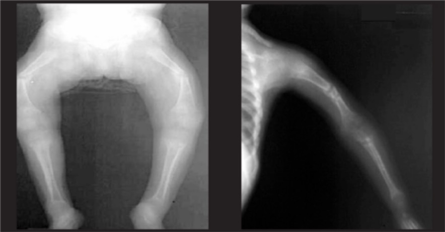
Rickets and osteomalacia are disorders of decreased bone mineralization Bone mineralization Calcium (Ca2+) and phosphate (PO43–) combine to form hydroxyapatite crystals on the bone matrix. Bones: Development and Ossification. Rickets affects the cartilage Cartilage Cartilage is a type of connective tissue derived from embryonic mesenchyme that is responsible for structural support, resilience, and the smoothness of physical actions. Perichondrium (connective tissue membrane surrounding cartilage) compensates for the absence of vasculature in cartilage by providing nutrition and support. Cartilage: Histology of the epiphyseal growth plates Growth Plates The area between the epiphysis and the diaphysis within which bone growth occurs. Osteosarcoma in children, while osteomalacia affects the sites of bone Bone Bone is a compact type of hardened connective tissue composed of bone cells, membranes, an extracellular mineralized matrix, and central bone marrow. The 2 primary types of bone are compact and spongy. Bones: Structure and Types turnover in children and adults. Although most cases of rickets and osteomalacia are due to vitamin D deficiency Vitamin D Deficiency A nutritional condition produced by a deficiency of vitamin D in the diet, insufficient production of vitamin D in the skin, inadequate absorption of vitamin D from the diet, or abnormal conversion of vitamin D to its bioactive metabolites. It is manifested clinically as rickets in children and osteomalacia in adults. Fat-soluble Vitamins and their Deficiencies, other genetic and nutritional disorders as well as medications can cause these disorders. Rickets commonly presents with skeletal deformities and growth abnormalities, while osteomalacia can present with bone Bone Bone is a compact type of hardened connective tissue composed of bone cells, membranes, an extracellular mineralized matrix, and central bone marrow. The 2 primary types of bone are compact and spongy. Bones: Structure and Types pain Pain An unpleasant sensation induced by noxious stimuli which are detected by nerve endings of nociceptive neurons. Pain: Types and Pathways, difficulty with ambulation and pathologic fractures. Diagnosis is made based on a combination of clinical findings, laboratory tests and imaging. Treatment includes vitamin D Vitamin D A vitamin that includes both cholecalciferols and ergocalciferols, which have the common effect of preventing or curing rickets in animals. It can also be viewed as a hormone since it can be formed in skin by action of ultraviolet rays upon the precursors, 7-dehydrocholesterol and ergosterol, and acts on vitamin D receptors to regulate calcium in opposition to parathyroid hormone. Fat-soluble Vitamins and their Deficiencies, calcium Calcium A basic element found in nearly all tissues. It is a member of the alkaline earth family of metals with the atomic symbol ca, atomic number 20, and atomic weight 40. Calcium is the most abundant mineral in the body and combines with phosphorus to form calcium phosphate in the bones and teeth. It is essential for the normal functioning of nerves and muscles and plays a role in blood coagulation (as factor IV) and in many enzymatic processes. Electrolytes, and phosphorus supplementation.

Last updated: May 13, 2024

Editorial responsibility: Stanley Oiseth, Lindsay Jones, Evelin Maza

Advertisement

Advertisement

Advertisement

Advertisement

Advertisement

Advertisement

Overview

Definition

Rickets and osteomalacia are disorders of decreased bone mineralization Bone mineralization Calcium (Ca2+) and phosphate (PO43–) combine to form hydroxyapatite crystals on the bone matrix. Bones: Development and Ossification. Rickets affects the cartilage Cartilage Cartilage is a type of connective tissue derived from embryonic mesenchyme that is responsible for structural support, resilience, and the smoothness of physical actions. Perichondrium (connective tissue membrane surrounding cartilage) compensates for the absence of vasculature in cartilage by providing nutrition and support. Cartilage: Histology of the epiphyseal growth plates Growth Plates The area between the epiphysis and the diaphysis within which bone growth occurs. Osteosarcoma in children, while osteomalacia affects the sites of bone Bone Bone is a compact type of hardened connective tissue composed of bone cells, membranes, an extracellular mineralized matrix, and central bone marrow. The 2 primary types of bone are compact and spongy. Bones: Structure and Types turnover in children and adults.

Epidemiology

Rickets:

  • In the United States, rickets mostly affects infants who have:
    • Chronic malabsorption Malabsorption General term for a group of malnutrition syndromes caused by failure of normal intestinal absorption of nutrients. Malabsorption and Maldigestion syndromes
    • Renal disease
    • Been exclusively breast-fed
      • Vitamin D deficiency Vitamin D Deficiency A nutritional condition produced by a deficiency of vitamin D in the diet, insufficient production of vitamin D in the skin, inadequate absorption of vitamin D from the diet, or abnormal conversion of vitamin D to its bioactive metabolites. It is manifested clinically as rickets in children and osteomalacia in adults. Fat-soluble Vitamins and their Deficiencies
      • Affecting dark-skinned infants
  • Worldwide prevalence Prevalence The total number of cases of a given disease in a specified population at a designated time. It is differentiated from incidence, which refers to the number of new cases in the population at a given time. Measures of Disease Frequency depends on:
    • Nutritional deprivation
    • Lack of sun exposure

Osteomalacia:

  • Mostly attributed to vitamin D deficiency Vitamin D Deficiency A nutritional condition produced by a deficiency of vitamin D in the diet, insufficient production of vitamin D in the skin, inadequate absorption of vitamin D from the diet, or abnormal conversion of vitamin D to its bioactive metabolites. It is manifested clinically as rickets in children and osteomalacia in adults. Fat-soluble Vitamins and their Deficiencies
  • Prevalence Prevalence The total number of cases of a given disease in a specified population at a designated time. It is differentiated from incidence, which refers to the number of new cases in the population at a given time. Measures of Disease Frequency is growing worldwide.
  • Commonly affects homebound elderly adults
  • Can co-exist with rickets in children

Etiology

Rickets and osteomalacia can be caused by deficiencies in vitamin D Vitamin D A vitamin that includes both cholecalciferols and ergocalciferols, which have the common effect of preventing or curing rickets in animals. It can also be viewed as a hormone since it can be formed in skin by action of ultraviolet rays upon the precursors, 7-dehydrocholesterol and ergosterol, and acts on vitamin D receptors to regulate calcium in opposition to parathyroid hormone. Fat-soluble Vitamins and their Deficiencies, calcium Calcium A basic element found in nearly all tissues. It is a member of the alkaline earth family of metals with the atomic symbol ca, atomic number 20, and atomic weight 40. Calcium is the most abundant mineral in the body and combines with phosphorus to form calcium phosphate in the bones and teeth. It is essential for the normal functioning of nerves and muscles and plays a role in blood coagulation (as factor IV) and in many enzymatic processes. Electrolytes, or phosphorus, or by abnormalities in bone Bone Bone is a compact type of hardened connective tissue composed of bone cells, membranes, an extracellular mineralized matrix, and central bone marrow. The 2 primary types of bone are compact and spongy. Bones: Structure and Types matrix deposition.

Vitamin D-related (most common):
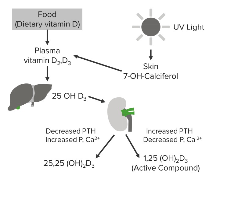
  • Vitamin D deficiency Vitamin D Deficiency A nutritional condition produced by a deficiency of vitamin D in the diet, insufficient production of vitamin D in the skin, inadequate absorption of vitamin D from the diet, or abnormal conversion of vitamin D to its bioactive metabolites. It is manifested clinically as rickets in children and osteomalacia in adults. Fat-soluble Vitamins and their Deficiencies
    • Dietary deprivation 
    • Lack of sunlight
  • Vitamin D Vitamin D A vitamin that includes both cholecalciferols and ergocalciferols, which have the common effect of preventing or curing rickets in animals. It can also be viewed as a hormone since it can be formed in skin by action of ultraviolet rays upon the precursors, 7-dehydrocholesterol and ergosterol, and acts on vitamin D receptors to regulate calcium in opposition to parathyroid hormone. Fat-soluble Vitamins and their Deficiencies malabsorption Malabsorption General term for a group of malnutrition syndromes caused by failure of normal intestinal absorption of nutrients. Malabsorption and Maldigestion:
    • Postgastrectomy
    • Gastric bypass Gastric bypass Surgical procedure in which the stomach is transected high on the body. The resulting small proximal gastric pouch is joined to any parts of the small intestine by an end-to-side surgical anastomosis, depending on the amounts of intestinal surface being bypasses. This procedure is used frequently in the treatment of morbid obesity by limiting the size of functional stomach, food intake, and food absorption. Gastroesophageal Reflux Disease (GERD)
    • Celiac disease Celiac disease Celiac disease (also known as celiac sprue or gluten enteropathy) is an autoimmune reaction to gliadin, which is a component of gluten. Celiac disease is closely associated with HLA-DQ2 and HLA-DQ8. The immune response is localized to the proximal small intestine and causes the characteristic histologic findings of villous atrophy, crypt hyperplasia, and intraepithelial lymphocytosis. Celiac Disease
    • Pancreatic insufficiency
    • Cholestyramine Cholestyramine A strongly basic anion exchange resin whose main constituent is polystyrene trimethylbenzylammonium cl(-) anion. Lipid Control Drugs treatment
    • Laxative Laxative Agents that produce a soft formed stool, and relax and loosen the bowels, typically used over a protracted period, to relieve constipation. Hypokalemia abuse
  • Impaired 1-hydroxylation of 25-hydroxyvitamin D:
  • Impaired target organ response to D3: vitamin D-dependent rickets type II
Vitamin d cycle

Schematic diagram depicting the vitamin D cycle

Image by Lecturio.

Hypophosphatemia Hypophosphatemia A condition of an abnormally low level of phosphates in the blood. Bartter Syndrome related:

  • X-linked X-linked Genetic diseases that are linked to gene mutations on the X chromosome in humans or the X chromosome in other species. Included here are animal models of human X-linked diseases. Common Variable Immunodeficiency (CVID) hypophosphatemia Hypophosphatemia A condition of an abnormally low level of phosphates in the blood. Bartter Syndrome 
  • Fibrous Fibrous Fibrocystic Change dysplasia
  • Antacid-induced osteomalacia
  • Chronic metabolic acidosis Acidosis A pathologic condition of acid accumulation or depletion of base in the body. The two main types are respiratory acidosis and metabolic acidosis, due to metabolic acid build up. Respiratory Acidosis
  • Tenofovir Tenofovir An adenine analog reverse transcriptase inhibitor with antiviral activity against HIV-1 and hepatitis b. It is used to treat HIV infections and chronic hepatitis b, in combination with other antiviral agents, due to the emergence of antiviral drug resistance when it is used alone. Anti-HIV Drugs
  • Cadmium
  • Paraproteinemia

Inhibitors of mineralization:

  • Etidronate Etidronate Bisphosphonates
  • Fluoride Fluoride Inorganic salts of hydrofluoric acid, hf, in which the fluorine atom is in the -1 oxidation state. Sodium and stannous salts are commonly used in dentifrices. Trace Elements
  • Aluminum 
  • Iron Iron A metallic element with atomic symbol fe, atomic number 26, and atomic weight 55. 85. It is an essential constituent of hemoglobins; cytochromes; and iron-binding proteins. It plays a role in cellular redox reactions and in the transport of oxygen. Trace Elements

Miscellaneous:

Pathophysiology

Rickets

  • Growth plate thickness is determined by:
    • Chondrocyte proliferation and hypertrophy Hypertrophy General increase in bulk of a part or organ due to cell enlargement and accumulation of fluids and secretions, not due to tumor formation, nor to an increase in the number of cells (hyperplasia). Cellular Adaptation
    • Vascular invasion of the growth plate
  • Vascular invasion requires mineralization of the growth plate cartilage Cartilage Cartilage is a type of connective tissue derived from embryonic mesenchyme that is responsible for structural support, resilience, and the smoothness of physical actions. Perichondrium (connective tissue membrane surrounding cartilage) compensates for the absence of vasculature in cartilage by providing nutrition and support. Cartilage: Histology.
  • In the absence of mineralization, the growth plate cartilage Cartilage Cartilage is a type of connective tissue derived from embryonic mesenchyme that is responsible for structural support, resilience, and the smoothness of physical actions. Perichondrium (connective tissue membrane surrounding cartilage) compensates for the absence of vasculature in cartilage by providing nutrition and support. Cartilage: Histology accumulates and becomes disorganized, and the growth plate thickens.
  • Accumulation of unmineralized osteoid Osteoid Bones: Development and Ossification in the metaphysis Metaphysis Bones: Structure and Types (right below the growth plate) eventually leads to bowing and deformation of bones.

Osteomalacia

  • At any one time, 7% of bone Bone Bone is a compact type of hardened connective tissue composed of bone cells, membranes, an extracellular mineralized matrix, and central bone marrow. The 2 primary types of bone are compact and spongy. Bones: Structure and Types surface is being actively remodeled.
  • Osteoclasts Osteoclasts A large multinuclear cell associated with the bone resorption. An odontoclast, also called cementoclast, is cytomorphologically the same as an osteoclast and is involved in cementum resorption. Bones: Development and Ossification resorb the old bone Bone Bone is a compact type of hardened connective tissue composed of bone cells, membranes, an extracellular mineralized matrix, and central bone marrow. The 2 primary types of bone are compact and spongy. Bones: Structure and Types while osteoblasts Osteoblasts Bone-forming cells which secrete an extracellular matrix. Hydroxyapatite crystals are then deposited into the matrix to form bone. Bones: Development and Ossification lay down new osteoid Osteoid Bones: Development and Ossification (organic matrix).
  • Osteomalacia can occur if:
    • New osteoid Osteoid Bones: Development and Ossification is qualitatively or quantitatively abnormal
    • Insufficient calcium Calcium A basic element found in nearly all tissues. It is a member of the alkaline earth family of metals with the atomic symbol ca, atomic number 20, and atomic weight 40. Calcium is the most abundant mineral in the body and combines with phosphorus to form calcium phosphate in the bones and teeth. It is essential for the normal functioning of nerves and muscles and plays a role in blood coagulation (as factor IV) and in many enzymatic processes. Electrolytes and phosphate Phosphate Inorganic salts of phosphoric acid. Electrolytes are in the extracellular fluid Extracellular fluid The fluid of the body that is outside of cells. It is the external environment for the cells. Body Fluid Compartments
    • Acidosis Acidosis A pathologic condition of acid accumulation or depletion of base in the body. The two main types are respiratory acidosis and metabolic acidosis, due to metabolic acid build up. Respiratory Acidosis is at the calcification site (renal disease)
    • Inadequate activity of alkaline phosphatase Alkaline Phosphatase An enzyme that catalyzes the conversion of an orthophosphoric monoester and water to an alcohol and orthophosphate. Osteosarcoma is present
    • Calcification is influenced by drugs (e.g., aluminum, fluoride Fluoride Inorganic salts of hydrofluoric acid, hf, in which the fluorine atom is in the -1 oxidation state. Sodium and stannous salts are commonly used in dentifrices. Trace Elements)

Clinical Presentation

Rickets

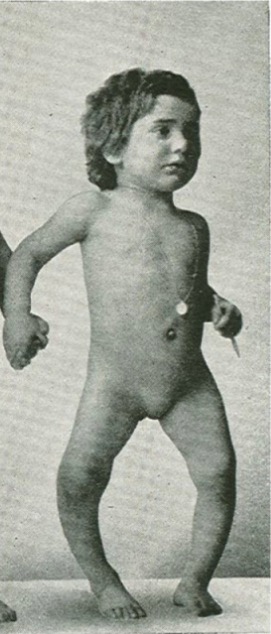
  • Delay in the time of closure of fontanelles Fontanelles Physical Examination of the Newborn and soft skull Skull The skull (cranium) is the skeletal structure of the head supporting the face and forming a protective cavity for the brain. The skull consists of 22 bones divided into the viscerocranium (facial skeleton) and the neurocranium. Skull: Anatomy bones (craniotabes)
  • Parietal Parietal One of a pair of irregularly shaped quadrilateral bones situated between the frontal bone and occipital bone, which together form the sides of the cranium. Skull: Anatomy and frontal bossing Frontal Bossing Hydrocephalus in Children
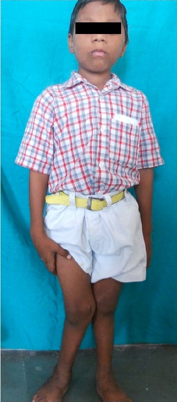
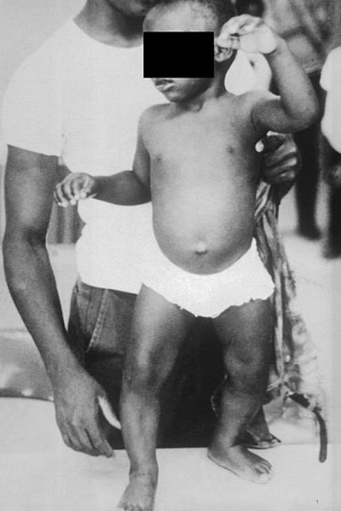
  • Genu varum Genu varum Genu varum is a deformation of the knee joint(s) that creates angulation of the lower limb(s) away from the midline in the coronal plane. Children ages 1-5 years are commonly affected. Genu Varum (toddlers) and Genu valgum Genu valgum Genu valgum is a deformation of the knee joint(s) that creates angulation of the lower limb(s) toward the midline in the coronal plane. Children ages 1-5 years are commonly affected. Genu Valgum (older children)
  • Deformity Deformity Examination of the Upper Limbs of the spine Spine The human spine, or vertebral column, is the most important anatomical and functional axis of the human body. It consists of 7 cervical vertebrae, 12 thoracic vertebrae, and 5 lumbar vertebrae and is limited cranially by the skull and caudally by the sacrum. Vertebral Column: Anatomy (kyphoscoliosis
  • Sulcus at the lower margin of the chest known as Harrison sulcus
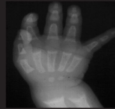
  • Bending primarily at the long bones Long bones Length greater than width. Bones: Structure and Types and widening of the wrist
  • Enlargement of costochondral junctions with “rib beading” ( rachitic rosary Rachitic rosary A row of bead-like prominence at the junction of a rib and its cartilage (enlarged costochondral joints), which resembles a rosary seen on chest x-ray. X-linked Hypophosphatemic Rickets)
  • Problems with the dental architecture
  • Greenstick fractures (linear fractures with intact periosteum Periosteum Thin outer membrane that surrounds a bone. It contains connective tissue, capillaries, nerves, and a number of cell types. Bones: Structure and Types)
  • Short stature

Osteomalacia

  • Fatigue Fatigue The state of weariness following a period of exertion, mental or physical, characterized by a decreased capacity for work and reduced efficiency to respond to stimuli. Fibromyalgia
  • Diffuse bone Bone Bone is a compact type of hardened connective tissue composed of bone cells, membranes, an extracellular mineralized matrix, and central bone marrow. The 2 primary types of bone are compact and spongy. Bones: Structure and Types pain Pain An unpleasant sensation induced by noxious stimuli which are detected by nerve endings of nociceptive neurons. Pain: Types and Pathways
  • Muscle weakness
  • Pathological fractures
  • Difficulty walking/ waddling gait Waddling Gait Duchenne Muscular Dystrophy
  • Can also present with signs of hypocalcemia Hypocalcemia Hypocalcemia, a serum calcium < 8.5 mg/dL, can result from various conditions. The causes may include hypoparathyroidism, drugs, disorders leading to vitamin D deficiency, and more. Calcium levels are regulated and affected by different elements such as dietary intake, parathyroid hormone (PTH), vitamin D, pH, and albumin. Presentation can range from an asymptomatic (mild deficiency) to a life-threatening condition (acute, significant deficiency). Hypocalcemia if associated with calcium Calcium A basic element found in nearly all tissues. It is a member of the alkaline earth family of metals with the atomic symbol ca, atomic number 20, and atomic weight 40. Calcium is the most abundant mineral in the body and combines with phosphorus to form calcium phosphate in the bones and teeth. It is essential for the normal functioning of nerves and muscles and plays a role in blood coagulation (as factor IV) and in many enzymatic processes. Electrolytes deficiency

Diagnosis and Management

Diagnosis

History:

  • Fractures
  • Bone Bone Bone is a compact type of hardened connective tissue composed of bone cells, membranes, an extracellular mineralized matrix, and central bone marrow. The 2 primary types of bone are compact and spongy. Bones: Structure and Types pain Pain An unpleasant sensation induced by noxious stimuli which are detected by nerve endings of nociceptive neurons. Pain: Types and Pathways
  • Family history Family History Adult Health Maintenance
  • Poor nutrition
  • Homebound lifestyle
  • Lack of sun exposure

Physical exam: skeletal deformities typical of rickets

Laboratory tests:

  • Laboratory values vary depending on the underlying deficiencies.
  • Calcipenic rickets:
    • Parathyroid Parathyroid The parathyroid glands are 2 pairs of small endocrine glands found in close proximity to the thyroid gland. The superior parathyroid glands are lodged within the parenchyma of the upper poles of the right and left thyroid lobes; the inferior parathyroid glands are close to the inferior tips or poles of the lobes. Parathyroid Glands: Anatomy hormone (PTH) 
    • Calcium Calcium A basic element found in nearly all tissues. It is a member of the alkaline earth family of metals with the atomic symbol ca, atomic number 20, and atomic weight 40. Calcium is the most abundant mineral in the body and combines with phosphorus to form calcium phosphate in the bones and teeth. It is essential for the normal functioning of nerves and muscles and plays a role in blood coagulation (as factor IV) and in many enzymatic processes. Electrolytes
    • Phosphate Phosphate Inorganic salts of phosphoric acid. Electrolytes
  • Phosphopenic rickets:
  • Vitamin D Vitamin D A vitamin that includes both cholecalciferols and ergocalciferols, which have the common effect of preventing or curing rickets in animals. It can also be viewed as a hormone since it can be formed in skin by action of ultraviolet rays upon the precursors, 7-dehydrocholesterol and ergosterol, and acts on vitamin D receptors to regulate calcium in opposition to parathyroid hormone. Fat-soluble Vitamins and their Deficiencies (in vitamin D deficiency Vitamin D Deficiency A nutritional condition produced by a deficiency of vitamin D in the diet, insufficient production of vitamin D in the skin, inadequate absorption of vitamin D from the diet, or abnormal conversion of vitamin D to its bioactive metabolites. It is manifested clinically as rickets in children and osteomalacia in adults. Fat-soluble Vitamins and their Deficiencies)
  • Alkaline phosphatase Alkaline Phosphatase An enzyme that catalyzes the conversion of an orthophosphoric monoester and water to an alcohol and orthophosphate. Osteosarcoma levels (except in hypophosphatasia)

X-ray X-ray Penetrating electromagnetic radiation emitted when the inner orbital electrons of an atom are excited and release radiant energy. X-ray wavelengths range from 1 pm to 10 nm. Hard x-rays are the higher energy, shorter wavelength x-rays. Soft x-rays or grenz rays are less energetic and longer in wavelength. The short wavelength end of the x-ray spectrum overlaps the gamma rays wavelength range. The distinction between gamma rays and x-rays is based on their radiation source. Pulmonary Function Tests imaging:
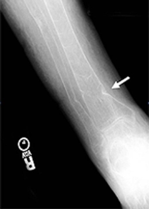
  • Rickets:
    • Epiphyseal widening
    • Metaphyseal cupping/fraying
    • Genu varum Genu varum Genu varum is a deformation of the knee joint(s) that creates angulation of the lower limb(s) away from the midline in the coronal plane. Children ages 1-5 years are commonly affected. Genu Varum or valgum (bowing of long bones Long bones Length greater than width. Bones: Structure and Types)
    • Rachitic rosary Rachitic rosary A row of bead-like prominence at the junction of a rib and its cartilage (enlarged costochondral joints), which resembles a rosary seen on chest x-ray. X-linked Hypophosphatemic Rickets 
    • Craniotabes
  • Osteomalacia:

Management

  • Vitamin D Vitamin D A vitamin that includes both cholecalciferols and ergocalciferols, which have the common effect of preventing or curing rickets in animals. It can also be viewed as a hormone since it can be formed in skin by action of ultraviolet rays upon the precursors, 7-dehydrocholesterol and ergosterol, and acts on vitamin D receptors to regulate calcium in opposition to parathyroid hormone. Fat-soluble Vitamins and their Deficiencies supplementation
  • Calcium-rich diet; calcium Calcium A basic element found in nearly all tissues. It is a member of the alkaline earth family of metals with the atomic symbol ca, atomic number 20, and atomic weight 40. Calcium is the most abundant mineral in the body and combines with phosphorus to form calcium phosphate in the bones and teeth. It is essential for the normal functioning of nerves and muscles and plays a role in blood coagulation (as factor IV) and in many enzymatic processes. Electrolytes and phosphate Phosphate Inorganic salts of phosphoric acid. Electrolytes supplementation 
  • Adequate exposure to sunlight 
  • Most deformities of rickets will correct with treatment.
  • If severe deformities have occurred, orthopedic correction is sometimes necessary.

Differential Diagnosis

  • Hyperparathyroidism Hyperparathyroidism Hyperparathyroidism is a condition associated with elevated blood levels of parathyroid hormone (PTH). Depending on the pathogenesis of this condition, hyperparathyroidism can be defined as primary, secondary or tertiary. Hyperparathyroidism: pathologically elevated PTH levels are summarized under the term hyperparathyroidism Hyperparathyroidism Hyperparathyroidism is a condition associated with elevated blood levels of parathyroid hormone (PTH). Depending on the pathogenesis of this condition, hyperparathyroidism can be defined as primary, secondary or tertiary. Hyperparathyroidism. Depending on the pathogenesis, distinctions can be made between 3 forms: primary, secondary, and tertiary hyperparathyroidism Tertiary Hyperparathyroidism Hyperparathyroidism. Long-standing hyperparathyroidism Hyperparathyroidism Hyperparathyroidism is a condition associated with elevated blood levels of parathyroid hormone (PTH). Depending on the pathogenesis of this condition, hyperparathyroidism can be defined as primary, secondary or tertiary. Hyperparathyroidism can result in osteopenia Osteopenia Osteoporosis and pathologic bone fractures Bone fractures Breaks in bones. Bones: Remodeling and Healing. Treatment approach depends on the underlying cause and involves parathyroidectomy Parathyroidectomy Excision of one or more of the parathyroid glands. Hyperparathyroidism for primary hyperparathyroidism Primary hyperparathyroidism A condition of abnormally elevated output of parathyroid hormone due to parathyroid hyperplasia or parathyroid neoplasms. It is characterized by the combination of hypercalcemia, phosphaturia, elevated renal 1, 25-dihydroxyvitamin d3 synthesis, and increased bone resorption. Hyperparathyroidism.
  • Chronic renal failure Renal failure Conditions in which the kidneys perform below the normal level in the ability to remove wastes, concentrate urine, and maintain electrolyte balance; blood pressure; and calcium metabolism. Renal insufficiency can be classified by the degree of kidney damage (as measured by the level of proteinuria) and reduction in glomerular filtration rate. Crush Syndrome: defined as an irreversible decrease of not only glomerular and tubular function, but also endocrine renal function. This condition presents as pathologically disturbed excretory and incretionary renal function. Chronic renal failure Renal failure Conditions in which the kidneys perform below the normal level in the ability to remove wastes, concentrate urine, and maintain electrolyte balance; blood pressure; and calcium metabolism. Renal insufficiency can be classified by the degree of kidney damage (as measured by the level of proteinuria) and reduction in glomerular filtration rate. Crush Syndrome can cause hypocalcemia Hypocalcemia Hypocalcemia, a serum calcium < 8.5 mg/dL, can result from various conditions. The causes may include hypoparathyroidism, drugs, disorders leading to vitamin D deficiency, and more. Calcium levels are regulated and affected by different elements such as dietary intake, parathyroid hormone (PTH), vitamin D, pH, and albumin. Presentation can range from an asymptomatic (mild deficiency) to a life-threatening condition (acute, significant deficiency). Hypocalcemia through decreased formation of active vitamin D Vitamin D A vitamin that includes both cholecalciferols and ergocalciferols, which have the common effect of preventing or curing rickets in animals. It can also be viewed as a hormone since it can be formed in skin by action of ultraviolet rays upon the precursors, 7-dehydrocholesterol and ergosterol, and acts on vitamin D receptors to regulate calcium in opposition to parathyroid hormone. Fat-soluble Vitamins and their Deficiencies, which in turn results in secondary hyperparathyroidism Secondary hyperparathyroidism Abnormally elevated parathyroid hormone secretion as a response to hypocalcemia. It is caused by chronic kidney failure or other abnormalities in the controls of bone and mineral metabolism, leading to various bone diseases, such as renal osteodystrophy. Hyperparathyroidism and leaking of calcium Calcium A basic element found in nearly all tissues. It is a member of the alkaline earth family of metals with the atomic symbol ca, atomic number 20, and atomic weight 40. Calcium is the most abundant mineral in the body and combines with phosphorus to form calcium phosphate in the bones and teeth. It is essential for the normal functioning of nerves and muscles and plays a role in blood coagulation (as factor IV) and in many enzymatic processes. Electrolytes from the bones. Treatment is mostly supportive. 
  • Blount’s disease: a pediatric orthopedic problem in which the leg Leg The lower leg, or just “leg” in anatomical terms, is the part of the lower limb between the knee and the ankle joint. The bony structure is composed of the tibia and fibula bones, and the muscles of the leg are grouped into the anterior, lateral, and posterior compartments by extensions of fascia. Leg: Anatomy is bowed outward or in a varus position. This disease is due to a dysfunctional growth of the posterior medial aspect of proximal physis of the tibia Tibia The second longest bone of the skeleton. It is located on the medial side of the lower leg, articulating with the fibula laterally, the talus distally, and the femur proximally. Knee Joint: Anatomy resulting in a lower limb deformity Deformity Examination of the Upper Limbs. Diagnosis is established based on clinical picture and imaging, and treatment involves bracing and sometimes surgical correction.
  • Achondroplasia: a genetic disorder that results in dwarfism. In those with the condition, the arms and legs are short, while the torso is typically of normal length. Those affected have an average adult height of 131 cm (4 ft 4 in) for males and 123 cm (4 ft) for females. Diagnosis is established clinically and confirmed by genetic testing Genetic Testing Detection of a mutation; genotype; karyotype; or specific alleles associated with genetic traits, heritable diseases, or predisposition to a disease, or that may lead to the disease in descendants. It includes prenatal genetic testing. Myotonic Dystrophies. Treatment is largely supportive.
  • Osteoporosis Osteoporosis Osteoporosis refers to a decrease in bone mass and density leading to an increased number of fractures. There are 2 forms of osteoporosis: primary, which is commonly postmenopausal or senile; and secondary, which is a manifestation of immobilization, underlying medical disorders, or long-term use of certain medications. Osteoporosis: a loss of bone Bone Bone is a compact type of hardened connective tissue composed of bone cells, membranes, an extracellular mineralized matrix, and central bone marrow. The 2 primary types of bone are compact and spongy. Bones: Structure and Types mass Mass Three-dimensional lesion that occupies a space within the breast Imaging of the Breast usually associated with aging and hormonal deficits. Most common in post-menopausal women. Presents with fragility fractures Fragility fractures Pelvic fractures are a disruption in the cortex of a pelvic bone involving iliac wing fractures, acetabular fractures, or those causing loss of integrity of the pelvic ring (the sacrum and the 2 innominate bones). Patients often present with a history of trauma or a fall, limb length discrepancy, intense pain on palpation, and mechanical instability. Pelvic Fractures. Diagnosis is based on clinical presentation and measurements of bone Bone Bone is a compact type of hardened connective tissue composed of bone cells, membranes, an extracellular mineralized matrix, and central bone marrow. The 2 primary types of bone are compact and spongy. Bones: Structure and Types mineral density. Treatment involves vitamin D Vitamin D A vitamin that includes both cholecalciferols and ergocalciferols, which have the common effect of preventing or curing rickets in animals. It can also be viewed as a hormone since it can be formed in skin by action of ultraviolet rays upon the precursors, 7-dehydrocholesterol and ergosterol, and acts on vitamin D receptors to regulate calcium in opposition to parathyroid hormone. Fat-soluble Vitamins and their Deficiencies and calcium Calcium A basic element found in nearly all tissues. It is a member of the alkaline earth family of metals with the atomic symbol ca, atomic number 20, and atomic weight 40. Calcium is the most abundant mineral in the body and combines with phosphorus to form calcium phosphate in the bones and teeth. It is essential for the normal functioning of nerves and muscles and plays a role in blood coagulation (as factor IV) and in many enzymatic processes. Electrolytes supplementation and bisphosphonates Bisphosphonates Bisphosphonates are pyrophosphate analogs most well-known for treating osteoporosis by preventing bone loss. Bisphosphonates end in the suffix “-dronate” or “-dronic acid” (e.g., alendronate, risedronate, pamidronate) and bind to hydroxyapatite crystals in bone, inhibiting osteoclast-induced bone resorption. Bisphosphonates.
  • Osteogenesis imperfecta Osteogenesis imperfecta Osteogenesis imperfecta (OI), or “brittle bone disease,” is a rare genetic connective tissue disorder characterized by severe bone fragility. Although OI is considered a single disease, OI includes over 16 genotypes and clinical phenotypes with differing symptom severity. Osteogenesis Imperfecta: a genetic disorder associated with extreme bone Bone Bone is a compact type of hardened connective tissue composed of bone cells, membranes, an extracellular mineralized matrix, and central bone marrow. The 2 primary types of bone are compact and spongy. Bones: Structure and Types fragility. The disease varies in severity, but usually presents in early childhood with multiple fractures and bone Bone Bone is a compact type of hardened connective tissue composed of bone cells, membranes, an extracellular mineralized matrix, and central bone marrow. The 2 primary types of bone are compact and spongy. Bones: Structure and Types deformities.  Osteogenesis imperfecta Osteogenesis imperfecta Osteogenesis imperfecta (OI), or “brittle bone disease,” is a rare genetic connective tissue disorder characterized by severe bone fragility. Although OI is considered a single disease, OI includes over 16 genotypes and clinical phenotypes with differing symptom severity. Osteogenesis Imperfecta is diagnosed clinically and with genetic testing Genetic Testing Detection of a mutation; genotype; karyotype; or specific alleles associated with genetic traits, heritable diseases, or predisposition to a disease, or that may lead to the disease in descendants. It includes prenatal genetic testing. Myotonic Dystrophies. Management includes bisphosphonates Bisphosphonates Bisphosphonates are pyrophosphate analogs most well-known for treating osteoporosis by preventing bone loss. Bisphosphonates end in the suffix “-dronate” or “-dronic acid” (e.g., alendronate, risedronate, pamidronate) and bind to hydroxyapatite crystals in bone, inhibiting osteoclast-induced bone resorption. Bisphosphonates and is mostly focused on fracture Fracture A fracture is a disruption of the cortex of any bone and periosteum and is commonly due to mechanical stress after an injury or accident. Open fractures due to trauma can be a medical emergency. Fractures are frequently associated with automobile accidents, workplace injuries, and trauma. Overview of Bone Fractures prevention.

References

  1. Bhan A., Rao A.D., Rao D.S. (2010). Osteomalacia as a result of vitamin D deficiency. Endocrinol Metab Clin North Am.
  2. Lowdon J. (2011). Rickets: concerns over the worldwide increase. J Fam Health Care.
  3. Carpenter T. (2020). Overview of rickets in children. Retrieved February 11, 2021, from https://www.uptodate.com/contents/overview-of-rickets-in-children
  4. Carpenter T. (2020). Etiology and treatment of calcipenic rickets in children. Retrieved February 11, 2021, from https://www.uptodate.com/contents/etiology-and-treatment-of-calcipenic-rickets-in-children
  5. Cohen A., Drake M.T. (2019). Clinical manifestations, diagnosis, and treatment of osteomalacia. Retrieved February 11, 2021, from https://www.uptodate.com/contents/clinical-manifestations-diagnosis-and-treatment-of-osteomalacia
  6. Schwarz S.M. (2017). Rickets. Retrieved February 11, 2021, from https://emedicine.medscape.com/article/985510-overview

Create your free account or log in to continue reading!

Sign up now and get free access to Lecturio with concept pages, medical videos, and questions for your medical education.

User Reviews

Unwrap New Skills This Holiday 🎄 Save 30% on all plans now!

Details