Advertisement

Advertisement

Advertisement

Advertisement

Olfaction: Anatomy

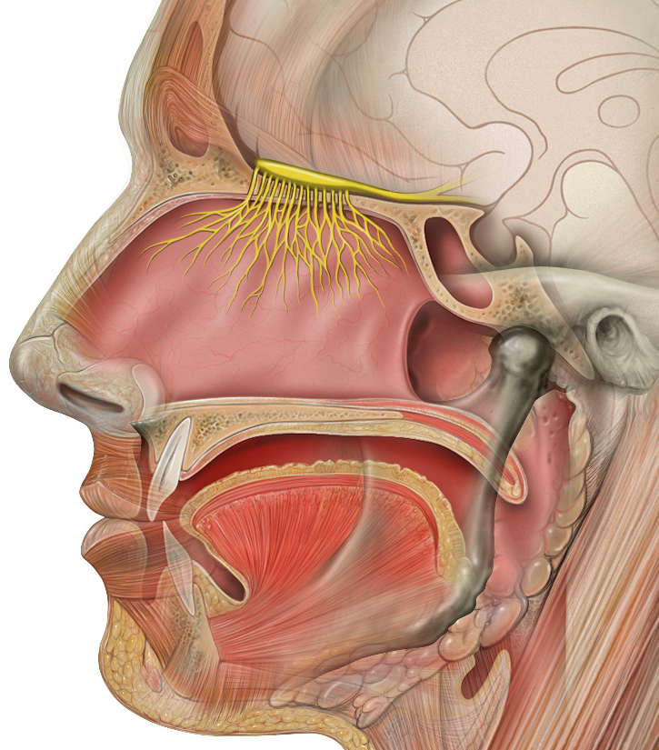
Olfaction represents an ancient, evolutionarily critical physiologic system. Humans have the ability to detect and discriminate at least 10,000 different odorants. The sense of smell, or olfaction, begins in a small area on the roof of the nasal cavity Nasal cavity The proximal portion of the respiratory passages on either side of the nasal septum. Nasal cavities, extending from the nares to the nasopharynx, are lined with ciliated nasal mucosa. Nose Anatomy (External & Internal), which is covered in specialized mucosa. From there, the olfactory nerve Olfactory nerve The 1st cranial nerve. The olfactory nerve conveys the sense of smell. It is formed by the axons of olfactory receptor neurons which project from the olfactory epithelium (in the nasal epithelium) to the olfactory bulb. Nose Anatomy (External & Internal) transmits the sensory Sensory Neurons which conduct nerve impulses to the central nervous system. Nervous System: Histology perception Perception The process by which the nature and meaning of sensory stimuli are recognized and interpreted. Psychiatric Assessment of smell via the olfactory pathway. This pathway is composed of the olfactory cells and bulb, the tractus and striae olfactoriae, and the primary olfactory cortex and amygdala Amygdala Almond-shaped group of basal nuclei anterior to the inferior horn of the lateral ventricle of the temporal lobe. The amygdala is part of the limbic system. Limbic System: Anatomy. Olfaction is responsible for the detection of hazards, pheromones, and food.

Last updated: Nov 19, 2024

Editorial responsibility: Stanley Oiseth, Lindsay Jones, Evelin Maza

Advertisement

Advertisement

Advertisement

Advertisement

Advertisement

Advertisement

Introduction

Structure of the nose Nose The nose is the human body’s primary organ of smell and functions as part of the upper respiratory system. The nose may be best known for inhaling oxygen and exhaling carbon dioxide, but it also contributes to other important functions, such as tasting. The anatomy of the nose can be divided into the external nose and the nasal cavity. Nose Anatomy (External & Internal)

  • The nasal passages are divided by the nasal septum Nasal septum The partition separating the two nasal cavities in the midplane. It is formed by the septal nasal cartilage, parts of skull bones, and membranous parts. Nose Anatomy (External & Internal) in the midline.
  • Each lateral nasal wall is formed by 3 turbinates Turbinates The scroll-like bony plates with curved margins on the lateral wall of the nasal cavity. Turbinates, also called nasal concha, increase the surface area of nasal cavity thus providing a mechanism for rapid warming and humidification of air as it passes to the lung. Nose Anatomy (External & Internal): inferior, middle, and superior.
  • The cribriform plate constitutes the roof of the nasal passage:
    • Part of the ethmoid bone Ethmoid bone A light and spongy (pneumatized) bone that lies between the orbital part of frontal bone and the anterior of sphenoid bone. Ethmoid bone separates the orbit from the ethmoid sinus. It consists of a horizontal plate, a perpendicular plate, and two lateral labyrinths. Orbit and Extraocular Muscles: Anatomy
    • Perforated by the olfactory foramina (approximately 20 foramina)

Location of the olfactory epithelium Epithelium The epithelium is a complex of specialized cellular organizations arranged into sheets and lining cavities and covering the surfaces of the body. The cells exhibit polarity, having an apical and a basal pole. Structures important for the epithelial integrity and function involve the basement membrane, the semipermeable sheet on which the cells rest, and interdigitations, as well as cellular junctions. Surface Epithelium: Histology

  • Along the cribriform plate
  • Along and medial to the superior turbinates Turbinates The scroll-like bony plates with curved margins on the lateral wall of the nasal cavity. Turbinates, also called nasal concha, increase the surface area of nasal cavity thus providing a mechanism for rapid warming and humidification of air as it passes to the lung. Nose Anatomy (External & Internal)
  • Can extend as far as the anterolateral middle turbinate and posterior nasal septum Nasal septum The partition separating the two nasal cavities in the midplane. It is formed by the septal nasal cartilage, parts of skull bones, and membranous parts. Nose Anatomy (External & Internal)
  • The location of the olfactory epithelium Epithelium The epithelium is a complex of specialized cellular organizations arranged into sheets and lining cavities and covering the surfaces of the body. The cells exhibit polarity, having an apical and a basal pole. Structures important for the epithelial integrity and function involve the basement membrane, the semipermeable sheet on which the cells rest, and interdigitations, as well as cellular junctions. Surface Epithelium: Histology varies among individuals.
  • Olfactory neurons Neurons The basic cellular units of nervous tissue. Each neuron consists of a body, an axon, and dendrites. Their purpose is to receive, conduct, and transmit impulses in the nervous system. Nervous System: Histology are lost with age, exposure to environmental toxins, and inflammation Inflammation Inflammation is a complex set of responses to infection and injury involving leukocytes as the principal cellular mediators in the body’s defense against pathogenic organisms. Inflammation is also seen as a response to tissue injury in the process of wound healing. The 5 cardinal signs of inflammation are pain, heat, redness, swelling, and loss of function. Inflammation.

Detection of smell

  • We detect smell by quickly inhaling air, which creates turbulent airflow in the nose Nose The nose is the human body’s primary organ of smell and functions as part of the upper respiratory system. The nose may be best known for inhaling oxygen and exhaling carbon dioxide, but it also contributes to other important functions, such as tasting. The anatomy of the nose can be divided into the external nose and the nasal cavity. Nose Anatomy (External & Internal).
  • This inhalation facilitates transfer of the odorants superiorly, where the olfactory epithelium Epithelium The epithelium is a complex of specialized cellular organizations arranged into sheets and lining cavities and covering the surfaces of the body. The cells exhibit polarity, having an apical and a basal pole. Structures important for the epithelial integrity and function involve the basement membrane, the semipermeable sheet on which the cells rest, and interdigitations, as well as cellular junctions. Surface Epithelium: Histology lies.
  • The odorants then diffuse into the mucus and are bound by proteins Proteins Linear polypeptides that are synthesized on ribosomes and may be further modified, crosslinked, cleaved, or assembled into complex proteins with several subunits. The specific sequence of amino acids determines the shape the polypeptide will take, during protein folding, and the function of the protein. Energy Homeostasis that transport them to the olfactory receptors Receptors Receptors are proteins located either on the surface of or within a cell that can bind to signaling molecules known as ligands (e.g., hormones) and cause some type of response within the cell. Receptors
  • Another way of detecting smell is through posterior transfer of odorants from the nasopharynx Nasopharynx The top portion of the pharynx situated posterior to the nose and superior to the soft palate. The nasopharynx is the posterior extension of the nasal cavities and has a respiratory function. Pharynx: Anatomy, which is important for the detection of flavor during eating and drinking.

Olfactory Epithelium and Nerve

Olfactory epithelium Epithelium The epithelium is a complex of specialized cellular organizations arranged into sheets and lining cavities and covering the surfaces of the body. The cells exhibit polarity, having an apical and a basal pole. Structures important for the epithelial integrity and function involve the basement membrane, the semipermeable sheet on which the cells rest, and interdigitations, as well as cellular junctions. Surface Epithelium: Histology

The olfactory epithelium Epithelium The epithelium is a complex of specialized cellular organizations arranged into sheets and lining cavities and covering the surfaces of the body. The cells exhibit polarity, having an apical and a basal pole. Structures important for the epithelial integrity and function involve the basement membrane, the semipermeable sheet on which the cells rest, and interdigitations, as well as cellular junctions. Surface Epithelium: Histology is a pseudostratified columnar epithelium Pseudostratified columnar epithelium Cells vary in height, with all cells resting on the basement membrane but only some reaching all the way to the apical surface. Surface Epithelium: Histology overlying a lamina propria Lamina propria Whipple’s Disease and consists of the following cell types:
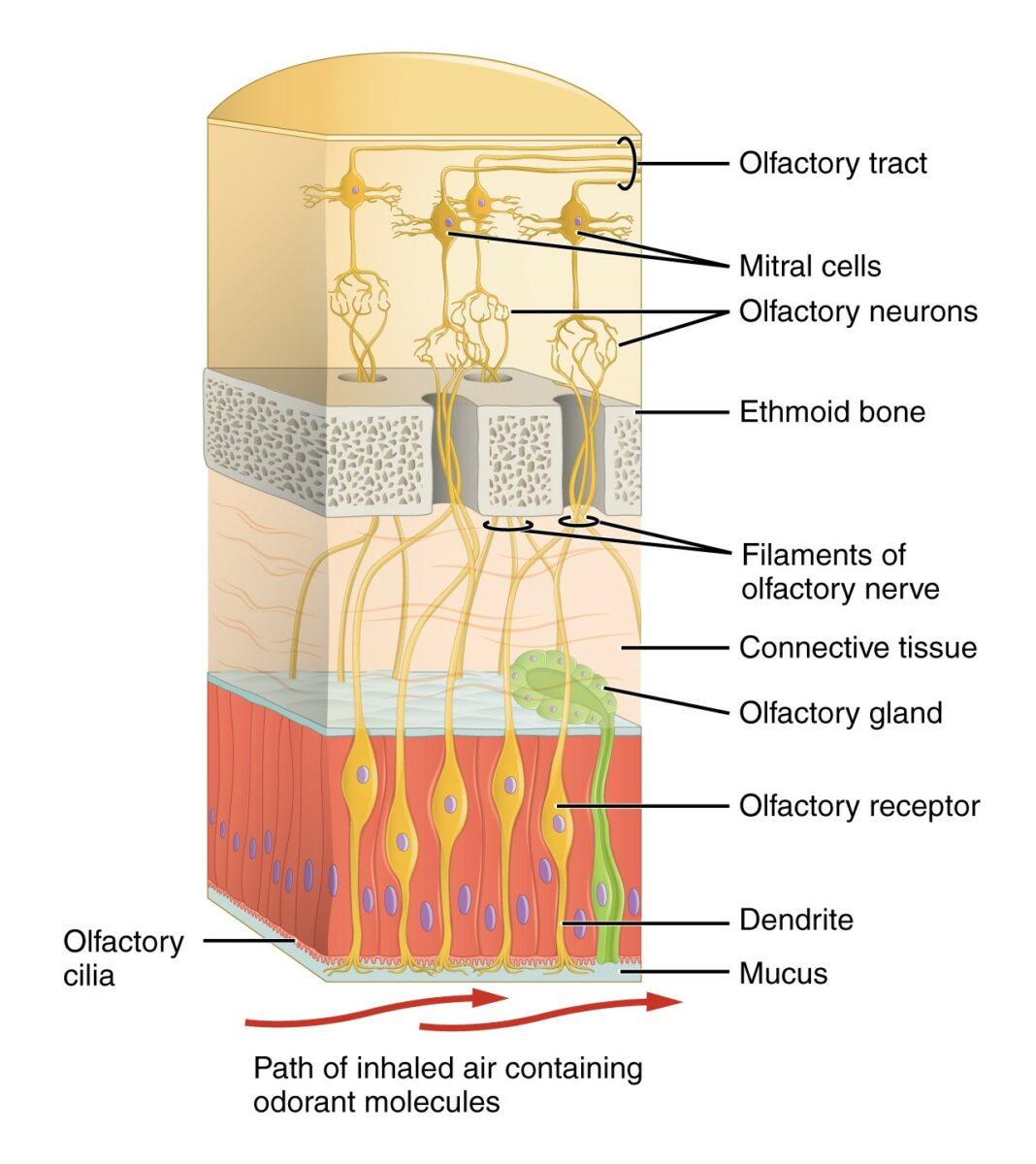
  • Basal cells:
    • Act as stem cells
    • Can differentiate into progenitors of the olfactory epithelium Epithelium The epithelium is a complex of specialized cellular organizations arranged into sheets and lining cavities and covering the surfaces of the body. The cells exhibit polarity, having an apical and a basal pole. Structures important for the epithelial integrity and function involve the basement membrane, the semipermeable sheet on which the cells rest, and interdigitations, as well as cellular junctions. Surface Epithelium: Histology
  • Sustentacular cells: support olfactory neuron function 
  • Olfactory gland cells (Bowman gland): function to carry secretions to the apical epithelial surface
  • Olfactory receptor Receptor Receptors are proteins located either on the surface of or within a cell that can bind to signaling molecules known as ligands (e.g., hormones) and cause some type of response within the cell. Receptors neurons Neurons The basic cellular units of nervous tissue. Each neuron consists of a body, an axon, and dendrites. Their purpose is to receive, conduct, and transmit impulses in the nervous system. Nervous System: Histology:
    • Located in an intermediate zone between basal and apical layers
    • Form the bulk of the epithelium Epithelium The epithelium is a complex of specialized cellular organizations arranged into sheets and lining cavities and covering the surfaces of the body. The cells exhibit polarity, having an apical and a basal pole. Structures important for the epithelial integrity and function involve the basement membrane, the semipermeable sheet on which the cells rest, and interdigitations, as well as cellular junctions. Surface Epithelium: Histology: humans have approximately 10 million to 20 million olfactory neurons Neurons The basic cellular units of nervous tissue. Each neuron consists of a body, an axon, and dendrites. Their purpose is to receive, conduct, and transmit impulses in the nervous system. Nervous System: Histology.
    • Bipolar Bipolar Nervous System: Histology cells:
      • Project a dendrite with a thickened end (the olfactory knob) that contains nonmotile sensory Sensory Neurons which conduct nerve impulses to the central nervous system. Nervous System: Histology cilia to the epithelial surface
      • Project another axon that transmits signals to the brain Brain The part of central nervous system that is contained within the skull (cranium). Arising from the neural tube, the embryonic brain is comprised of three major parts including prosencephalon (the forebrain); mesencephalon (the midbrain); and rhombencephalon (the hindbrain). The developed brain consists of cerebrum; cerebellum; and other structures in the brain stem. Nervous System: Anatomy, Structure, and Classification after crossing the cribriform plate
    • Express olfactory receptors Receptors Receptors are proteins located either on the surface of or within a cell that can bind to signaling molecules known as ligands (e.g., hormones) and cause some type of response within the cell. Receptors:
      • Constitute one of the largest families of G-protein–coupled receptors Receptors Receptors are proteins located either on the surface of or within a cell that can bind to signaling molecules known as ligands (e.g., hormones) and cause some type of response within the cell. Receptors
      • Controlled by as many as 800 genes Genes A category of nucleic acid sequences that function as units of heredity and which code for the basic instructions for the development, reproduction, and maintenance of organisms. DNA Types and Structure in humans
Diagram depicting the 1st structures of the olfactory system

Diagram depicting the 1st structures of the olfactory system:
Odorants are perceived by specialized cilia within the nasal mucosa, which send an electrical signal via the olfactory nerve through the ethmoid bone to the olfactory bulb and tract.
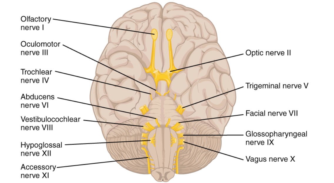
Image: “(b) The olfactory receptor neurons are within the olfactory epithelium” by OpenStax College. License: CC BY 4.0, cropped by Lecturio.

Olfactory nerve Olfactory nerve The 1st cranial nerve. The olfactory nerve conveys the sense of smell. It is formed by the axons of olfactory receptor neurons which project from the olfactory epithelium (in the nasal epithelium) to the olfactory bulb. Nose Anatomy (External & Internal)

  • Axons Axons Nerve fibers that are capable of rapidly conducting impulses away from the neuron cell body. Nervous System: Histology from the olfactory neurons Neurons The basic cellular units of nervous tissue. Each neuron consists of a body, an axon, and dendrites. Their purpose is to receive, conduct, and transmit impulses in the nervous system. Nervous System: Histology form nerve bundles (fila olfactoria). Many such bundles coalesce to create the olfactory nerve Olfactory nerve The 1st cranial nerve. The olfactory nerve conveys the sense of smell. It is formed by the axons of olfactory receptor neurons which project from the olfactory epithelium (in the nasal epithelium) to the olfactory bulb. Nose Anatomy (External & Internal).
  • Enclosed by dura and arachnoid mater Arachnoid mater A delicate membrane enveloping the brain and spinal cord. It lies between the pia mater and the dura mater. It is separated from the pia mater by the subarachnoid cavity which is filled with cerebrospinal fluid. Meninges: Anatomy
  • Shortest of the 12 cranial nerves Cranial nerves There are 12 pairs of cranial nerves (CNs), which run from the brain to various parts of the head, neck, and trunk. The CNs can be sensory or motor or both. The CNs are named and numbered in Roman numerals according to their location, from the front to the back of the brain. The 12 Cranial Nerves: Overview and Functions and unique in that it can partially regenerate
  • Crosses the cribriform plate and synapses with neurons Neurons The basic cellular units of nervous tissue. Each neuron consists of a body, an axon, and dendrites. Their purpose is to receive, conduct, and transmit impulses in the nervous system. Nervous System: Histology in the olfactory bulb

Olfactory Pathway

Overview

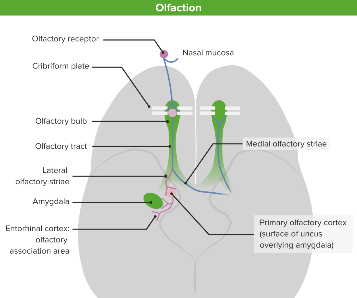
Odors are first detected at the olfactory bulb, where the information is received and transmitted posteriorly along the olfactory tract to the olfactory cortex.
Olfactory bulb

  • The main relay station of the olfactory pathway
  • Lies on top of the cribriform plate
  • The olfactory bulb consists of:
    • Glomeruli: dense tangles of axonal and dendritic branches 
    • Mitral cells: Each cell extends a primary dendrite into a single glomerulus, which divides into a tuft of branches onto which the primary olfactory axons Axons Nerve fibers that are capable of rapidly conducting impulses away from the neuron cell body. Nervous System: Histology synapse Synapse The junction between 2 neurons is called a synapse. The synapse allows a neuron to pass an electrical or chemical signal to another neuron or target effector cell. Synapses and Neurotransmission.

Olfactory tract

  • Consists of nerve fibers Nerve Fibers Slender processes of neurons, including the axons and their glial envelopes (myelin sheath). Nerve fibers conduct nerve impulses to and from the central nervous system. Nervous System: Histology created by the axons Axons Nerve fibers that are capable of rapidly conducting impulses away from the neuron cell body. Nervous System: Histology of the mitral cells of the olfactory bulb
  • The tract passes underneath the medial frontal lobe Frontal lobe The part of the cerebral hemisphere anterior to the central sulcus, and anterior and superior to the lateral sulcus. Cerebral Cortex: Anatomy inside the olfactory groove.
  • The olfactory tract ultimately divides into 2 striae:
    • The lateral stria terminates in the primary olfactory cortex of the temporal lobe Temporal lobe Lower lateral part of the cerebral hemisphere responsible for auditory, olfactory, and semantic processing. It is located inferior to the lateral fissure and anterior to the occipital lobe. Cerebral Cortex: Anatomy.
    • The medial stria:
      • Passes through the anterior commissure to the contralateral olfactory tract
      • Terminates in limbic structures to contribute to the emotional responses brought on by the smell
Schematic diagram of the olfactory pathway from the nasal mucosa to the primary and secondary olfactory cortex

Schematic diagram of the olfactory pathway from the nasal mucosa to the primary and secondary olfactory cortex:
Note the ipsilateral and contralateral projections of the middle and lateral olfactory striae.

Image by Lecturio.

Olfactory cortex

The olfactory cortex is unique in receiving direct sensory Sensory Neurons which conduct nerve impulses to the central nervous system. Nervous System: Histology inputs from the mitral cells of the olfactory bulb. This process differs from the classic sensory Sensory Neurons which conduct nerve impulses to the central nervous system. Nervous System: Histology pathway, where sensory Sensory Neurons which conduct nerve impulses to the central nervous system. Nervous System: Histology information is 1st relayed in the thalamus Thalamus The thalamus is a large, ovoid structure in the dorsal part of the diencephalon that is located between the cerebral cortex and midbrain. It consists of several interconnected nuclei of grey matter separated by the laminae of white matter. The thalamus is the main conductor of information that passes between the cerebral cortex and the periphery, spinal cord, or brain stem. Thalamus: Anatomy before reaching the neocortex Neocortex The largest portion of the cerebral cortex in which the neurons are arranged in six layers in the mammalian brain: molecular, external granular, external pyramidal, internal granular, internal pyramidal and multiform layers. Cerebral Cortex: Anatomy.
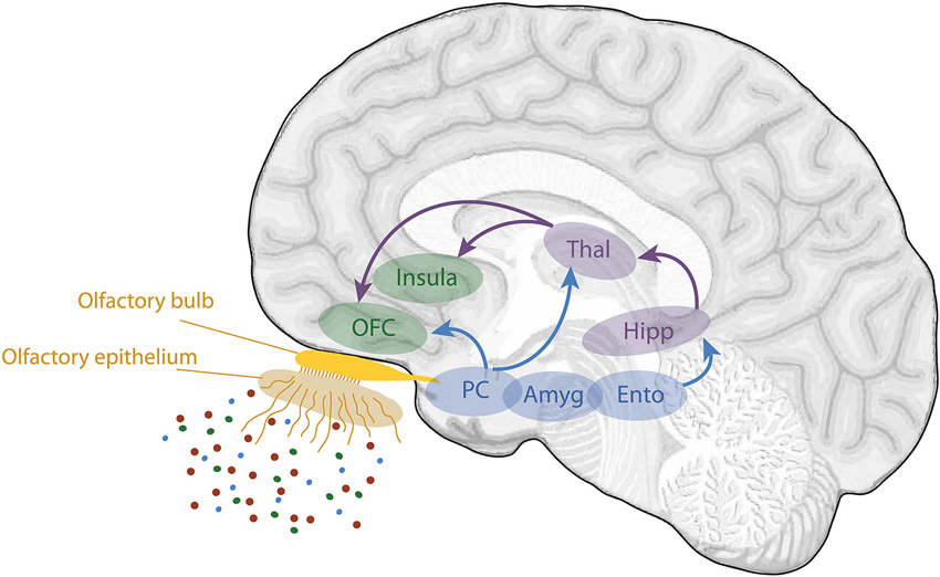
  • Located on the base of the frontal lobe Frontal lobe The part of the cerebral hemisphere anterior to the central sulcus, and anterior and superior to the lateral sulcus. Cerebral Cortex: Anatomy and medial aspect of the temporal lobe Temporal lobe Lower lateral part of the cerebral hemisphere responsible for auditory, olfactory, and semantic processing. It is located inferior to the lateral fissure and anterior to the occipital lobe. Cerebral Cortex: Anatomy 
  • Divided into several structures along the anterior–posterior axis:
    • Anterior olfactory nucleus Nucleus Within a eukaryotic cell, a membrane-limited body which contains chromosomes and one or more nucleoli (cell nucleolus). The nuclear membrane consists of a double unit-type membrane which is perforated by a number of pores; the outermost membrane is continuous with the endoplasmic reticulum. A cell may contain more than one nucleus. The Cell: Organelles 
    • Olfactory tubercle
    • Piriform cortex:
    • Olfactory amygdala Amygdala Almond-shaped group of basal nuclei anterior to the inferior horn of the lateral ventricle of the temporal lobe. The amygdala is part of the limbic system. Limbic System: Anatomy:
      • Located within the temporal lobe Temporal lobe Lower lateral part of the cerebral hemisphere responsible for auditory, olfactory, and semantic processing. It is located inferior to the lateral fissure and anterior to the occipital lobe. Cerebral Cortex: Anatomy, in front of the temporal horn of the lateral ventricle
      • Includes the prepiriform and periamygdaloid areas
      • Associated with emotional responses to smell, such as fear
    • Lateral entorhinal cortex
  • From the olfactory cortex, olfactory information is secondarily relayed via the mediodorsal nucleus Nucleus Within a eukaryotic cell, a membrane-limited body which contains chromosomes and one or more nucleoli (cell nucleolus). The nuclear membrane consists of a double unit-type membrane which is perforated by a number of pores; the outermost membrane is continuous with the endoplasmic reticulum. A cell may contain more than one nucleus. The Cell: Organelles of the thalamus Thalamus The thalamus is a large, ovoid structure in the dorsal part of the diencephalon that is located between the cerebral cortex and midbrain. It consists of several interconnected nuclei of grey matter separated by the laminae of white matter. The thalamus is the main conductor of information that passes between the cerebral cortex and the periphery, spinal cord, or brain stem. Thalamus: Anatomy to the:
    • Insular cortex:
      • Located inside the Sylvian fissure Fissure A crack or split that extends into the dermis Generalized and Localized Rashes
      • Believed to be the site where olfactory and taste information integrate to produce the sense of flavor
    • Orbitofrontal cortex:
      • Located on the base of the frontal lobe Frontal lobe The part of the cerebral hemisphere anterior to the central sulcus, and anterior and superior to the lateral sulcus. Cerebral Cortex: Anatomy
      • Lesions of this cortical region can result in an inability to distinguish different odors.
  • Odor information is also sent to the orbitofrontal cortex and portions of the hypothalamus Hypothalamus The hypothalamus is a collection of various nuclei within the diencephalon in the center of the brain. The hypothalamus plays a vital role in endocrine regulation as the primary regulator of the pituitary gland, and it is the major point of integration between the central nervous and endocrine systems. Hypothalamus and brain Brain The part of central nervous system that is contained within the skull (cranium). Arising from the neural tube, the embryonic brain is comprised of three major parts including prosencephalon (the forebrain); mesencephalon (the midbrain); and rhombencephalon (the hindbrain). The developed brain consists of cerebrum; cerebellum; and other structures in the brain stem. Nervous System: Anatomy, Structure, and Classification stem to trigger Trigger The type of signal that initiates the inspiratory phase by the ventilator Invasive Mechanical Ventilation appetite, salivation, and gastric contraction.
Schematic view of the human olfactory system

Schematic view of the human olfactory system:
The primary and secondary olfactory cortices are represented in blue and green, respectively.
Amyg: amygdala
Ento: entorhinal cortex
Hipp: hippocampus
OFC: orbitofrontal cortex
PC: piriform cortex
Thal: thalamus

Image: “Schematic view of the human olfactory system” by Saive, Royet, and Plailly (adapted from Royet et al.). License: CC BY 3.0

Clinical Relevance

  • Anosmia Anosmia Complete or severe loss of the subjective sense of smell. Loss of smell may be caused by many factors such as a cold, allergy, olfactory nerve diseases, viral respiratory tract infections (e.g., COVID-19), aging and various neurological disorders (e.g., Alzheimer disease). Cranial Nerve Palsies: temporary or permanent loss of the sense of smell. Anosmia Anosmia Complete or severe loss of the subjective sense of smell. Loss of smell may be caused by many factors such as a cold, allergy, olfactory nerve diseases, viral respiratory tract infections (e.g., COVID-19), aging and various neurological disorders (e.g., Alzheimer disease). Cranial Nerve Palsies can be due to numerous factors, including inflammation Inflammation Inflammation is a complex set of responses to infection and injury involving leukocytes as the principal cellular mediators in the body’s defense against pathogenic organisms. Inflammation is also seen as a response to tissue injury in the process of wound healing. The 5 cardinal signs of inflammation are pain, heat, redness, swelling, and loss of function. Inflammation within the nasal mucosa Nasal mucosa The mucous lining of the nasal cavity, including lining of the nostril (vestibule) and the olfactory mucosa. Nasal mucosa consists of ciliated cells, goblet cells, brush cells, small granule cells, basal cells (stem cells) and glands containing both mucous and serous cells. Nose Anatomy (External & Internal) ( rhinitis Rhinitis Inflammation of the nasal mucosa, the mucous membrane lining the nasal cavities. Rhinitis) from allergy Allergy An abnormal adaptive immune response that may or may not involve antigen-specific IgE Type I Hypersensitivity Reaction or postviral infection; blockage of nasal passages; trauma to the orbit or nasal cavity Nasal cavity The proximal portion of the respiratory passages on either side of the nasal septum. Nasal cavities, extending from the nares to the nasopharynx, are lined with ciliated nasal mucosa. Nose Anatomy (External & Internal), such as fracture Fracture A fracture is a disruption of the cortex of any bone and periosteum and is commonly due to mechanical stress after an injury or accident. Open fractures due to trauma can be a medical emergency. Fractures are frequently associated with automobile accidents, workplace injuries, and trauma. Overview of Bone Fractures of the cribriform plate, which may lead to disruption of the olfactory nerve Olfactory nerve The 1st cranial nerve. The olfactory nerve conveys the sense of smell. It is formed by the axons of olfactory receptor neurons which project from the olfactory epithelium (in the nasal epithelium) to the olfactory bulb. Nose Anatomy (External & Internal); a pathologic lesion (i.e., tumor Tumor Inflammation or inflammation Inflammation Inflammation is a complex set of responses to infection and injury involving leukocytes as the principal cellular mediators in the body’s defense against pathogenic organisms. Inflammation is also seen as a response to tissue injury in the process of wound healing. The 5 cardinal signs of inflammation are pain, heat, redness, swelling, and loss of function. Inflammation) of the temporal lobe Temporal lobe Lower lateral part of the cerebral hemisphere responsible for auditory, olfactory, and semantic processing. It is located inferior to the lateral fissure and anterior to the occipital lobe. Cerebral Cortex: Anatomy; or degenerative CNS disease such as Alzheimer, Huntington, or Parkinson disease Parkinson disease Parkinson’s disease (PD) is a chronic, progressive neurodegenerative disorder. Although the cause is unknown, several genetic and environmental risk factors are currently being studied. Individuals present clinically with resting tremor, bradykinesia, rigidity, and postural instability. Parkinson’s Disease. Treatment depends on the reversibility of the underlying condition. 
  • Dysosmia Dysosmia Cranial Nerve Palsies: qualitative alteration or distortion Distortion Defense Mechanisms of the perception Perception The process by which the nature and meaning of sensory stimuli are recognized and interpreted. Psychiatric Assessment of smell. Dysosmia Dysosmia Cranial Nerve Palsies can be classified as parosmia and phantosmia. Parosmia is an unpleasant odor perception Perception The process by which the nature and meaning of sensory stimuli are recognized and interpreted. Psychiatric Assessment. Phantosmia is the perception Perception The process by which the nature and meaning of sensory stimuli are recognized and interpreted. Psychiatric Assessment of an odor when no odorant is present. Dysosmia Dysosmia Cranial Nerve Palsies can be associated with prior head trauma Head trauma Head trauma occurs when external forces are directed to the skull and brain structures, resulting in damage to the skull, brain, and intracranial structures. Head injuries can be classified as open (penetrating) or closed (blunt), and primary (from the initial trauma) or secondary (indirect brain injury), and range from mild to severe and life-threatening. Head Trauma, chronic nasal congestion, and chronic sinusitis Sinusitis Sinusitis refers to inflammation of the mucosal lining of the paranasal sinuses. The condition usually occurs concurrently with inflammation of the nasal mucosa (rhinitis), a condition known as rhinosinusitis. Acute sinusitis is due to an upper respiratory infection caused by a viral, bacterial, or fungal agent. Sinusitis. Dysosmia Dysosmia Cranial Nerve Palsies is relatively common and has been reported in as many as 6% of adults. There is no established treatment, although medications such as antiepileptics, migraine Migraine Migraine headache is a primary headache disorder and is among the most prevalent disorders in the world. Migraine is characterized by episodic, moderate to severe headaches that may be associated with increased sensitivity to light and sound, as well as nausea and/or vomiting. Migraine Headache medications, and benzodiazepines Benzodiazepines Benzodiazepines work on the gamma-aminobutyric acid type A (GABAA) receptor to produce inhibitory effects on the CNS. Benzodiazepines do not mimic GABA, the main inhibitory neurotransmitter in humans, but instead potentiate GABA activity. Benzodiazepines have been used with various degrees of success. In extreme cases, surgical resection of the olfactory mucosa is offered.

References

  1. Lafreniere, D. (2021). Taste and olfactory disorders in adults: evaluation and management. UpToDate. Retrieved September 14, 2021, from https://www.uptodate.com/contents/taste-and-olfactory-disorders-in-adults-evaluation-and-management
  2. Klinger, E. (2017). Development and organization of the evolutionarily conserved three-layered olfactory cortex. eNeuro 4(1):ENEURO.0193-16.2016. https://doi.org/10.1523/eneuro.0193-16.2016 
  3. Pinto, J.M. (2011). Olfaction. Proc Am Thorac Soc 8:46–52. https://doi.org/10.1513/pats.201005-035RN 
  4. Purves, D., Augustine, G.J., Fitzpatrick, D., et al. (2001). The olfactory bulb. In: Neuroscience, 2nd ed. Sunderland, MA: Sinauer Associates.

Create your free account or log in to continue reading!

Sign up now and get free access to Lecturio with concept pages, medical videos, and questions for your medical education.

User Reviews

Unwrap New Skills This Holiday 🎄 Save 30% on all plans now!

Details