Advertisement

Advertisement

Advertisement
Advertisement

Nocardia/Nocardiosis

Nocardia is a branching, filamentous, gram-positive Gram-Positive Penicillins bacilli Bacilli Shigella. It is partially acid fast Acid fast Tuberculosis due to the presence of mycolic acids Mycolic acids Long fatty acids found in the cell walls of some actinobacteria, including Mycobacterium tuberculosis. Mycobacterium in the cell wall Cell wall The outermost layer of a cell in most plants; bacteria; fungi; and algae. The cell wall is usually a rigid structure that lies external to the cell membrane, and provides a protective barrier against physical or chemical agents. Cell Types: Eukaryotic versus Prokaryotic. Nocardia is a ubiquitous soil organism that most commonly affects immunocompromised immunocompromised A human or animal whose immunologic mechanism is deficient because of an immunodeficiency disorder or other disease or as the result of the administration of immunosuppressive drugs or radiation. Gastroenteritis patients Patients Individuals participating in the health care system for the purpose of receiving therapeutic, diagnostic, or preventive procedures. Clinician–Patient Relationship. Nocardia is transmitted via inhalation of aerosolized bacteria Bacteria Bacteria are prokaryotic single-celled microorganisms that are metabolically active and divide by binary fission. Some of these organisms play a significant role in the pathogenesis of diseases. Bacteriology or less commonly, via direct contact with wounds. Nocardia causes opportunistic infections Infections Invasion of the host organism by microorganisms or their toxins or by parasites that can cause pathological conditions or diseases. Chronic Granulomatous Disease, primarily pulmonary infections Infections Invasion of the host organism by microorganisms or their toxins or by parasites that can cause pathological conditions or diseases. Chronic Granulomatous Disease ( pneumonia Pneumonia Pneumonia or pulmonary inflammation is an acute or chronic inflammation of lung tissue. Causes include infection with bacteria, viruses, or fungi. In more rare cases, pneumonia can also be caused through toxic triggers through inhalation of toxic substances, immunological processes, or in the course of radiotherapy. Pneumonia, abscess Abscess Accumulation of purulent material in tissues, organs, or circumscribed spaces, usually associated with signs of infection. Chronic Granulomatous Disease, or cavitary lesions), which may spread to form brain Brain The part of central nervous system that is contained within the skull (cranium). Arising from the neural tube, the embryonic brain is comprised of three major parts including prosencephalon (the forebrain); mesencephalon (the midbrain); and rhombencephalon (the hindbrain). The developed brain consists of cerebrum; cerebellum; and other structures in the brain stem. Nervous System: Anatomy, Structure, and Classification abscesses. In immunocompetent patients Patients Individuals participating in the health care system for the purpose of receiving therapeutic, diagnostic, or preventive procedures. Clinician–Patient Relationship, Nocardia can cause a cutaneous infection. Treatment for nocardiosis is with trimethoprim Trimethoprim The sulfonamides are a class of antimicrobial drugs inhibiting folic acid synthesize in pathogens. The prototypical drug in the class is sulfamethoxazole. Although not technically sulfonamides, trimethoprim, dapsone, and pyrimethamine are also important antimicrobial agents inhibiting folic acid synthesis. The agents are often combined with sulfonamides, resulting in a synergistic effect. Sulfonamides and Trimethoprim sulfamethoxazole Sulfamethoxazole A bacteriostatic antibacterial agent that interferes with folic acid synthesis in susceptible bacteria. Its broad spectrum of activity has been limited by the development of resistance. Sulfonamides and Trimethoprim and/or surgical intervention as indicated.

Last updated: Dec 29, 2023

Editorial responsibility: Stanley Oiseth, Lindsay Jones, Evelin Maza

Advertisement

Advertisement

Advertisement

Advertisement

Advertisement

Advertisement

Classification

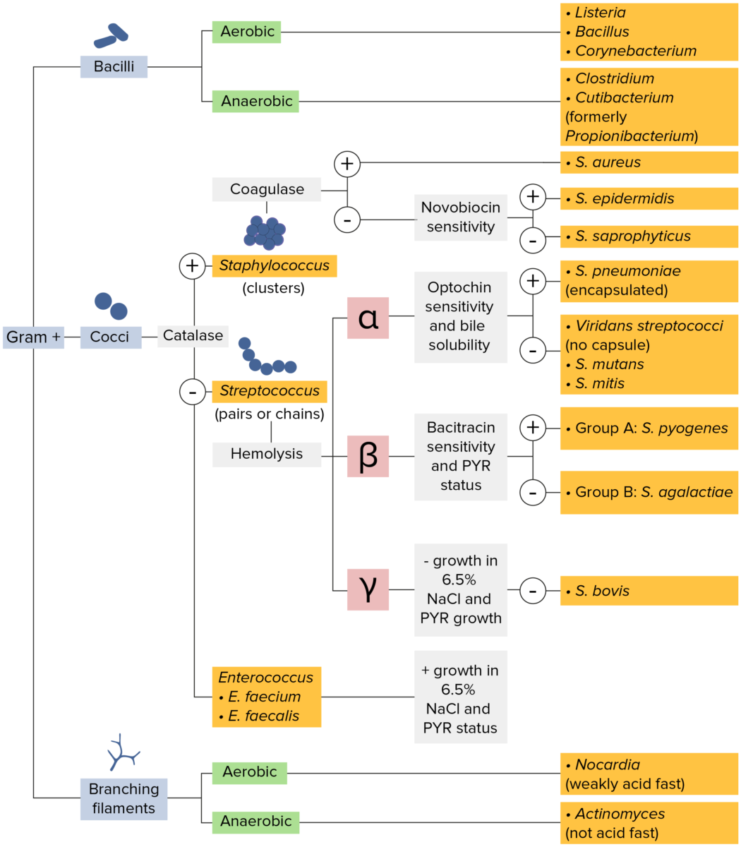
Microbiology flowchart gram-positive bacteria classification

Gram-positive bacteria:
Most bacteria can be classified according to a lab procedure called Gram staining.
Bacteria with cell walls that have a thick layer of peptidoglycan retain the crystal violet stain utilized in Gram staining but are not affected by the safranin counterstain. These bacteria appear as purple-blue on the stain, indicating that they are gram positive. The bacteria can be further classified according to morphology (branching filaments, bacilli, and cocci in clusters or chains) and their ability to grow in the presence of oxygen (aerobic versus anaerobic). The cocci can also be further identified. Staphylococci can be narrowed down on the basis of the presence of the enzyme coagulase and on their sensitivity to the antibiotic novobiocin. Streptococci are grown on blood agar and classified on the basis of which form of hemolysis they employ (α, β, or γ). Streptococci are further narrowed on the basis of their response to the pyrrolidonyl-β-naphthylamide (PYR) test, their sensitivity to specific antimicrobials (optochin and bacitracin), and their ability to grow on sodium chloride (NaCl) media.
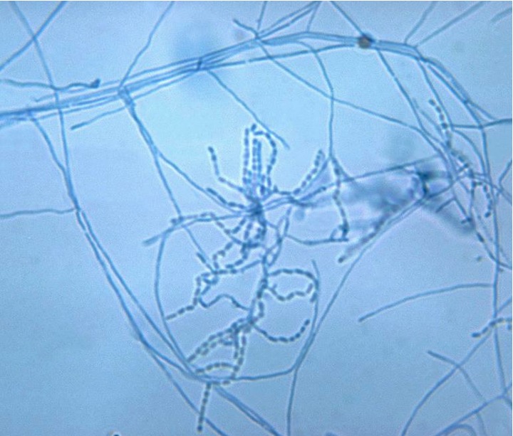
Image by Lecturio. License: CC BY-NC-SA 4.0

General Characteristics

Nocardia

  • General characteristics:
    • Structure:
      • Branching, filamentous rods
      • Filaments fragment readily into coccobacillary forms.
    • Gram stain Gram stain Klebsiella: gram positive
    • Other stain(s): weakly acid fast Acid fast Tuberculosis due to mycolic acids Mycolic acids Long fatty acids found in the cell walls of some actinobacteria, including Mycobacterium tuberculosis. Mycobacterium in the cell wall Cell wall The outermost layer of a cell in most plants; bacteria; fungi; and algae. The cell wall is usually a rigid structure that lies external to the cell membrane, and provides a protective barrier against physical or chemical agents. Cell Types: Eukaryotic versus Prokaryotic
    • Enzyme produced: urease positive, catalase positive
    • Spore Spore The reproductive elements of lower organisms, such as bacteria; fungi; and cryptogamic plants. Microsporidia/Microsporidiosis formation: non-spore forming
    • Oxygen requirement: obligate aerobe
  • Culture medium: grows on most ordinary laboratory media (such as blood agar) 
  • Nocardiosis (disease caused):
    • Frequently an opportunistic infection, manifesting as a pulmonary or disseminated disease.
    • Localized disease occurs, and usually presents as a cutaneous syndrome.
Stain nocardia species

This slide with gram-positive aerobic Nocardia asteroides reveals chains of bacteria among aerial mycelia.

Image by Lecturio.
Clinically relevant species

  • Nocardia asteroides(N. asteroides) has been divided based antimicrobial susceptibility patterns:
    • N. abscessus complex
    • N. brevicatena-paucivorans complex
    • N. nova complex
    • N. transvalensis complex
    • N. farcinica
    • N. cyriacigeorgica
  • Nocardia brasiliensis (N. brasiliensis)

Epidemiology

  • Incidence Incidence The number of new cases of a given disease during a given period in a specified population. It also is used for the rate at which new events occur in a defined population. It is differentiated from prevalence, which refers to all cases in the population at a given time. Measures of Disease Frequency of nocardiosis: 500–1,000 cases per year in the United States
  • Infection affects men > women
  • All ages are susceptible to infection.

Related videos

Pathogenesis

Reservoir Reservoir Animate or inanimate sources which normally harbor disease-causing organisms and thus serve as potential sources of disease outbreaks. Reservoirs are distinguished from vectors (disease vectors) and carriers, which are agents of disease transmission rather than continuing sources of potential disease outbreaks. Humans may serve both as disease reservoirs and carriers. Escherichia coli and transmission

  • Reservoir Reservoir Animate or inanimate sources which normally harbor disease-causing organisms and thus serve as potential sources of disease outbreaks. Reservoirs are distinguished from vectors (disease vectors) and carriers, which are agents of disease transmission rather than continuing sources of potential disease outbreaks. Humans may serve both as disease reservoirs and carriers. Escherichia coli(s): soil, decaying organic matter, water
  • Transmission:
    • Inhalation:
    • Environmental exposure through trauma or wound contamination:
      • Mainly with N. brasiliensis 
      • Infections Infections Invasion of the host organism by microorganisms or their toxins or by parasites that can cause pathological conditions or diseases. Chronic Granulomatous Disease affect the skin Skin The skin, also referred to as the integumentary system, is the largest organ of the body. The skin is primarily composed of the epidermis (outer layer) and dermis (deep layer). The epidermis is primarily composed of keratinocytes that undergo rapid turnover, while the dermis contains dense layers of connective tissue. Skin: Structure and Functions and lymph nodes Lymph Nodes They are oval or bean shaped bodies (1 – 30 mm in diameter) located along the lymphatic system. Lymphatic Drainage System: Anatomy.

Virulence Virulence The degree of pathogenicity within a group or species of microorganisms or viruses as indicated by case fatality rates and/or the ability of the organism to invade the tissues of the host. The pathogenic capacity of an organism is determined by its virulence factors. Proteus

  • Mechanisms of Nocardia spp:
    • Resistance Resistance Physiologically, the opposition to flow of air caused by the forces of friction. As a part of pulmonary function testing, it is the ratio of driving pressure to the rate of air flow. Ventilation: Mechanics of Breathing to phagocytosis Phagocytosis The engulfing and degradation of microorganisms; other cells that are dead, dying, or pathogenic; and foreign particles by phagocytic cells (phagocytes). Innate Immunity: Phagocytes and Antigen Presentation: when bacterial growth is in log phase
    • Resistance Resistance Physiologically, the opposition to flow of air caused by the forces of friction. As a part of pulmonary function testing, it is the ratio of driving pressure to the rate of air flow. Ventilation: Mechanics of Breathing to hydrolysis Hydrolysis The process of cleaving a chemical compound by the addition of a molecule of water. Proteins and Peptides: If phagocytosis Phagocytosis The engulfing and degradation of microorganisms; other cells that are dead, dying, or pathogenic; and foreign particles by phagocytic cells (phagocytes). Innate Immunity: Phagocytes and Antigen Presentation occurs, the fusion of the phagosome and lysosome is stopped.
    • Resistance Resistance Physiologically, the opposition to flow of air caused by the forces of friction. As a part of pulmonary function testing, it is the ratio of driving pressure to the rate of air flow. Ventilation: Mechanics of Breathing to antimicrobial action of neutrophils Neutrophils Granular leukocytes having a nucleus with three to five lobes connected by slender threads of chromatin, and cytoplasm containing fine inconspicuous granules and stainable by neutral dyes. Innate Immunity: Phagocytes and Antigen Presentation:
      • Superoxide dismutase Superoxide dismutase An oxidoreductase that catalyzes the reaction between superoxides and hydrogen to yield molecular oxygen and hydrogen peroxide. The enzyme protects the cell against dangerous levels of superoxide. Myeloperoxidase Deficiency from the cell surface
      • Increased catalase 
  • N. farcinica:
    • More virulent than other species
    • With antimicrobial resistance Resistance Physiologically, the opposition to flow of air caused by the forces of friction. As a part of pulmonary function testing, it is the ratio of driving pressure to the rate of air flow. Ventilation: Mechanics of Breathing
    • Associated with disseminated infection
  • L forms:
    • Microbial variants without a structurally intact cell wall Cell wall The outermost layer of a cell in most plants; bacteria; fungi; and algae. The cell wall is usually a rigid structure that lies external to the cell membrane, and provides a protective barrier against physical or chemical agents. Cell Types: Eukaryotic versus Prokaryotic
    • Found to persist in animal models (in cerebrospinal fluid Cerebrospinal Fluid A watery fluid that is continuously produced in the choroid plexus and circulates around the surface of the brain; spinal cord; and in the cerebral ventricles. Ventricular System: Anatomy)
    • Believed to be related to relapse Relapse Relapsing Fever or development of neurologic signs even after treatment

Disease process

  • Nocardia is introduced after a local injury to the skin Skin The skin, also referred to as the integumentary system, is the largest organ of the body. The skin is primarily composed of the epidermis (outer layer) and dermis (deep layer). The epidermis is primarily composed of keratinocytes that undergo rapid turnover, while the dermis contains dense layers of connective tissue. Skin: Structure and Functions and infection occurs:
    • In immunocompetent patients Patients Individuals participating in the health care system for the purpose of receiving therapeutic, diagnostic, or preventive procedures. Clinician–Patient Relationship: remains localized
    • In immunocompromised immunocompromised A human or animal whose immunologic mechanism is deficient because of an immunodeficiency disorder or other disease or as the result of the administration of immunosuppressive drugs or radiation. Gastroenteritis patients Patients Individuals participating in the health care system for the purpose of receiving therapeutic, diagnostic, or preventive procedures. Clinician–Patient Relationship: with potential to spread to other organs
  • Pleuropulmonary nocardiosis:
    • Infection follows after inhalation of contaminated aerosolized droplets Droplets Varicella-Zoster Virus/Chickenpox
    • Majority of patients Patients Individuals participating in the health care system for the purpose of receiving therapeutic, diagnostic, or preventive procedures. Clinician–Patient Relationship are immunocompromised immunocompromised A human or animal whose immunologic mechanism is deficient because of an immunodeficiency disorder or other disease or as the result of the administration of immunosuppressive drugs or radiation. Gastroenteritis.
Pathogenesis of nocardia

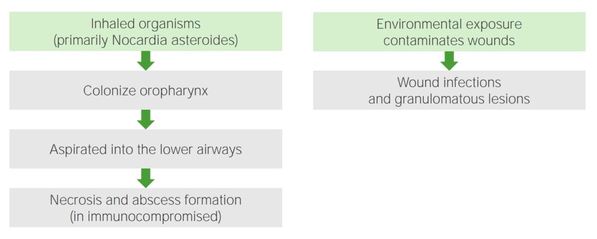
Pathogenesis of Nocardia:
Left: Inhaled organisms colonize the oropharynx and are passively aspirated into the lower airways, resulting in necrosis and abscess formation (in immunocompromised patients).
Right: Environmental exposure and contamination of wounds lead to wound infections or granulomatous lesions.
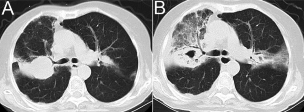
Image by Lecturio.

Host risk factors

  • Frequently the immunocompromised immunocompromised A human or animal whose immunologic mechanism is deficient because of an immunodeficiency disorder or other disease or as the result of the administration of immunosuppressive drugs or radiation. Gastroenteritis, particularly with impaired cell-mediated immunity Cell-mediated immunity Manifestations of the immune response which are mediated by antigen-sensitized T-lymphocytes via lymphokines or direct cytotoxicity. This takes place in the absence of circulating antibody or where antibody plays a subordinate role. Squamous Cell Carcinoma (SCC):
    • Human immunodeficiency Immunodeficiency Chédiak-Higashi Syndrome virus Virus Viruses are infectious, obligate intracellular parasites composed of a nucleic acid core surrounded by a protein capsid. Viruses can be either naked (non-enveloped) or enveloped. The classification of viruses is complex and based on many factors, including type and structure of the nucleoid and capsid, the presence of an envelope, the replication cycle, and the host range. Virology ( HIV HIV Anti-HIV Drugs) syndrome: usually in patients Patients Individuals participating in the health care system for the purpose of receiving therapeutic, diagnostic, or preventive procedures. Clinician–Patient Relationship with < 250 CD4+ T lymphocytes T lymphocytes Lymphocytes responsible for cell-mediated immunity. Two types have been identified – cytotoxic (t-lymphocytes, cytotoxic) and helper T-lymphocytes (t-lymphocytes, helper-inducer). They are formed when lymphocytes circulate through the thymus gland and differentiate to thymocytes. When exposed to an antigen, they divide rapidly and produce large numbers of new T cells sensitized to that antigen. T cells: Types and Functions/μL
    • Transplant recipients 
    • Chronic glucocorticoid use
    • Chronic immunosuppression
    • Malignancy Malignancy Hemothorax
  • Others:
    • Alcoholism Alcoholism A primary, chronic disease with genetic, psychosocial, and environmental factors influencing its development and manifestations. The disease is often progressive and fatal. It is characterized by impaired control over drinking, preoccupation with the drug alcohol, use of alcohol despite adverse consequences, and distortions in thinking, most notably denial. Each of these symptoms may be continuous or periodic. Wernicke Encephalopathy and Korsakoff Syndrome
    • Diabetes Diabetes Diabetes mellitus (DM) is a metabolic disease characterized by hyperglycemia and dysfunction of the regulation of glucose metabolism by insulin. Type 1 DM is diagnosed mostly in children and young adults as the result of autoimmune destruction of β cells in the pancreas and the resulting lack of insulin. Type 2 DM has a significant association with obesity and is characterized by insulin resistance. Diabetes Mellitus
    • Chronic obstructive pulmonary disease Pulmonary disease Diseases involving the respiratory system. Blastomyces/Blastomycosis
    • Inflammatory bowel disease

Clinical Presentation

Pulmonary nocardiosis

  • Predominant clinical presentation
  • Presents with fever Fever Fever is defined as a measured body temperature of at least 38°C (100.4°F). Fever is caused by circulating endogenous and/or exogenous pyrogens that increase levels of prostaglandin E2 in the hypothalamus. Fever is commonly associated with chills, rigors, sweating, and flushing of the skin. Fever, night sweats Night sweats Tuberculosis, dyspnea Dyspnea Dyspnea is the subjective sensation of breathing discomfort. Dyspnea is a normal manifestation of heavy physical or psychological exertion, but also may be caused by underlying conditions (both pulmonary and extrapulmonary). Dyspnea, weight loss Weight loss Decrease in existing body weight. Bariatric Surgery, cough, chest pain Pain An unpleasant sensation induced by noxious stimuli which are detected by nerve endings of nociceptive neurons. Pain: Types and Pathways, weight loss Weight loss Decrease in existing body weight. Bariatric Surgery 
  • Seen mostly in the immunocompromised immunocompromised A human or animal whose immunologic mechanism is deficient because of an immunodeficiency disorder or other disease or as the result of the administration of immunosuppressive drugs or radiation. Gastroenteritis
  • Chest X-ray X-ray Penetrating electromagnetic radiation emitted when the inner orbital electrons of an atom are excited and release radiant energy. X-ray wavelengths range from 1 pm to 10 nm. Hard x-rays are the higher energy, shorter wavelength x-rays. Soft x-rays or grenz rays are less energetic and longer in wavelength. The short wavelength end of the x-ray spectrum overlaps the gamma rays wavelength range. The distinction between gamma rays and x-rays is based on their radiation source. Pulmonary Function Tests/computed tomography (CT) scan: infiltrates, cavitary lesions, abscess Abscess Accumulation of purulent material in tissues, organs, or circumscribed spaces, usually associated with signs of infection. Chronic Granulomatous Disease, or pleural effusion Pleural Effusion Pleural effusion refers to the accumulation of fluid between the layers of the parietal and visceral pleura. Common causes of this condition include infection, malignancy, autoimmune disorders, or volume overload. Clinical manifestations include chest pain, cough, and dyspnea. Pleural Effusion
  • May mimic tuberculosis Tuberculosis Tuberculosis (TB) is an infectious disease caused by Mycobacterium tuberculosis complex bacteria. The bacteria usually attack the lungs but can also damage other parts of the body. Approximately 30% of people around the world are infected with this pathogen, with the majority harboring a latent infection. Tuberculosis spreads through the air when a person with active pulmonary infection coughs or sneezes. Tuberculosis but with negative purified protein derivative (PPD) test
Copd and nocardia spp

Computed tomography (CT) scan imaging of the lung in a patient with chronic obstructive pulmonary disease and Nocardia infection:
A: right upper lobar consolidation
B: computed tomography images after 12 days, showing cavitary consolidation and ground glass opacity
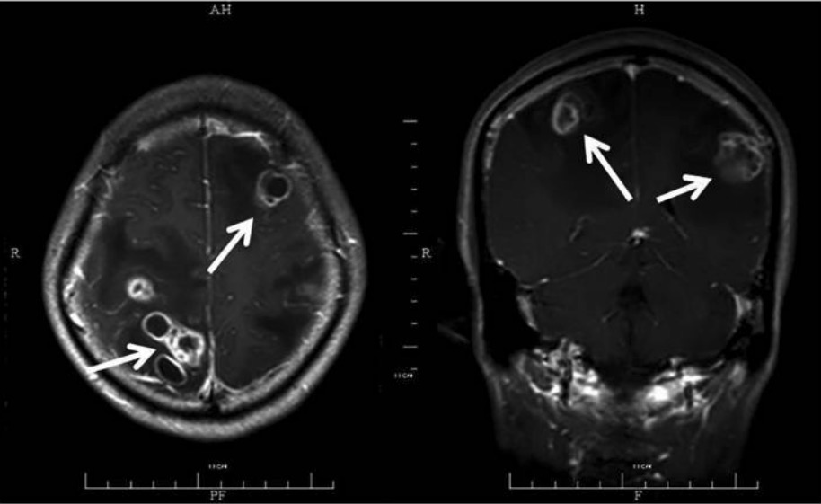
Image: “COPD and Nocardia spp.” by the Department of Respiratory Diseases, The First Affiliated Hospital, College of Medicine, Zhejiang University, Hangzhou, Zhejiang, People’s Republic of China. License: CC BY 4.0.

Disseminated nocardiosis

  • 2 or more non-contiguous sites are affected.
  • Infections Infections Invasion of the host organism by microorganisms or their toxins or by parasites that can cause pathological conditions or diseases. Chronic Granulomatous Disease may spread to the central nervous system Central nervous system The main information-processing organs of the nervous system, consisting of the brain, spinal cord, and meninges. Nervous System: Anatomy, Structure, and Classification (CNS), causing brain Brain The part of central nervous system that is contained within the skull (cranium). Arising from the neural tube, the embryonic brain is comprised of three major parts including prosencephalon (the forebrain); mesencephalon (the midbrain); and rhombencephalon (the hindbrain). The developed brain consists of cerebrum; cerebellum; and other structures in the brain stem. Nervous System: Anatomy, Structure, and Classification abscesses.
  • Slow-growing mass Mass Three-dimensional lesion that occupies a space within the breast Imaging of the Breast(es) causing headaches, fever Fever Fever is defined as a measured body temperature of at least 38°C (100.4°F). Fever is caused by circulating endogenous and/or exogenous pyrogens that increase levels of prostaglandin E2 in the hypothalamus. Fever is commonly associated with chills, rigors, sweating, and flushing of the skin. Fever, seizures Seizures A seizure is abnormal electrical activity of the neurons in the cerebral cortex that can manifest in numerous ways depending on the region of the brain affected. Seizures consist of a sudden imbalance that occurs between the excitatory and inhibitory signals in cortical neurons, creating a net excitation. The 2 major classes of seizures are focal and generalized. Seizures, and neurologic deficits Neurologic Deficits High-Risk Headaches
  • Signs of meningitis Meningitis Meningitis is inflammation of the meninges, the protective membranes of the brain, and spinal cord. The causes of meningitis are varied, with the most common being bacterial or viral infection. The classic presentation of meningitis is a triad of fever, altered mental status, and nuchal rigidity. Meningitis may be present.
Disseminated central nervous system nocardiosis

Magnetic resonance imaging (MRI) with contrast showing multiple abscesses (arrows): The patient has disseminated CNS nocardiosis, with bilateral involvement of the brain. Multiple ring lesions with surrounding edema are shown.

Image: “45-year-old male” by the State Key Laboratory for Diagnosis and Treatment of Infectious Diseases, The First Affiliated Hospital, School of Medicine, Zhejiang University, Hangzhou, Zhejiang 310003, P.R. China. License: CC BY 3.0.
Cutaneous nocardiosis

  • Commonly from N. brasiliensis
  • Occurs after trauma (gardening, surgery, animal bite)
  • Affects even the immunocompetent
  • Chronic, slowly progressing lesions
  • Presentation:
    • Primary cutaneous infection:
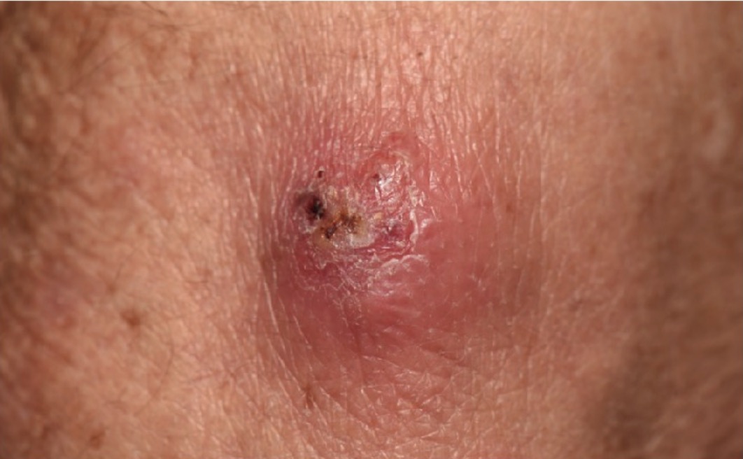
      • Cellulitis Cellulitis Cellulitis is a common infection caused by bacteria that affects the dermis and subcutaneous tissue of the skin. It is frequently caused by Staphylococcus aureus and Streptococcus pyogenes. The skin infection presents as an erythematous and edematous area with warmth and tenderness. Cellulitis or a single/multiple erythematous nodule Nodule Chalazion(s) at the site of trauma
      • May drain purulent material
    • Lymphocutaneous nocardiosis:
      • Cutaneous infection with lymphadenitis Lymphadenitis Inflammation of the lymph nodes. Peritonsillar Abscess, possibly ulcerating and/or draining purulent material 
      • Similar to how Sporothrix schenckii infection presents (thus also called “sporotrichoid nocardiosis”)
    • Mycetoma:
      • Progressive and chronic deep skin Skin The skin, also referred to as the integumentary system, is the largest organ of the body. The skin is primarily composed of the epidermis (outer layer) and dermis (deep layer). The epidermis is primarily composed of keratinocytes that undergo rapid turnover, while the dermis contains dense layers of connective tissue. Skin: Structure and Functions infection 
      • May extend to the fascia Fascia Layers of connective tissue of variable thickness. The superficial fascia is found immediately below the skin; the deep fascia invests muscles, nerves, and other organs. Cellulitis, muscle, and bone Bone Bone is a compact type of hardened connective tissue composed of bone cells, membranes, an extracellular mineralized matrix, and central bone marrow. The 2 primary types of bone are compact and spongy. Bones: Structure and Types
      • May cause deformities and sinus tracts
      • May be a form of disseminated infection in immunocompromised immunocompromised A human or animal whose immunologic mechanism is deficient because of an immunodeficiency disorder or other disease or as the result of the administration of immunosuppressive drugs or radiation. Gastroenteritis patients Patients Individuals participating in the health care system for the purpose of receiving therapeutic, diagnostic, or preventive procedures. Clinician–Patient Relationship
Nocardia brasiliensis infection

Image of cutaneous nocardiosis: fluctuant, purulent, and crusted nodule noted in a patient receiving long-term immunosuppressive therapy. Tissue cultures revealed N. brasiliensis.

Image: “Lesional skin” by Alison M. Fernandes et al. License: CC BY 4.0.
Diagnosis and Management

Diagnosis

  • Culture:
    • Grows on ordinary laboratory media after 3 to 5 days incubation Incubation The amount time between exposure to an infectious agent and becoming symptomatic. Rabies Virus in air
    • Specimens: sputum, pus, spinal fluid, or biopsy Biopsy Removal and pathologic examination of specimens from the living body. Ewing Sarcoma material
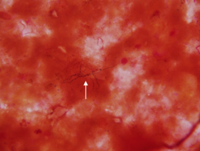
  • Microscopic examination: filamentous rods, with beading and branching morphology
  • Imaging studies are dictated by the clinical presentation:
    • Chest X-ray X-ray Penetrating electromagnetic radiation emitted when the inner orbital electrons of an atom are excited and release radiant energy. X-ray wavelengths range from 1 pm to 10 nm. Hard x-rays are the higher energy, shorter wavelength x-rays. Soft x-rays or grenz rays are less energetic and longer in wavelength. The short wavelength end of the x-ray spectrum overlaps the gamma rays wavelength range. The distinction between gamma rays and x-rays is based on their radiation source. Pulmonary Function Tests/CT scan of the chest: in pulmonary nocardiosis
    • CT scan of the brain Brain The part of central nervous system that is contained within the skull (cranium). Arising from the neural tube, the embryonic brain is comprised of three major parts including prosencephalon (the forebrain); mesencephalon (the midbrain); and rhombencephalon (the hindbrain). The developed brain consists of cerebrum; cerebellum; and other structures in the brain stem. Nervous System: Anatomy, Structure, and Classification/MRI: in patients Patients Individuals participating in the health care system for the purpose of receiving therapeutic, diagnostic, or preventive procedures. Clinician–Patient Relationship with neurologic symptoms
Nocardia brain biopsy

Nocardia (white arrow) as found on a brain biopsy

Image: “Nocardia as found on a brain biopsy” by Doc James. License: Public domain, edited by Lecturio.

Management

  • Trimethoprim-sulfamethoxazole
  • Newer β-lactams ( imipenem Imipenem Semisynthetic thienamycin that has a wide spectrum of antibacterial activity against gram-negative and gram-positive aerobic and anaerobic bacteria, including many multiresistant strains. It is stable to beta-lactamases. Clinical studies have demonstrated high efficacy in the treatment of infections of various body systems. Its effectiveness is enhanced when it is administered in combination with cilastatin, a renal dipeptidase inhibitor. Carbapenems and Aztreonam, meropenem Meropenem A thienamycin derivative antibacterial agent that is more stable to renal dehydropeptidase I than imipenem, but does not need to be given with an enzyme inhibitor such as cilastatin. It is used in the treatment of bacterial infections, including infections in immunocompromised patients. Carbapenems and Aztreonam, cefotaxime Cefotaxime Semisynthetic broad-spectrum cephalosporin. Cephalosporins) may be effective.
  • Surgical drainage for abscess Abscess Accumulation of purulent material in tissues, organs, or circumscribed spaces, usually associated with signs of infection. Chronic Granulomatous Disease

Comparisons

Actinomyces Actinomyces Actinomyces is an anaerobic, gram-positive, branching, filamentous rod. Actinomyces israelii is the most common species involved in human disease. The organism is commonly found as part of the normal flora in the oral cavity, gastrointestinal tract, and reproductive tract. Actinomyces/Actinomycosis and Nocardia

Table: Gram-positive Gram-Positive Penicillins, branching, filamentous rods that must be distinguished
Differentiating factors Actinomyces Actinomyces Actinomyces is an anaerobic, gram-positive, branching, filamentous rod. Actinomyces israelii is the most common species involved in human disease. The organism is commonly found as part of the normal flora in the oral cavity, gastrointestinal tract, and reproductive tract. Actinomyces/Actinomycosis Nocardia
Oxygen requirement Anaerobic Aerobic
Acid-fast stain Acid-Fast Stain Meningitis in Children Not acid fast Acid fast Tuberculosis Acid fast Acid fast Tuberculosis (partially)
Reservoir Reservoir Animate or inanimate sources which normally harbor disease-causing organisms and thus serve as potential sources of disease outbreaks. Reservoirs are distinguished from vectors (disease vectors) and carriers, which are agents of disease transmission rather than continuing sources of potential disease outbreaks. Humans may serve both as disease reservoirs and carriers. Escherichia coli/habitat Normal oral, gastrointestinal, and reproductive flora Found in soil, water, decaying organic matter
Infections Infections Invasion of the host organism by microorganisms or their toxins or by parasites that can cause pathological conditions or diseases. Chronic Granulomatous Disease caused
  • Orofacial abscesses, associated with dental caries or facial trauma
  • Sinus tracts
  • Pelvic inflammatory disease Pelvic inflammatory disease Pelvic inflammatory disease (PID) is defined as a polymicrobial infection of the upper female reproductive system. The disease can affect the uterus, fallopian tubes, ovaries, and adjacent structures. Pelvic inflammatory disease is closely linked with sexually transmitted diseases, most commonly caused by Chlamydia trachomatis, Neisseria gonorrhoeae, and Gardnerella vaginalis. Pelvic Inflammatory Disease
  • Pulmonary infection in immunocompromised immunocompromised A human or animal whose immunologic mechanism is deficient because of an immunodeficiency disorder or other disease or as the result of the administration of immunosuppressive drugs or radiation. Gastroenteritis patients Patients Individuals participating in the health care system for the purpose of receiving therapeutic, diagnostic, or preventive procedures. Clinician–Patient Relationship (can spread hematogenously and cause brain Brain The part of central nervous system that is contained within the skull (cranium). Arising from the neural tube, the embryonic brain is comprised of three major parts including prosencephalon (the forebrain); mesencephalon (the midbrain); and rhombencephalon (the hindbrain). The developed brain consists of cerebrum; cerebellum; and other structures in the brain stem. Nervous System: Anatomy, Structure, and Classification abscesses)
  • Cutaneous lesions in immunocompetent patients Patients Individuals participating in the health care system for the purpose of receiving therapeutic, diagnostic, or preventive procedures. Clinician–Patient Relationship
Management Treatment with penicillin Penicillin Rheumatic Fever Treatment with trimethoprim-sulfamethoxazole
Treatment mnemonic: SNAP (sulfonamides-Nocardia, Actinomyces-penicillin)

Mycobacterium Mycobacterium Mycobacterium is a genus of the family Mycobacteriaceae in the phylum Actinobacteria. Mycobacteria comprise more than 150 species of facultative intracellular bacilli that are mostly obligate aerobes. Mycobacteria are responsible for multiple human infections including serious diseases, such as tuberculosis (M. tuberculosis), leprosy (M. leprae), and M. avium complex infections. Mycobacterium and Nocardia

Table: Acid-fast rods comparison
Differentiating factors Mycobacterium Mycobacterium Mycobacterium is a genus of the family Mycobacteriaceae in the phylum Actinobacteria. Mycobacteria comprise more than 150 species of facultative intracellular bacilli that are mostly obligate aerobes. Mycobacteria are responsible for multiple human infections including serious diseases, such as tuberculosis (M. tuberculosis), leprosy (M. leprae), and M. avium complex infections. Mycobacterium Nocardia
Oxygen requirement Aerobic Aerobic
Spore Spore The reproductive elements of lower organisms, such as bacteria; fungi; and cryptogamic plants. Microsporidia/Microsporidiosis formation Non-spore forming Non-spore forming
Acid-fast stain Acid-Fast Stain Meningitis in Children Acid fast Acid fast Tuberculosis Partially acid fast Acid fast Tuberculosis
Structure Straight or slightly curved rods Branching filamentous rods
PPD* test PPD* positive PPD* negative
*PPD: purified protein derivative

Differential Diagnosis

  • Sporotrichosis: infection caused by Sporothrix schenckii. Lymphocutaneous infection is the most common form and presents like cutaneous nocardiosis (inoculation of soil through the skin Skin The skin, also referred to as the integumentary system, is the largest organ of the body. The skin is primarily composed of the epidermis (outer layer) and dermis (deep layer). The epidermis is primarily composed of keratinocytes that undergo rapid turnover, while the dermis contains dense layers of connective tissue. Skin: Structure and Functions with erythematous, nodular lesion(s) along lymphatic channels Channels The Cell: Cell Membrane). Culture of aspirated material helps distinguish the organisms.
  • Mycobacterial infection: pulmonary infection from Mycobacterium tuberculosis Mycobacterium tuberculosis Tuberculosis (TB) is an infectious disease caused by Mycobacterium tuberculosis complex bacteria. The bacteria usually attack the lungs but can also damage other parts of the body. Approximately 30% of people around the world are infected with this pathogen, with the majority harboring a latent infection. Tuberculosis spreads through the air when a person with active pulmonary infection coughs or sneezes. Tuberculosis (M. tuberculosis Tuberculosis Tuberculosis (TB) is an infectious disease caused by Mycobacterium tuberculosis complex bacteria. The bacteria usually attack the lungs but can also damage other parts of the body. Approximately 30% of people around the world are infected with this pathogen, with the majority harboring a latent infection. Tuberculosis spreads through the air when a person with active pulmonary infection coughs or sneezes. Tuberculosis) also presents with fever Fever Fever is defined as a measured body temperature of at least 38°C (100.4°F). Fever is caused by circulating endogenous and/or exogenous pyrogens that increase levels of prostaglandin E2 in the hypothalamus. Fever is commonly associated with chills, rigors, sweating, and flushing of the skin. Fever, night sweats Night sweats Tuberculosis, weight loss Weight loss Decrease in existing body weight. Bariatric Surgery, cough, hemoptysis Hemoptysis Hemoptysis is defined as the expectoration of blood originating in the lower respiratory tract. Hemoptysis is a consequence of another disease process and can be classified as either life threatening or non-life threatening. Hemoptysis can result in significant morbidity and mortality due to both drowning (reduced gas exchange as the lungs fill with blood) and hemorrhagic shock. Hemoptysis, and dyspnea Dyspnea Dyspnea is the subjective sensation of breathing discomfort. Dyspnea is a normal manifestation of heavy physical or psychological exertion, but also may be caused by underlying conditions (both pulmonary and extrapulmonary). Dyspnea. Both are acid-fast organisms, but Nocardia is only partially acid fast Acid fast Tuberculosis and is seen as a branching rod (resembling fungal hyphae Hyphae Microscopic threadlike filaments in fungi that are filled with a layer of protoplasm. Collectively, the hyphae make up the mycelium. Mycology). Recent infection with M. tuberculosis Tuberculosis Tuberculosis (TB) is an infectious disease caused by Mycobacterium tuberculosis complex bacteria. The bacteria usually attack the lungs but can also damage other parts of the body. Approximately 30% of people around the world are infected with this pathogen, with the majority harboring a latent infection. Tuberculosis spreads through the air when a person with active pulmonary infection coughs or sneezes. Tuberculosis results in a positive PPD test PPD test One of several skin tests to determine past or present tuberculosis infection. A purified protein derivative of the tubercle bacilli, called tuberculin, is introduced into the skin by scratch, puncture, or interdermal injection. Tuberculosis.
  • Brain Brain The part of central nervous system that is contained within the skull (cranium). Arising from the neural tube, the embryonic brain is comprised of three major parts including prosencephalon (the forebrain); mesencephalon (the midbrain); and rhombencephalon (the hindbrain). The developed brain consists of cerebrum; cerebellum; and other structures in the brain stem. Nervous System: Anatomy, Structure, and Classification metastasis Metastasis The transfer of a neoplasm from one organ or part of the body to another remote from the primary site. Grading, Staging, and Metastasis: Both brain Brain The part of central nervous system that is contained within the skull (cranium). Arising from the neural tube, the embryonic brain is comprised of three major parts including prosencephalon (the forebrain); mesencephalon (the midbrain); and rhombencephalon (the hindbrain). The developed brain consists of cerebrum; cerebellum; and other structures in the brain stem. Nervous System: Anatomy, Structure, and Classification metastasis Metastasis The transfer of a neoplasm from one organ or part of the body to another remote from the primary site. Grading, Staging, and Metastasis and CNS nocardiosis present insidiously and imaging may show multiple lesions in one or both cerebral hemispheres. History helps in diagnosing these conditions; disseminated nocardiosis usually comes from a lung infection Lung infection Pneumonia or pulmonary inflammation is an acute or chronic inflammation of lung tissue. Causes include infection with bacteria, viruses, or fungi. In more rare cases, pneumonia can also be caused through toxic triggers through inhalation of toxic substances, immunological processes, or in the course of radiotherapy. Pneumonia. Pulmonary investigation with imaging and cultures also help differentiate nocardiosis.

References

  1. Beaman, B., Black, C., Doughty, F., & Beaman, L. (1985) Role of superoxide dismutase and catalase as determinants of pathogenicity of Nocardia asteroides: importance in resistance to microbicidal activities of human polymorphonuclear neutrophils. Infect Immun. 1985 Jan;47(1):135-41. doi: 10.1128/IAI.47.1.135-141.
  2. Filice, G.A. (2018). Nocardiosis. In Jameson, J., Fauci, A.S., Kasper, D.L., Hauser, S.L., Longo, D.L., & Loscalzo, J. (Eds.), Harrison’s Principles of Internal Medicine, 20e. McGraw-Hill. 
  3. Kurdgelashvili, G. (2018). Nocardiosis. Medscape. Retrieved 25 Nov 2020, from https://emedicine.medscape.com/article/224123-overview
  4. Long, S., Prober, C., & Fischer, M. (2018). Nocardia species. Principles and Practice of Pediatric Infectious Diseases. (5th ed, p. 812). Elsevier, Inc.
  5. Riedel, S., Hobden, J.A., Miller, S., Morse, S.A., Mietzner, T.A., Detrick, B., Mitchell, T.G., Sakanari, J.A., Hotez, P., & Mejia, R. (Eds.) (2019). Aerobic non–spore-forming gram-positive bacilli: Corynebacterium, Listeria, Erysipelothrix, Nocardia, and related pathogens. Jawetz, Melnick, & Adelberg’s Medical Microbiology, 28e. McGraw-Hill. 
  6. Ryan, K.J. (Ed.) (2017). Actinomyces and Nocardia. Sherris Medical Microbiology, 7e. McGraw-Hill. 
  7. Spellman, D., Sexton, D., & Mitty, J. (2019). Microbiology, epidemiology and pathogenesis of nocardiosis. UpToDate. Retrieved 25 Nov 2020, from https://www.uptodate.com/contents/microbiology-epidemiology-and-pathogenesis-of-nocardiosis

Create your free account or log in to continue reading!

Sign up now and get free access to Lecturio with concept pages, medical videos, and questions for your medical education.

User Reviews

Unwrap New Skills This Holiday 🎄 Save 30% on all plans now!

Details