Advertisement

Advertisement

Advertisement

Advertisement

Necrotizing Enterocolitis

Necrotizing enterocolitis Enterocolitis Inflammation of the mucosa of both the small intestine and the large intestine. Etiology includes ischemia, infections, allergic, and immune responses. Yersinia spp./Yersiniosis (NEC) is an intestinal inflammatory process that can lead to mucosal injury and necrosis Necrosis The death of cells in an organ or tissue due to disease, injury or failure of the blood supply. Ischemic Cell Damage. The condition is multifactorial, with underlying risk factors that include prematurity Prematurity Neonatal Respiratory Distress Syndrome and formula feeding Formula feeding Formulations for the nutrition of infants that are substituted for breast milk. Infant Care. The clinical presentation varies in severity from feeding intolerance, acute findings on abdominal exam, and systemic symptoms. The diagnosis is based on a clinical suspicion, abnormal abdominal radiographs, and supporting abnormal laboratory results. Management consists of supportive medical care Medical care Conflict of Interest for milder stages and bowel rest and surgical intervention for more advanced stages. Necrotizing enterocolitis Enterocolitis Inflammation of the mucosa of both the small intestine and the large intestine. Etiology includes ischemia, infections, allergic, and immune responses. Yersinia spp./Yersiniosis and its complications carry a high risk of morbidity Morbidity The proportion of patients with a particular disease during a given year per given unit of population. Measures of Health Status and mortality Mortality All deaths reported in a given population. Measures of Health Status.

Last updated: Dec 1, 2022

Editorial responsibility: Stanley Oiseth, Lindsay Jones, Evelin Maza

Advertisement

Advertisement

Advertisement

Advertisement

Advertisement

Advertisement

Overview

Definition

Necrotizing enterocolitis Enterocolitis Inflammation of the mucosa of both the small intestine and the large intestine. Etiology includes ischemia, infections, allergic, and immune responses. Yersinia spp./Yersiniosis (NEC) is an intestinal inflammatory condition associated with focal or diffuse ulceration Ulceration Corneal Abrasions, Erosion, and Ulcers and necrosis Necrosis The death of cells in an organ or tissue due to disease, injury or failure of the blood supply. Ischemic Cell Damage that primarily affects the terminal ileum Ileum The distal and narrowest portion of the small intestine, between the jejunum and the ileocecal valve of the large intestine. Small Intestine: Anatomy and colon Colon The large intestines constitute the last portion of the digestive system. The large intestine consists of the cecum, appendix, colon (with ascending, transverse, descending, and sigmoid segments), rectum, and anal canal. The primary function of the colon is to remove water and compact the stool prior to expulsion from the body via the rectum and anal canal. Colon, Cecum, and Appendix: Anatomy.

Epidemiology

  • Necrotizing enterocolitis Enterocolitis Inflammation of the mucosa of both the small intestine and the large intestine. Etiology includes ischemia, infections, allergic, and immune responses. Yersinia spp./Yersiniosis is the most common cause of GI emergencies and intestinal perforation Intestinal perforation Perforated viscus or GI perforation represents a condition in which the integrity of the GI wall is lost with subsequent leakage of enteric contents into the peritoneal cavity, resulting in peritonitis. The causes of perforated viscus include trauma, bowel ischemia, infections, or ulcerative conditions, all of which ultimately lead to a full-thickness disruption of the intestinal wall. Perforated Viscus in premature infants Premature infants A human infant born before 37 weeks of gestation. Sudden Infant Death Syndrome (SIDS).
  • Incidence Incidence The number of new cases of a given disease during a given period in a specified population. It also is used for the rate at which new events occur in a defined population. It is differentiated from prevalence, which refers to all cases in the population at a given time. Measures of Disease Frequency: 0.3–2.4 per 1000 live births
  • Prevalence Prevalence The total number of cases of a given disease in a specified population at a designated time. It is differentiated from incidence, which refers to the number of new cases in the population at a given time. Measures of Disease Frequency: 2%–5% of preterm infants admitted to the NICU

Etiology

  • No single identified cause
  • Multifactorial, with underlying risk factors leading to intestinal inflammatory process:
    • Prematurity Prematurity Neonatal Respiratory Distress Syndrome: < 32 weeks
    • Low birth weight: < 1500 grams
    • Formula feeding Formula feeding Formulations for the nutrition of infants that are substituted for breast milk. Infant Care (particularly hyperosmolar feedings)
    • Aggressive advancement of feeding volume
    • Perinatal stress
      • Shock Shock Shock is a life-threatening condition associated with impaired circulation that results in tissue hypoxia. The different types of shock are based on the underlying cause: distributive (↑ cardiac output (CO), ↓ systemic vascular resistance (SVR)), cardiogenic (↓ CO, ↑ SVR), hypovolemic (↓ CO, ↑ SVR), obstructive (↓ CO), and mixed. Types of Shock
      • Asphyxia Asphyxia A pathological condition caused by lack of oxygen, manifested in impending or actual cessation of life. Drowning
      • Sepsis Sepsis Systemic inflammatory response syndrome with a proven or suspected infectious etiology. When sepsis is associated with organ dysfunction distant from the site of infection, it is called severe sepsis. When sepsis is accompanied by hypotension despite adequate fluid infusion, it is called septic shock. Sepsis and Septic Shock
    • Nursery outbreak
    • Maternal illicit drug use (especially of cocaine Cocaine An alkaloid ester extracted from the leaves of plants including coca. It is a local anesthetic and vasoconstrictor and is clinically used for that purpose, particularly in the eye, ear, nose, and throat. It also has powerful central nervous system effects similar to the amphetamines and is a drug of abuse. Cocaine, like amphetamines, acts by multiple mechanisms on brain catecholaminergic neurons; the mechanism of its reinforcing effects is thought to involve inhibition of dopamine uptake. Local Anesthetics)
    • Congenital Congenital Chorioretinitis GI defects
    • Congenital Congenital Chorioretinitis heart disease

Pathophysiology and Clinical Presentation

Pathophysiology

  • Multifactorial, with several possible contributing factors:
    • Genetic predisposition
    • Intestinal immaturity
    • Imbalance in intestinal microvascular tone
    • Abnormal intestinal microbial colonization Colonization Bacteriology
    • ↑ Immunoreactivity of intestinal mucosa Intestinal Mucosa Lining of the intestines, consisting of an inner epithelium, a middle lamina propria, and an outer muscularis mucosae. In the small intestine, the mucosa is characterized by a series of folds and abundance of absorptive cells (enterocytes) with microvilli. Small Intestine: Anatomy
  • Possible mechanism:
    • Initial ischemic or reperfusion injury Reperfusion injury Adverse functional, metabolic, or structural changes in ischemic tissues resulting from the restoration of blood flow to the tissue (reperfusion), including swelling; hemorrhage; necrosis; and damage from free radicals. The most common instance is myocardial reperfusion injury. Ischemic Cell Damage → activation of proinflammatory mediators
    • Damage to the intestinal lining → ↑ intestinal permeability → allows bacterial invasion
    • ↑ Feedings → proliferation of luminal bacteria Bacteria Bacteria are prokaryotic single-celled microorganisms that are metabolically active and divide by binary fission. Some of these organisms play a significant role in the pathogenesis of diseases. Bacteriology
    • Penetration Penetration X-rays of bacteria Bacteria Bacteria are prokaryotic single-celled microorganisms that are metabolically active and divide by binary fission. Some of these organisms play a significant role in the pathogenesis of diseases. Bacteriology into the intestinal wall → production of hydrogen gas → pneumatosis intestinalis and gas in the portal vein Portal vein A short thick vein formed by union of the superior mesenteric vein and the splenic vein. Liver: Anatomy
    • Continued inflammation Inflammation Inflammation is a complex set of responses to infection and injury involving leukocytes as the principal cellular mediators in the body’s defense against pathogenic organisms. Inflammation is also seen as a response to tissue injury in the process of wound healing. The 5 cardinal signs of inflammation are pain, heat, redness, swelling, and loss of function. Inflammation and intestinal wall destruction → ischemia Ischemia A hypoperfusion of the blood through an organ or tissue caused by a pathologic constriction or obstruction of its blood vessels, or an absence of blood circulation. Ischemic Cell Damage and necrosis Necrosis The death of cells in an organ or tissue due to disease, injury or failure of the blood supply. Ischemic Cell Damage
    • Eventual bowel perforation Bowel perforation Perforated viscus or GI perforation represents a condition in which the integrity of the GI wall is lost with subsequent leakage of enteric contents into the peritoneal cavity, resulting in peritonitis. The causes of perforated viscus include trauma, bowel ischemia, infections, or ulcerative conditions, all of which ultimately lead to a full-thickness disruption of the intestinal wall. Perforated Viscus peritonitis Peritonitis Inflammation of the peritoneum lining the abdominal cavity as the result of infectious, autoimmune, or chemical processes. Primary peritonitis is due to infection of the peritoneal cavity via hematogenous or lymphatic spread and without intra-abdominal source. Secondary peritonitis arises from the abdominal cavity itself through rupture or abscess of intra-abdominal organs. Penetrating Abdominal Injury
  • Most commonly affected sites:
    • Terminal ileum Ileum The distal and narrowest portion of the small intestine, between the jejunum and the ileocecal valve of the large intestine. Small Intestine: Anatomy
    • Proximal colon Colon The large intestines constitute the last portion of the digestive system. The large intestine consists of the cecum, appendix, colon (with ascending, transverse, descending, and sigmoid segments), rectum, and anal canal. The primary function of the colon is to remove water and compact the stool prior to expulsion from the body via the rectum and anal canal. Colon, Cecum, and Appendix: Anatomy

Clinical presentation

  • Time of symptom onset varies by gestational age Gestational age The age of the conceptus, beginning from the time of fertilization. In clinical obstetrics, the gestational age is often estimated as the time from the last day of the last menstruation which is about 2 weeks before ovulation and fertilization. Pregnancy: Diagnosis, Physiology, and Care:
    • Inverse relationship between gestational age Gestational age The age of the conceptus, beginning from the time of fertilization. In clinical obstetrics, the gestational age is often estimated as the time from the last day of the last menstruation which is about 2 weeks before ovulation and fertilization. Pregnancy: Diagnosis, Physiology, and Care and timing of presentation
    • Preterm: typically in the 2nd to 4th week of life
    • Term or near term: typically in the 1st week of life
  • Clinical course:
    • Can progress rapidly, within hours
    • Starts as vague signs of feeding intolerance and mild systemic symptoms:
      • Vomiting Vomiting The forcible expulsion of the contents of the stomach through the mouth. Hypokalemia
      • Increased residual after feeds
      • Temperature instability
      • Decreased activity
      • Fatigue Fatigue The state of weariness following a period of exertion, mental or physical, characterized by a decreased capacity for work and reduced efficiency to respond to stimuli. Fibromyalgia
    • Progresses to acute abdominal symptoms:
    • Severe systemic involvement:
      • Apnea and respiratory failure Respiratory failure Respiratory failure is a syndrome that develops when the respiratory system is unable to maintain oxygenation and/or ventilation. Respiratory failure may be acute or chronic and is classified as hypoxemic, hypercapnic, or a combination of the two. Respiratory Failure
      • Hypotension Hypotension Hypotension is defined as low blood pressure, specifically < 90/60 mm Hg, and is most commonly a physiologic response. Hypotension may be mild, serious, or life threatening, depending on the cause. Hypotension and shock Shock Shock is a life-threatening condition associated with impaired circulation that results in tissue hypoxia. The different types of shock are based on the underlying cause: distributive (↑ cardiac output (CO), ↓ systemic vascular resistance (SVR)), cardiogenic (↓ CO, ↑ SVR), hypovolemic (↓ CO, ↑ SVR), obstructive (↓ CO), and mixed. Types of Shock
      • Unresponsiveness

Diagnosis

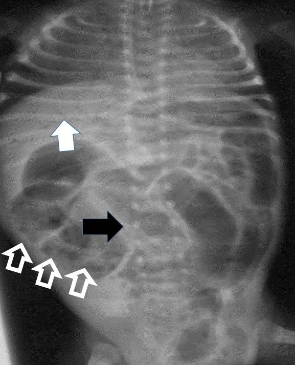
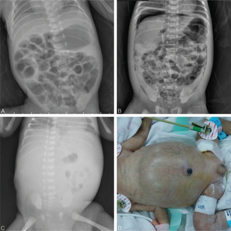
The diagnosis of NEC is made primarily by imaging.

Abdominal X-ray X-ray Penetrating electromagnetic radiation emitted when the inner orbital electrons of an atom are excited and release radiant energy. X-ray wavelengths range from 1 pm to 10 nm. Hard x-rays are the higher energy, shorter wavelength x-rays. Soft x-rays or grenz rays are less energetic and longer in wavelength. The short wavelength end of the x-ray spectrum overlaps the gamma rays wavelength range. The distinction between gamma rays and x-rays is based on their radiation source. Pulmonary Function Tests

Abdominal X-ray X-ray Penetrating electromagnetic radiation emitted when the inner orbital electrons of an atom are excited and release radiant energy. X-ray wavelengths range from 1 pm to 10 nm. Hard x-rays are the higher energy, shorter wavelength x-rays. Soft x-rays or grenz rays are less energetic and longer in wavelength. The short wavelength end of the x-ray spectrum overlaps the gamma rays wavelength range. The distinction between gamma rays and x-rays is based on their radiation source. Pulmonary Function Tests is the method of choice to establish the diagnosis of NEC.
  • Views:
    • Anteroposterior
    • Left lateral decubitus or supine cross-table lateral
  • Findings:
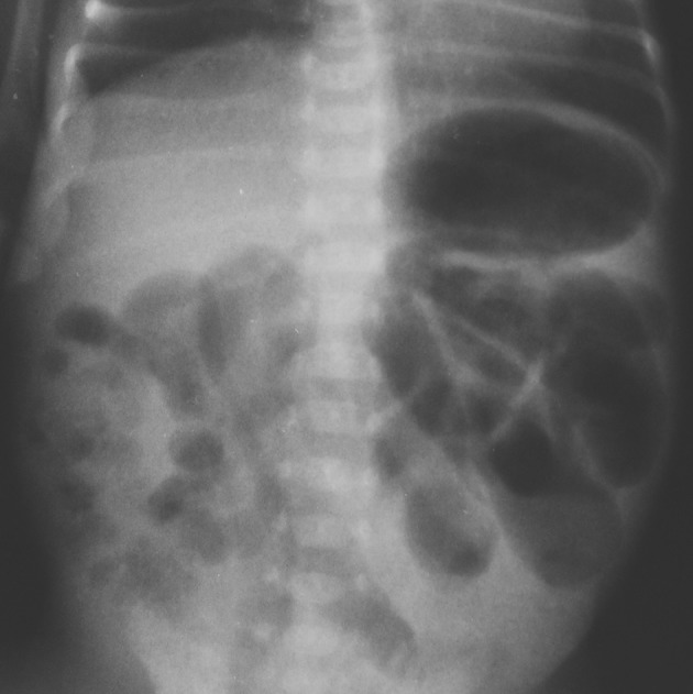
    • Thickened bowel loops
    • Ileus Ileus A condition caused by the lack of intestinal peristalsis or intestinal motility without any mechanical obstruction. This interference of the flow of intestinal contents often leads to intestinal obstruction. Ileus may be classified into postoperative, inflammatory, metabolic, neurogenic, and drug-induced. Small Bowel Obstruction
    • Fixed, dilated bowel loops (does not change with serial imaging studies)
    • Intraperitoneal Intraperitoneal Peritoneum: Anatomy fluid ( ascites Ascites Ascites is the pathologic accumulation of fluid within the peritoneal cavity that occurs due to an osmotic and/or hydrostatic pressure imbalance secondary to portal hypertension (cirrhosis, heart failure) or non-portal hypertension (hypoalbuminemia, malignancy, infection). Ascites):
      • Opacification of the abdomen
      • Absence of intestinal gas (worrisome finding)
    • Pneumatosis intestinalis:
      • Pathognomonic
      • “Train-track” lucency within bowel walls
      • Due to entrapment of air produced by gas-forming bacteria Bacteria Bacteria are prokaryotic single-celled microorganisms that are metabolically active and divide by binary fission. Some of these organisms play a significant role in the pathogenesis of diseases. Bacteriology
    • Portal venous gas Portal Venous Gas Imaging of the Intestines (pneumatosis hepatis)
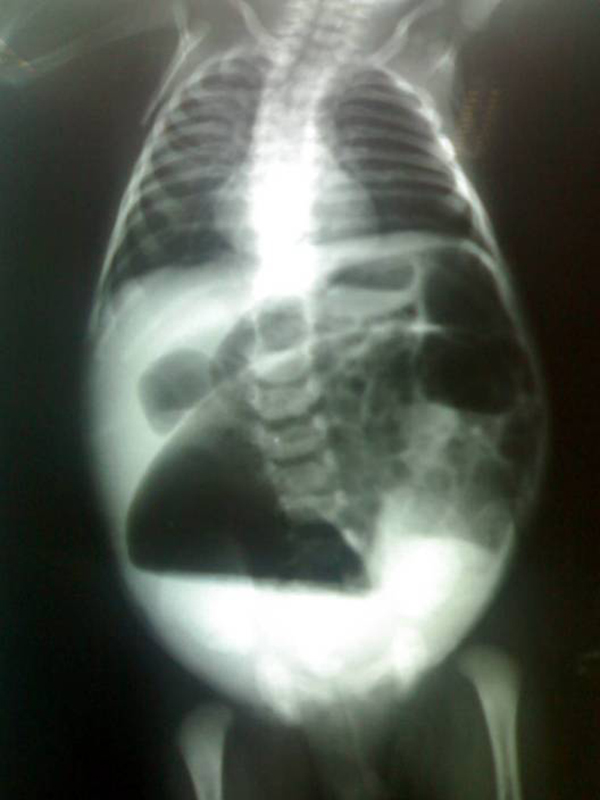
    • Pneumoperitoneum Pneumoperitoneum A condition with trapped gas or air in the peritoneal cavity, usually secondary to perforation of the internal organs such as the lung and the gastrointestinal tract, or to recent surgery. Pneumoperitoneum may be purposely introduced to aid radiological examination. Perforated Viscus:
      • Large, hyperlucent area above the liver Liver The liver is the largest gland in the human body. The liver is found in the superior right quadrant of the abdomen and weighs approximately 1.5 kilograms. Its main functions are detoxification, metabolism, nutrient storage (e.g., iron and vitamins), synthesis of coagulation factors, formation of bile, filtration, and storage of blood. Liver: Anatomy indicating free air below diaphragm Diaphragm The diaphragm is a large, dome-shaped muscle that separates the thoracic cavity from the abdominal cavity. The diaphragm consists of muscle fibers and a large central tendon, which is divided into right and left parts. As the primary muscle of inspiration, the diaphragm contributes 75% of the total inspiratory muscle force. Diaphragm: Anatomy
      • Indicative of bowel perforation Bowel perforation Perforated viscus or GI perforation represents a condition in which the integrity of the GI wall is lost with subsequent leakage of enteric contents into the peritoneal cavity, resulting in peritonitis. The causes of perforated viscus include trauma, bowel ischemia, infections, or ulcerative conditions, all of which ultimately lead to a full-thickness disruption of the intestinal wall. Perforated Viscus
Dilated bowel loops on an abdominal radiograph

Dilated bowel loops on an abdominal radiograph in patient with necrotizing enterocolitis

Image: “A report of a rare congenital malformation in a Nepalese child with congenital pouch colon: a case report” by Shakya VC, Agrawal CS, Koirala R, Khaniya S, Poudel P, Adhikary S. License: CC BY 3.0
Ultrasonography

  • Less commonly used
  • Can be used as an adjunct if radiography yields nonspecific findings
  • Findings:
    • Changes in bowel wall thickness and echogenicity
    • Pneumatosis intestinalis
    • Portal venous gas Portal Venous Gas Imaging of the Intestines
    • Changes in bowel wall perfusion
    • Ascites Ascites Ascites is the pathologic accumulation of fluid within the peritoneal cavity that occurs due to an osmotic and/or hydrostatic pressure imbalance secondary to portal hypertension (cirrhosis, heart failure) or non-portal hypertension (hypoalbuminemia, malignancy, infection). Ascites
    • Absent peristalsis Peristalsis A movement, caused by sequential muscle contraction, that pushes the contents of the intestines or other tubular organs in one direction. Gastrointestinal Motility
    • Abdominal free air

Laboratory evaluation

The following studies are nonspecific, but they may support the diagnosis and demonstrate the severity of the disease.

  • Positive stool occult blood test
  • CBC:
    • Anemia Anemia Anemia is a condition in which individuals have low Hb levels, which can arise from various causes. Anemia is accompanied by a reduced number of RBCs and may manifest with fatigue, shortness of breath, pallor, and weakness. Subtypes are classified by the size of RBCs, chronicity, and etiology. Anemia: Overview and Types
    • Leukocytosis Leukocytosis A transient increase in the number of leukocytes in a body fluid. West Nile Virus with left shift Left Shift Yersinia pestis/Plague or leukopenia
    • Thrombocytopenia Thrombocytopenia Thrombocytopenia occurs when the platelet count is < 150,000 per microliter. The normal range for platelets is usually 150,000-450,000/µL of whole blood. Thrombocytopenia can be a result of decreased production, increased destruction, or splenic sequestration of platelets. Patients are often asymptomatic until platelet counts are < 50,000/µL. Thrombocytopenia
  • Blood cultures:
    • Obtained before giving antibiotics
    • Most do not grow any organisms
  • Metabolic panel:
    • Hyponatremia Hyponatremia Hyponatremia is defined as a decreased serum sodium (sNa+) concentration less than 135 mmol/L. Serum sodium is the greatest contributor to plasma osmolality, which is very tightly controlled via antidiuretic hormone (ADH) release from the hypothalamus and by the thirst mechanism. Hyponatremia
    • Bicarbonate Bicarbonate Inorganic salts that contain the -HCO3 radical. They are an important factor in determining the ph of the blood and the concentration of bicarbonate ions is regulated by the kidney. Levels in the blood are an index of the alkali reserve or buffering capacity. Electrolytes → metabolic acidosis Acidosis A pathologic condition of acid accumulation or depletion of base in the body. The two main types are respiratory acidosis and metabolic acidosis, due to metabolic acid build up. Respiratory Acidosis
    • ↑ Lactic acid
  • Blood gas:
    • Confirms a metabolic acidosis Acidosis A pathologic condition of acid accumulation or depletion of base in the body. The two main types are respiratory acidosis and metabolic acidosis, due to metabolic acid build up. Respiratory Acidosis
    • Evaluate for hypoventilation (↑CO2)
  • Coagulation:
  • Inflammatory markers:

Modified Bell’s staging Staging Methods which attempt to express in replicable terms the extent of the neoplasm in the patient. Grading, Staging, and Metastasis criteria for NEC

The following table provides staging Staging Methods which attempt to express in replicable terms the extent of the neoplasm in the patient. Grading, Staging, and Metastasis criteria used to enhance recognition of NEC and assess its severity.

Stage Classification Clinical signs Radiologic signs
IA Suspected NEC
  • Abdominal distention Abdominal distention Megacolon
  • Emesis or gastric residuals
  • Lethargy Lethargy A general state of sluggishness, listless, or uninterested, with being tired, and having difficulty concentrating and doing simple tasks. It may be related to depression or drug addiction. Hyponatremia
  • Apnea
  • Bradycardia Bradycardia Bradyarrhythmia is a rhythm in which the heart rate is less than 60/min. Bradyarrhythmia can be physiologic, without symptoms or hemodynamic change. Pathologic bradyarrhythmia results in reduced cardiac output and hemodynamic instability causing syncope, dizziness, or dyspnea. Bradyarrhythmias
  • Temperature instability
  • Heme-positive stool
Normal to mild intestinal dilation
IB In addition to the above findings: hematochezia Hematochezia Gastrointestinal Bleeding
IIA
  • Definite NEC
  • Mildly ill
In addition to the above findings:
  • Absent bowel sounds
  • +/– Abdominal tenderness
  • Intestinal dilation
  • Ileus Ileus A condition caused by the lack of intestinal peristalsis or intestinal motility without any mechanical obstruction. This interference of the flow of intestinal contents often leads to intestinal obstruction. Ileus may be classified into postoperative, inflammatory, metabolic, neurogenic, and drug-induced. Small Bowel Obstruction
  • Pneumatosis intestinalis
IIB
  • Definite NEC
  • Moderately ill
In addition to the above findings:
  • Abdominal tenderness
  • Metabolic acidosis Acidosis A pathologic condition of acid accumulation or depletion of base in the body. The two main types are respiratory acidosis and metabolic acidosis, due to metabolic acid build up. Respiratory Acidosis
  • Thrombocytopenia Thrombocytopenia Thrombocytopenia occurs when the platelet count is < 150,000 per microliter. The normal range for platelets is usually 150,000-450,000/µL of whole blood. Thrombocytopenia can be a result of decreased production, increased destruction, or splenic sequestration of platelets. Patients are often asymptomatic until platelet counts are < 50,000/µL. Thrombocytopenia
  • +/– Abdominal cellulitis Cellulitis Cellulitis is a common infection caused by bacteria that affects the dermis and subcutaneous tissue of the skin. It is frequently caused by Staphylococcus aureus and Streptococcus pyogenes. The skin infection presents as an erythematous and edematous area with warmth and tenderness. Cellulitis
  • +/– RLQ mass Mass Three-dimensional lesion that occupies a space within the breast Imaging of the Breast
In addition to the above findings:
  • Portal venous gas Portal Venous Gas Imaging of the Intestines
  • +/– Ascites Ascites Ascites is the pathologic accumulation of fluid within the peritoneal cavity that occurs due to an osmotic and/or hydrostatic pressure imbalance secondary to portal hypertension (cirrhosis, heart failure) or non-portal hypertension (hypoalbuminemia, malignancy, infection). Ascites
IIIA
  • Advanced NEC
  • Severely ill
  • Bowel intact
In addition to the above findings:
  • Marked distention and tenderness
  • Generalized peritonitis Peritonitis Inflammation of the peritoneum lining the abdominal cavity as the result of infectious, autoimmune, or chemical processes. Primary peritonitis is due to infection of the peritoneal cavity via hematogenous or lymphatic spread and without intra-abdominal source. Secondary peritonitis arises from the abdominal cavity itself through rupture or abscess of intra-abdominal organs. Penetrating Abdominal Injury
  • Hypotension Hypotension Hypotension is defined as low blood pressure, specifically < 90/60 mm Hg, and is most commonly a physiologic response. Hypotension may be mild, serious, or life threatening, depending on the cause. Hypotension
  • Severe apnea
  • Combined metabolic and respiratory acidosis Acidosis A pathologic condition of acid accumulation or depletion of base in the body. The two main types are respiratory acidosis and metabolic acidosis, due to metabolic acid build up. Respiratory Acidosis
  • Disseminated intravascular coagulation Disseminated intravascular coagulation Disseminated intravascular coagulation (DIC) is a condition characterized by systemic bodywide activation of the coagulation cascade. This cascade results in both widespread microvascular thrombi contributing to multiple organ dysfunction and consumption of clotting factors and platelets, leading to hemorrhage. Disseminated Intravascular Coagulation ( DIC DIC Disseminated intravascular coagulation (DIC) is a condition characterized by systemic bodywide activation of the coagulation cascade. This cascade results in both widespread microvascular thrombi contributing to multiple organ dysfunction and consumption of clotting factors and platelets, leading to hemorrhage. Disseminated Intravascular Coagulation)
  • Neutropenia Neutropenia Neutrophils are an important component of the immune system and play a significant role in the eradication of infections. Low numbers of circulating neutrophils, referred to as neutropenia, predispose the body to recurrent infections or sepsis, though patients can also be asymptomatic. Neutropenia
In addition to the above findings: ascites Ascites Ascites is the pathologic accumulation of fluid within the peritoneal cavity that occurs due to an osmotic and/or hydrostatic pressure imbalance secondary to portal hypertension (cirrhosis, heart failure) or non-portal hypertension (hypoalbuminemia, malignancy, infection). Ascites
IIIB
  • Advanced NEC
  • Severely ill
  • Bowel perforated
In addition to the above findings: pneumoperitoneum Pneumoperitoneum A condition with trapped gas or air in the peritoneal cavity, usually secondary to perforation of the internal organs such as the lung and the gastrointestinal tract, or to recent surgery. Pneumoperitoneum may be purposely introduced to aid radiological examination. Perforated Viscus

Management

Early and aggressive treatment is necessary.

Medical management:

  • Indicated for stages I and II
  • Resuscitation Resuscitation The restoration to life or consciousness of one apparently dead. . Neonatal Respiratory Distress Syndrome:
  • Initial interventions:
    • Discontinue enteral feedings immediately.
    • Gastric decompression using intermittent nasogastric suction Nasogastric suction Metabolic Alkalosis
    • Broad-spectrum Broad-Spectrum Fluoroquinolones IV antibiotics:
      • Can be modified later based on culture results
      • Duration of therapy is dependent on stage.
    • Correct electrolyte abnormalities.
    • Correct coagulopathy.
    • Total parenteral nutrition Parenteral nutrition The administering of nutrients for assimilation and utilization by a patient who cannot maintain adequate nutrition by enteral feeding alone. Nutrients are administered by a route other than the alimentary canal (e.g., intravenously, subcutaneously). Central Venous Catheter (TPN)
  • Monitoring:
    • Serial clinical exams every 2 hours
    • Serial abdominal radiography every 6–12 hours

Surgical intervention:

  • Indications:
    • Perforation Perforation A pathological hole in an organ, blood vessel or other soft part of the body, occurring in the absence of external force. Esophagitis
    • Clinical worsening despite medical therapy
  • Options:
    • Primary peritoneal drainage
    • Laparotomy Laparotomy Incision into the side of the abdomen between the ribs and pelvis. Laparotomy and Laparoscopy with necrotic bowel resection and enterostomy

Prevention

  • Antenatal corticosteroids Corticosteroids Chorioretinitis for < 34 weeks of gestation
  • Preferential use of breast milk for feedings
  • Standardized feeding protocols:
    • Timed initiation of low-volume trophic feedings
    • Slow advancement of feedings
  • Avoidance of agents that reduce gastric acidity
  • Avoidance of prolonged courses of antibiotics

Additional complications

  • Peritonitis Peritonitis Inflammation of the peritoneum lining the abdominal cavity as the result of infectious, autoimmune, or chemical processes. Primary peritonitis is due to infection of the peritoneal cavity via hematogenous or lymphatic spread and without intra-abdominal source. Secondary peritonitis arises from the abdominal cavity itself through rupture or abscess of intra-abdominal organs. Penetrating Abdominal Injury
  • Sepsis Sepsis Systemic inflammatory response syndrome with a proven or suspected infectious etiology. When sepsis is associated with organ dysfunction distant from the site of infection, it is called severe sepsis. When sepsis is accompanied by hypotension despite adequate fluid infusion, it is called septic shock. Sepsis and Septic Shock occurs in 20%–30%.
  • Death:
  • Intestinal strictures and obstruction
  • Parenteral nutrition-associated liver Liver The liver is the largest gland in the human body. The liver is found in the superior right quadrant of the abdomen and weighs approximately 1.5 kilograms. Its main functions are detoxification, metabolism, nutrient storage (e.g., iron and vitamins), synthesis of coagulation factors, formation of bile, filtration, and storage of blood. Liver: Anatomy disease
  • Short bowel syndrome Short bowel syndrome Short bowel syndrome is a malabsorptive condition most commonly associated with extensive intestinal resection for etiologies such as Crohn’s disease, bowel obstruction, trauma, radiation therapy, and vascular insufficiency. The short length of bowel results in insufficient surface area for fluid and electrolyte absorption. Short Bowel Syndrome:
    • Necrotizing enterocolitis Enterocolitis Inflammation of the mucosa of both the small intestine and the large intestine. Etiology includes ischemia, infections, allergic, and immune responses. Yersinia spp./Yersiniosis is the most common cause.
    • Due to shortened bowel after surgical resection
    • Causes malabsorption Malabsorption General term for a group of malnutrition syndromes caused by failure of normal intestinal absorption of nutrients. Malabsorption and Maldigestion and nutritional deficiencies
  • Neurodevelopmental disorders

Differential Diagnosis

  • Spontaneous intestinal perforation Intestinal perforation Perforated viscus or GI perforation represents a condition in which the integrity of the GI wall is lost with subsequent leakage of enteric contents into the peritoneal cavity, resulting in peritonitis. The causes of perforated viscus include trauma, bowel ischemia, infections, or ulcerative conditions, all of which ultimately lead to a full-thickness disruption of the intestinal wall. Perforated Viscus (SIP): an isolated perforation Perforation A pathological hole in an organ, blood vessel or other soft part of the body, occurring in the absence of external force. Esophagitis without demonstrable cause in an otherwise normal small bowel Small bowel The small intestine is the longest part of the GI tract, extending from the pyloric orifice of the stomach to the ileocecal junction. The small intestine is the major organ responsible for chemical digestion and absorption of nutrients. It is divided into 3 segments: the duodenum, the jejunum, and the ileum. Small Intestine: Anatomy: In contrast to NEC, SIP typically presents in the 1st week of life and is not dependent on the risk factor of enteral feedings. Spontaneous intestinal perforation Intestinal perforation Perforated viscus or GI perforation represents a condition in which the integrity of the GI wall is lost with subsequent leakage of enteric contents into the peritoneal cavity, resulting in peritonitis. The causes of perforated viscus include trauma, bowel ischemia, infections, or ulcerative conditions, all of which ultimately lead to a full-thickness disruption of the intestinal wall. Perforated Viscus presents with abdominal distention Abdominal distention Megacolon and vomiting Vomiting The forcible expulsion of the contents of the stomach through the mouth. Hypokalemia. Exam may show a bluish discoloration of the abdomen. The condition is diagnosed by abdominal radiography showing pneumoperitoneum Pneumoperitoneum A condition with trapped gas or air in the peritoneal cavity, usually secondary to perforation of the internal organs such as the lung and the gastrointestinal tract, or to recent surgery. Pneumoperitoneum may be purposely introduced to aid radiological examination. Perforated Viscus but not pneumatosis intestinalis. Spontaneous intestinal perforation Intestinal perforation Perforated viscus or GI perforation represents a condition in which the integrity of the GI wall is lost with subsequent leakage of enteric contents into the peritoneal cavity, resulting in peritonitis. The causes of perforated viscus include trauma, bowel ischemia, infections, or ulcerative conditions, all of which ultimately lead to a full-thickness disruption of the intestinal wall. Perforated Viscus is managed surgically.
  • Intestinal obstruction Intestinal obstruction Any impairment, arrest, or reversal of the normal flow of intestinal contents toward the anal canal. Ascaris/Ascariasis: In the neonatal period, obstruction may be due to various underlying causes, including meconium Meconium The thick green-to-black mucilaginous material found in the intestines of a full-term fetus. It consists of secretions of the intestinal glands; bile pigments; fatty acids; amniotic fluid; and intrauterine debris. It constitutes the first stools passed by a newborn. Prenatal and Postnatal Physiology of the Neonate ileus Ileus A condition caused by the lack of intestinal peristalsis or intestinal motility without any mechanical obstruction. This interference of the flow of intestinal contents often leads to intestinal obstruction. Ileus may be classified into postoperative, inflammatory, metabolic, neurogenic, and drug-induced. Small Bowel Obstruction, intestinal atresias, Hirschsprung’s disease, intussusception Intussusception Intussusception occurs when a part of the intestine (intussusceptum) telescopes into another part (intussuscipiens) of the intestine. The condition can cause obstruction and, if untreated, progress to bowel ischemia. Intussusception is most common in the pediatric population, but is occasionally encountered in adults. Intussusception, and volvulus Volvulus A volvulus is the twisting or axial rotation of a portion of the bowel around its mesentery. The most common site of volvulus in adults is the colon; most frequently the sigmoid volvulus. Patients typically present with symptoms of bowel obstruction such as abdominal pain, distension, vomiting, and constipation/obstipation. Volvulus. Obstruction will present as abdominal distention Abdominal distention Megacolon, vomiting Vomiting The forcible expulsion of the contents of the stomach through the mouth. Hypokalemia, and possibly delayed passage of meconium Meconium The thick green-to-black mucilaginous material found in the intestines of a full-term fetus. It consists of secretions of the intestinal glands; bile pigments; fatty acids; amniotic fluid; and intrauterine debris. It constitutes the first stools passed by a newborn. Prenatal and Postnatal Physiology of the Neonate. Abdominal imaging will provide the diagnosis. Management is surgical.
  • Infectious enteritis Enteritis Inflammation of any segment of the small intestine. Lactose Intolerance: a viral or bacterial infection of the intestines that can also present with vomiting Vomiting The forcible expulsion of the contents of the stomach through the mouth. Hypokalemia and bloody stools: The diagnosis is often clinical, and bacterial causes may be confirmed by stool culture. Management is supportive.
  • Anal fissure Fissure A crack or split that extends into the dermis Generalized and Localized Rashes: a tear or ulceration Ulceration Corneal Abrasions, Erosion, and Ulcers of the anal canal that may lead to blood in stool with none of the other associated clinical findings of NEC: Diagnosis is by visualization on exam. Management is generally supportive.
  • Cow’s milk protein intolerance: an abnormal response by the immune system Immune system The body’s defense mechanism against foreign organisms or substances and deviant native cells. It includes the humoral immune response and the cell-mediated response and consists of a complex of interrelated cellular, molecular, and genetic components. Primary Lymphatic Organs to cow’s milk; the most common food allergy Allergy An abnormal adaptive immune response that may or may not involve antigen-specific IgE Type I Hypersensitivity Reaction in early life: This intolerance typically presents later than NEC and can progress to bloody stools and vomiting Vomiting The forcible expulsion of the contents of the stomach through the mouth. Hypokalemia. The diagnosis is clinical and supported by improvement in symptoms with avoidance of cow’s milk. Management includes transition to a protein hydrolysate or elemental amino acid Amino acid Amino acids (AAs) are composed of a central carbon atom attached to a carboxyl group, an amino group, a hydrogen atom, and a side chain (R group). Basics of Amino Acids formula.

References

  1. Neu J, Walker W. (2011). Necrotizing enterocolitis. New England Journal of Medicine 364:255–264. https://dx.doi.org/10.1056%2FNEJMra1005408
  2. Bell MJ, Ternberg JL, et al. (1978). Neonatal necrotizing enterocolitis: Therapeutic decisions based upon clinical staging. Annals of Surgery 187:1–7. https://dx.doi.org/10.1097%2F00000658-197801000-00001
  3. Charu T, Sandals G, Jayaswal S, Shah H. (2015). Spontaneous Intestinal Perforation in Neonates. Journal of Neonatal Surgery 4(2):14. https://www.ncbi.nlm.nih.gov/pmc/articles/PMC4447467/
  4. Kim J. Neonatal necrotizing enterocolitis: Clinical features and diagnosis. Retrieved March 5, 2021, from: https://www.uptodate.com/contents/neonatal-necrotizing-enterocolitis-clinical-features-and-diagnosis
  5. Kim J. Neonatal necrotizing enterocolitis: Management. Retrieved March 5, 2021, from: https://www.uptodate.com/contents/neonatal-necrotizing-enterocolitis-management
  6. Cochran WJ. (2020). Necrotizing enterocolitis. MSD Manual Professional Version. Retrieved March 6, 2021, from https://www.msdmanuals.com/professional/pediatrics/gastrointestinal-disorders-in-neonates-and-infants/necrotizing-enterocolitis
  7. Springer SC, Annibale DJ. (2017). Necrotizing enterocolitis. In Aslam M (Ed.). Medscape. Retrieved March 6, 2021, from https://emedicine.medscape.com/article/977956-overview
  8. Ginglen JG. (2020). Necrotizing enterocolitis. StatPearls. Retrieved March 6, 2021, from https://www.ncbi.nlm.nih.gov/books/NBK513357/

Create your free account or log in to continue reading!

Sign up now and get free access to Lecturio with concept pages, medical videos, and questions for your medical education.

User Reviews

Unwrap New Skills This Holiday 🎄 Save 30% on all plans now!

Details