Advertisement

Advertisement

Advertisement

Advertisement

Multitrauma

Multitrauma occurs when 2 or more traumatic injuries occur in at least 2 areas of the body. A systematic management approach is necessary for individuals who have undergone trauma to maximize outcomes and reduce the risk of undiscovered injuries. Assessment of multitrauma starts with a primary survey Primary Survey Thoracic Trauma in Children followed by the A-B-C-D-E scheme, involving securing of the airway Airway ABCDE Assessment (A), and evaluating breathing (B), circulation Circulation The movement of the blood as it is pumped through the cardiovascular system. ABCDE Assessment (C), recognition of neurologic deficits Neurologic Deficits High-Risk Headaches or disability Disability Determination of the degree of a physical, mental, or emotional handicap. The diagnosis is applied to legal qualification for benefits and income under disability insurance and to eligibility for social security and workman's compensation benefits. ABCDE Assessment (D), and exposure to environmental control (E). Once the primary survey Primary Survey Thoracic Trauma in Children is completed, a secondary survey Secondary Survey ABCDE Assessment is performed to obtain pertinent history and nature of the trauma based on a thorough examination and diagnostic studies. The A-B-C-D-E approach is crucial for the overall stabilization, treatment, and identification Identification Defense Mechanisms of any missed injuries.

Last updated: Jan 24, 2023

Editorial responsibility: Stanley Oiseth, Lindsay Jones, Evelin Maza

Advertisement

Advertisement

Advertisement

Advertisement

Advertisement

Advertisement

Overview

Definition

Multitrauma or polytrauma is defined as 2 or more severe injuries in at least 2 areas of the body (with at least 1 life-threatening injury).

Epidemiology

Trauma is a leading cause of mortality Mortality All deaths reported in a given population. Measures of Health Status and morbidity Morbidity The proportion of patients with a particular disease during a given year per given unit of population. Measures of Health Status globally.

  • Worldwide, motor Motor Neurons which send impulses peripherally to activate muscles or secretory cells. Nervous System: Histology vehicle fatality is the leading cause of accidental death in the 18–29-year-old age group.
  • Homicide is the leading cause of death in non-Hispanic Black boys in their teens.

Hemorrhage is the most common preventable cause of mortality Mortality All deaths reported in a given population. Measures of Health Status in trauma.

Factors associated with poor outcomes in trauma:

  • Old age
  • Obesity Obesity Obesity is a condition associated with excess body weight, specifically with the deposition of excessive adipose tissue. Obesity is considered a global epidemic. Major influences come from the western diet and sedentary lifestyles, but the exact mechanisms likely include a mixture of genetic and environmental factors. Obesity
  • Major comorbidities Comorbidities The presence of co-existing or additional diseases with reference to an initial diagnosis or with reference to the index condition that is the subject of study. Comorbidity may affect the ability of affected individuals to function and also their survival; it may be used as a prognostic indicator for length of hospital stay, cost factors, and outcome or survival. St. Louis Encephalitis Virus

Primary Survey

Primary survey Primary Survey Thoracic Trauma in Children is the initial evaluation used to identify and manage life-threatening injuries in an individual with trauma. The survey consists of 5 assessments performed in a stepwise manner: airway, breathing, circulation, disability, and exposure.
Airway Airway ABCDE Assessment

  • Purpose:
  • Causes of airway Airway ABCDE Assessment compromise:
    • Traumatic injury
    • By the tongue Tongue The tongue, on the other hand, is a complex muscular structure that permits tasting and facilitates the process of mastication and communication. The blood supply of the tongue originates from the external carotid artery, and the innervation is through cranial nerves. Lips and Tongue: Anatomy of the affected individual
    • Foreign body Foreign Body Foreign Body Aspiration (e.g., swallowing Swallowing The act of taking solids and liquids into the gastrointestinal tract through the mouth and throat. Gastrointestinal Motility an external object)
    • Vomit, blood, and secretions
    • Localized swelling Swelling Inflammation due to infection or anaphylaxis Anaphylaxis An acute hypersensitivity reaction due to exposure to a previously encountered antigen. The reaction may include rapidly progressing urticaria, respiratory distress, vascular collapse, systemic shock, and death. Type I Hypersensitivity Reaction
  • Airway Airway ABCDE Assessment assessment:
  • Affected individuals can speak normally → airway Airway ABCDE Assessment is intact
  • Signs of an unprotected airway Airway ABCDE Assessment:
    • Paradoxical chest and abdominal movements
    • Cyanosis Cyanosis A bluish or purplish discoloration of the skin and mucous membranes due to an increase in the amount of deoxygenated hemoglobin in the blood or a structural defect in the hemoglobin molecule. Pulmonary Examination
    • Abnormal breathing sounds (e.g., snoring, stridor Stridor Laryngomalacia and Tracheomalacia, gurgling, expiratory wheezing Expiratory Wheezing Wheezing)
    • Presence of subcutaneous emphysema Subcutaneous emphysema Presence of air or gas in the subcutaneous tissues of the body. Mallory-Weiss Syndrome (Mallory-Weiss Tear)
    • Glasgow Coma Coma Coma is defined as a deep state of unarousable unresponsiveness, characterized by a score of 3 points on the GCS. A comatose state can be caused by a multitude of conditions, making the precise epidemiology and prognosis of coma difficult to determine. Coma Scale Scale Dermatologic Examination ( GCS GCS A scale that assesses the response to stimuli in patients with craniocerebral injuries. The parameters are eye opening, motor response, and verbal response. Coma) < 9
  • Airway Airway ABCDE Assessment management:
  • If maneuvers fail, establish a definitive airway Airway ABCDE Assessment:
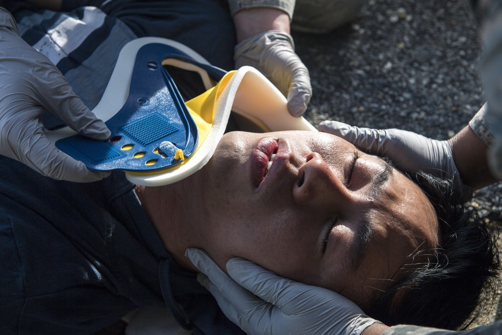
    • Endotracheal (orotracheal) intubation Intubation Peritonsillar Abscess:
      • Insertion of a tube into the trachea Trachea The trachea is a tubular structure that forms part of the lower respiratory tract. The trachea is continuous superiorly with the larynx and inferiorly becomes the bronchial tree within the lungs. The trachea consists of a support frame of semicircular, or C-shaped, rings made out of hyaline cartilage and reinforced by collagenous connective tissue. Trachea: Anatomy through the mouth (less commonly through the nose Nose The nose is the human body’s primary organ of smell and functions as part of the upper respiratory system. The nose may be best known for inhaling oxygen and exhaling carbon dioxide, but it also contributes to other important functions, such as tasting. The anatomy of the nose can be divided into the external nose and the nasal cavity. Nose Anatomy (External & Internal))
      • 1st-line procedure
    • Cricothyrotomy Cricothyrotomy ABCDE Assessment:
      • Incision of the membrane between the thyroid Thyroid The thyroid gland is one of the largest endocrine glands in the human body. The thyroid gland is a highly vascular, brownish-red gland located in the visceral compartment of the anterior region of the neck. Thyroid Gland: Anatomy cartilage Cartilage Cartilage is a type of connective tissue derived from embryonic mesenchyme that is responsible for structural support, resilience, and the smoothness of physical actions. Perichondrium (connective tissue membrane surrounding cartilage) compensates for the absence of vasculature in cartilage by providing nutrition and support. Cartilage: Histology and cricoid cartilage Cartilage Cartilage is a type of connective tissue derived from embryonic mesenchyme that is responsible for structural support, resilience, and the smoothness of physical actions. Perichondrium (connective tissue membrane surrounding cartilage) compensates for the absence of vasculature in cartilage by providing nutrition and support. Cartilage: Histology
      • In severe traumatic injuries, if intubation Intubation Peritonsillar Abscess fails or if the airway Airway ABCDE Assessment is severely swollen ( anaphylaxis Anaphylaxis An acute hypersensitivity reaction due to exposure to a previously encountered antigen. The reaction may include rapidly progressing urticaria, respiratory distress, vascular collapse, systemic shock, and death. Type I Hypersensitivity Reaction)
      • Easier to perform than tracheostomy Tracheostomy Surgical formation of an opening into the trachea through the neck, or the opening so created. Laryngomalacia and Tracheomalacia
      • Temporary procedure
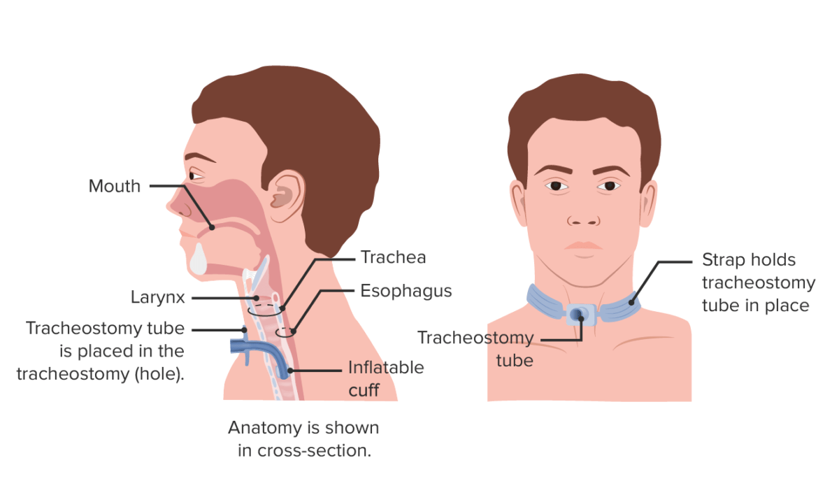
    • Tracheotomy Tracheotomy Surgical incision of the trachea. Basic Procedures:
      • Incision to the trachea Trachea The trachea is a tubular structure that forms part of the lower respiratory tract. The trachea is continuous superiorly with the larynx and inferiorly becomes the bronchial tree within the lungs. The trachea consists of a support frame of semicircular, or C-shaped, rings made out of hyaline cartilage and reinforced by collagenous connective tissue. Trachea: Anatomy and insertion of a tracheal tube
      • Preferred in pediatric individuals (< 8 years of age) as the cricoid is smaller
Neck collar attachment for multitrauma patient

Application of the backboard and C-collar by emergency medical services (EMS) in the field:
If the affected individual arrives in a personal vehicle, the C-collar is applied during the airway (A) portion of the primary survey.
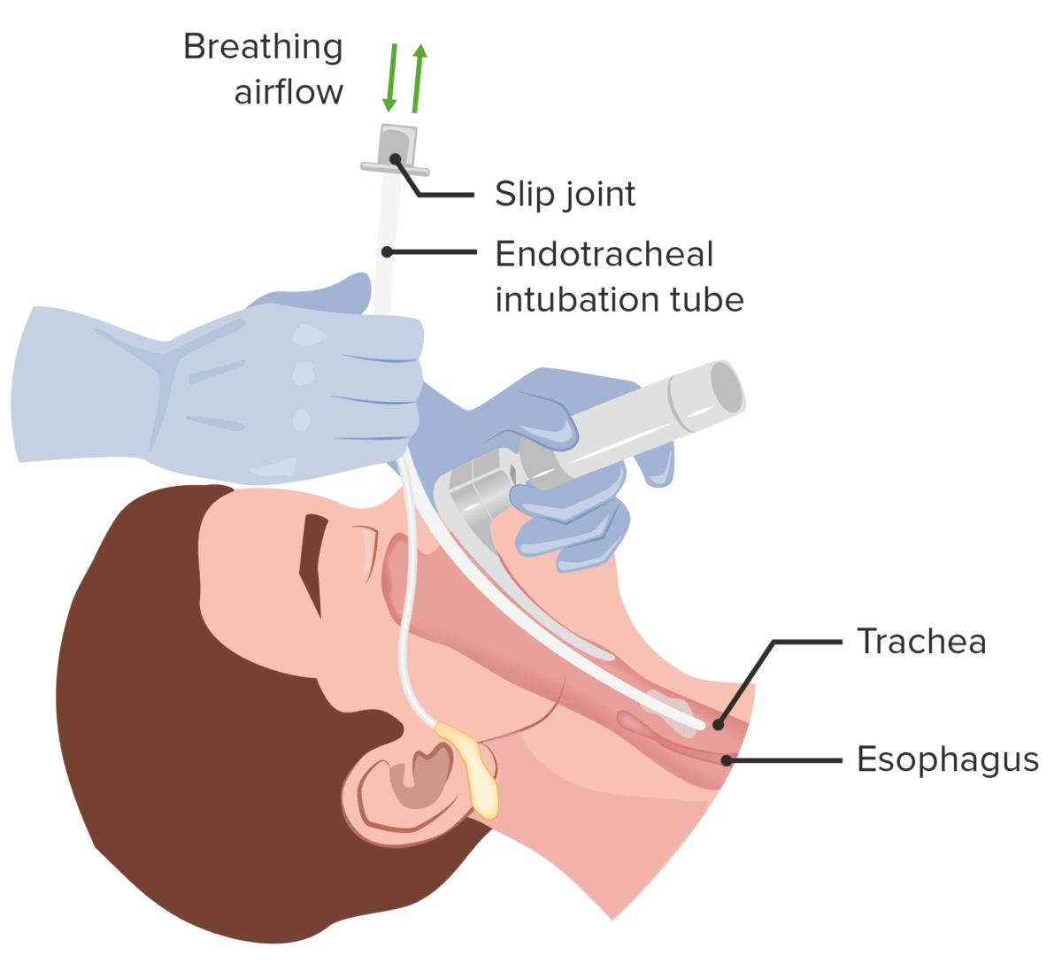
Image: “Neck collar attachment for multitrauma patient” by Senior Airman Janiqua P. Robinson. License: CC0 1.0

Breathing

  • Purpose:
    • To detect signs of respiratory distress
    • To maintain sufficient oxygenation and ventilation Ventilation The total volume of gas inspired or expired per unit of time, usually measured in liters per minute. Ventilation: Mechanics of Breathing
    • If breathing problems occur at this step, it may be necessary to perform procedures ( thoracostomy Thoracostomy Surgical procedure involving the creation of an opening (stoma) into the chest cavity for drainage; used in the treatment of pleural effusion; pneumothorax; hemothorax; and empyema. Hemothorax) to correct the problem.
  • Breathing assessment:
    • Listen to breath sounds.
    • Determine the respiratory rate Respiratory rate The number of times an organism breathes with the lungs (respiration) per unit time, usually per minute. Pulmonary Examination: 12–20 breaths/minutes is normal.
    • If the respiratory rate Respiratory rate The number of times an organism breathes with the lungs (respiration) per unit time, usually per minute. Pulmonary Examination is ↓ or ↑, consider airway Airway ABCDE Assessment assistance
    • 40% of individuals who have had a cardiac arrest Cardiac arrest Cardiac arrest is the sudden, complete cessation of cardiac output with hemodynamic collapse. Patients present as pulseless, unresponsive, and apneic. Rhythms associated with cardiac arrest are ventricular fibrillation/tachycardia, asystole, or pulseless electrical activity. Cardiac Arrest may exhibit agonal breathing Agonal Breathing ABCDE Assessment (series of noisy gasps).
  • Signs of respiratory distress:
    • Inspection Inspection Dermatologic Examination:
    • Percussion Percussion Act of striking a part with short, sharp blows as an aid in diagnosing the condition beneath the sound obtained. Pulmonary Examination:
      • Hyperresonance ( pneumothorax Pneumothorax A pneumothorax is a life-threatening condition in which air collects in the pleural space, causing partial or full collapse of the lung. A pneumothorax can be traumatic or spontaneous. Patients present with a sudden onset of sharp chest pain, dyspnea, and diminished breath sounds on exam. Pneumothorax)
      • Dullness ( hemothorax Hemothorax A hemothorax is a collection of blood in the pleural cavity. Hemothorax most commonly occurs due to damage to the intercostal arteries or from a lung laceration following chest trauma. Hemothorax can also occur as a complication of disease, or hemothorax may be spontaneous or iatrogenic. Hemothorax)
    • Palpation Palpation Application of fingers with light pressure to the surface of the body to determine consistency of parts beneath in physical diagnosis; includes palpation for determining the outlines of organs. Dermatologic Examination:
    • Auscultation: ↓ air entry during auscultation
    • Oxygen saturation Oxygen Saturation Basic Procedures: < 88%
    • ↓ End-tidal CO2 (capnography)
  • Breathing management:
    • Depends on the cause
    • Return to A ( airway Airway ABCDE Assessment) to establish a definitive airway Airway ABCDE Assessment if there is respiratory distress.
    • Tube thoracostomy Tube Thoracostomy Surgical procedure involving the creation of an opening (stoma) into the chest cavity for drainage; used in the treatment of pleural effusion; pneumothorax; hemothorax; and empyema. Thoracic Surgery (small incision on the chest wall Chest wall The chest wall consists of skin, fat, muscles, bones, and cartilage. The bony structure of the chest wall is composed of the ribs, sternum, and thoracic vertebrae. The chest wall serves as armor for the vital intrathoracic organs and provides the stability necessary for the movement of the shoulders and arms. Chest Wall: Anatomy and insertion of a chest tube) is needed in:
      • Tension pneumothorax Tension Pneumothorax Pneumothorax, open pneumothorax Open Pneumothorax Pneumothorax
      • Flail chest Flail chest Flail chest is a life-threatening traumatic injury that occurs when 3 or more contiguous ribs are fractured in 2 or more different locations. Patients present with chest pain, tachypnea, hypoxia, and paradoxical chest wall movement. Flail Chest
      • Massive hemothorax Hemothorax A hemothorax is a collection of blood in the pleural cavity. Hemothorax most commonly occurs due to damage to the intercostal arteries or from a lung laceration following chest trauma. Hemothorax can also occur as a complication of disease, or hemothorax may be spontaneous or iatrogenic. Hemothorax
    • Pericardiocentesis Pericardiocentesis Puncture and aspiration of fluid from the pericardium. Cardiac Surgery (insertion of a needle and small catheter into the pericardial sac to drain excess fluid) is necessary for cardiac tamponade Tamponade Pericardial effusion, usually of rapid onset, exceeding ventricular filling pressures and causing collapse of the heart with a markedly reduced cardiac output. Pericarditis.

Circulation Circulation The movement of the blood as it is pumped through the cardiovascular system. ABCDE Assessment

  • Purpose:
    • To determine the effectiveness of cardiac output Cardiac output The volume of blood passing through the heart per unit of time. It is usually expressed as liters (volume) per minute so as not to be confused with stroke volume (volume per beat). Cardiac Mechanics
    • To secure adequate tissue perfusion
    • To treat external bleeding
  • Causes of poor circulation Circulation The movement of the blood as it is pumped through the cardiovascular system. ABCDE Assessment:
    • Shock Shock Shock is a life-threatening condition associated with impaired circulation that results in tissue hypoxia. The different types of shock are based on the underlying cause: distributive (↑ cardiac output (CO), ↓ systemic vascular resistance (SVR)), cardiogenic (↓ CO, ↑ SVR), hypovolemic (↓ CO, ↑ SVR), obstructive (↓ CO), and mixed. Types of Shock (including hypovolemia Hypovolemia Sepsis in Children, septic, or anaphylactic shock Shock Shock is a life-threatening condition associated with impaired circulation that results in tissue hypoxia. The different types of shock are based on the underlying cause: distributive (↑ cardiac output (CO), ↓ systemic vascular resistance (SVR)), cardiogenic (↓ CO, ↑ SVR), hypovolemic (↓ CO, ↑ SVR), obstructive (↓ CO), and mixed. Types of Shock)
    • Traumatic injury to arteries Arteries Arteries are tubular collections of cells that transport oxygenated blood and nutrients from the heart to the tissues of the body. The blood passes through the arteries in order of decreasing luminal diameter, starting in the largest artery (the aorta) and ending in the small arterioles. Arteries are classified into 3 types: large elastic arteries, medium muscular arteries, and small arteries and arterioles. Arteries: Histology ( compartment syndrome Compartment Syndrome Compartment syndrome is a surgical emergency usually occurring secondary to trauma. The condition is marked by increased pressure within a compartment that compromises the circulation and function of the tissues within that space. Compartment Syndrome, arterial lacerations, crush injury Crush injury Excessive compression of parts of the body that causes muscle swelling, fracture, and/or neurological disturbances in the affected areas. Crush injury with systemic manifestations is referred to as crush syndrome. Crush Syndrome)
    • Cardiac arrhythmias
    • Heart failure Heart Failure A heterogeneous condition in which the heart is unable to pump out sufficient blood to meet the metabolic need of the body. Heart failure can be caused by structural defects, functional abnormalities (ventricular dysfunction), or a sudden overload beyond its capacity. Chronic heart failure is more common than acute heart failure which results from sudden insult to cardiac function, such as myocardial infarction. Total Anomalous Pulmonary Venous Return (TAPVR)
    • Pulmonary embolism Pulmonary Embolism Pulmonary embolism (PE) is a potentially fatal condition that occurs as a result of intraluminal obstruction of the main pulmonary artery or its branches. The causative factors include thrombi, air, amniotic fluid, and fat. In PE, gas exchange is impaired due to the decreased return of deoxygenated blood to the lungs. Pulmonary Embolism
  • Circulation Circulation The movement of the blood as it is pumped through the cardiovascular system. ABCDE Assessment assessment:
    • Nonbreathing (apneic) individuals: CPR CPR The artificial substitution of heart and lung action as indicated for heart arrest resulting from electric shock, drowning, respiratory arrest, or other causes. The two major components of cardiopulmonary resuscitation are artificial ventilation and closed-chest cardiac massage. Cardiac Arrest
    • Individuals who are breathing:
      • Blood pressure assessment: indication of the effectiveness of the cardiac output Cardiac output The volume of blood passing through the heart per unit of time. It is usually expressed as liters (volume) per minute so as not to be confused with stroke volume (volume per beat). Cardiac Mechanics, considered ↓ if systolic blood pressure < 90 mm MM Multiple myeloma (MM) is a malignant condition of plasma cells (activated B lymphocytes) primarily seen in the elderly. Monoclonal proliferation of plasma cells results in cytokine-driven osteoclastic activity and excessive secretion of IgG antibodies. Multiple Myeloma Hg
      • Check skin Skin The skin, also referred to as the integumentary system, is the largest organ of the body. The skin is primarily composed of the epidermis (outer layer) and dermis (deep layer). The epidermis is primarily composed of keratinocytes that undergo rapid turnover, while the dermis contains dense layers of connective tissue. Skin: Structure and Functions (cold, clammy skin Skin The skin, also referred to as the integumentary system, is the largest organ of the body. The skin is primarily composed of the epidermis (outer layer) and dermis (deep layer). The epidermis is primarily composed of keratinocytes that undergo rapid turnover, while the dermis contains dense layers of connective tissue. Skin: Structure and Functions is an indication of hypovolemia Hypovolemia Sepsis in Children).
      • Check capillary refill time Capillary Refill Time Cardiovascular Examination (abnormal if > 2 seconds).
      • Check urinary output (< 0.5 mL/kg/h is considered low).
      • Check HR (↑ HR (> 100/minute) could mean shock Shock Shock is a life-threatening condition associated with impaired circulation that results in tissue hypoxia. The different types of shock are based on the underlying cause: distributive (↑ cardiac output (CO), ↓ systemic vascular resistance (SVR)), cardiogenic (↓ CO, ↑ SVR), hypovolemic (↓ CO, ↑ SVR), obstructive (↓ CO), and mixed. Types of Shock).
  • Circulation Circulation The movement of the blood as it is pumped through the cardiovascular system. ABCDE Assessment management:
    • Direct pressure to control any external hemorrhage
    • 2 large-bore IV lines to be placed in all affected individuals. If unfeasible, alternatives include:
      • Central access into femoral, jugular, subclavian veins Veins Veins are tubular collections of cells, which transport deoxygenated blood and waste from the capillary beds back to the heart. Veins are classified into 3 types: small veins/venules, medium veins, and large veins. Each type contains 3 primary layers: tunica intima, tunica media, and tunica adventitia. Veins: Histology
      • Intraosseous access
      • Percutaneous and cutdown catheters in the saphenous vein of the lower limb (less common)
    • Consider mass Mass Three-dimensional lesion that occupies a space within the breast Imaging of the Breast transfusion blood protocol.
    • Signs of shock Shock Shock is a life-threatening condition associated with impaired circulation that results in tissue hypoxia. The different types of shock are based on the underlying cause: distributive (↑ cardiac output (CO), ↓ systemic vascular resistance (SVR)), cardiogenic (↓ CO, ↑ SVR), hypovolemic (↓ CO, ↑ SVR), obstructive (↓ CO), and mixed. Types of Shock (pale, cool, and moist skin Skin The skin, also referred to as the integumentary system, is the largest organ of the body. The skin is primarily composed of the epidermis (outer layer) and dermis (deep layer). The epidermis is primarily composed of keratinocytes that undergo rapid turnover, while the dermis contains dense layers of connective tissue. Skin: Structure and Functions, hypotension Hypotension Hypotension is defined as low blood pressure, specifically < 90/60 mm Hg, and is most commonly a physiologic response. Hypotension may be mild, serious, or life threatening, depending on the cause. Hypotension) may not appear initially until the affected individual loses up to 30% of blood.
    • Check the need for reversal of anticoagulation Anticoagulation Pulmonary Hypertension Drugs.

Disability Disability Determination of the degree of a physical, mental, or emotional handicap. The diagnosis is applied to legal qualification for benefits and income under disability insurance and to eligibility for social security and workman’s compensation benefits. ABCDE Assessment

Disability Disability Determination of the degree of a physical, mental, or emotional handicap. The diagnosis is applied to legal qualification for benefits and income under disability insurance and to eligibility for social security and workman’s compensation benefits. ABCDE Assessment assessment:

  • Examine pupils: Pupil Pupil The pupil is the space within the eye that permits light to project onto the retina. Anatomically located in front of the lens, the pupil’s size is controlled by the surrounding iris. The pupil provides insight into the function of the central and autonomic nervous systems. Pupil: Physiology and Abnormalities dilation suggests ipsilateral brain Brain The part of central nervous system that is contained within the skull (cranium). Arising from the neural tube, the embryonic brain is comprised of three major parts including prosencephalon (the forebrain); mesencephalon (the midbrain); and rhombencephalon (the hindbrain). The developed brain consists of cerebrum; cerebellum; and other structures in the brain stem. Nervous System: Anatomy, Structure, and Classification mass Mass Three-dimensional lesion that occupies a space within the breast Imaging of the Breast or blood collection causing compression Compression Blunt Chest Trauma of the 3rd cranial nerve.
  • Motor Motor Neurons which send impulses peripherally to activate muscles or secretory cells. Nervous System: Histology and sensory Sensory Neurons which conduct nerve impulses to the central nervous system. Nervous System: Histology examination
  • Assess the level of consciousness and mental status using GCS GCS A scale that assesses the response to stimuli in patients with craniocerebral injuries. The parameters are eye opening, motor response, and verbal response. Coma:
    • The affected individual receives a score for the best response in each area.
    • Scores in each area are combined to achieve a total score of 3–15.
    • ↑ Number → the better the prognosis Prognosis A prediction of the probable outcome of a disease based on a individual’s condition and the usual course of the disease as seen in similar situations. Non-Hodgkin Lymphomas
    • A score ≤ 9 indicates coma Coma Coma is defined as a deep state of unarousable unresponsiveness, characterized by a score of 3 points on the GCS. A comatose state can be caused by a multitude of conditions, making the precise epidemiology and prognosis of coma difficult to determine. Coma and the need for endotracheal intubation Intubation Peritonsillar Abscess.
  • Life-threatening neurological injuries include:
    • Penetrating cranial injury
    • Intracranial hemorrhage Intracranial hemorrhage Subarachnoid hemorrhage (SAH) is a type of cerebrovascular accident (stroke) resulting from intracranial hemorrhage into the subarachnoid space between the arachnoid and the pia mater layers of the meninges surrounding the brain. Most sahs originate from a saccular aneurysm in the circle of willis but may also occur as a result of trauma, uncontrolled hypertension, vasculitis, anticoagulant use, or stimulant use. Subarachnoid Hemorrhage
    • Diffuse axonal injury Diffuse axonal injury A relatively common sequela of blunt head injury, characterized by a global disruption of axons throughout the brain. Associated clinical features may include neurobehavioral manifestations; persistent vegetative state; dementia; and other disorders. Head Trauma
    • High spinal cord Spinal cord The spinal cord is the major conduction pathway connecting the brain to the body; it is part of the CNS. In cross section, the spinal cord is divided into an H-shaped area of gray matter (consisting of synapsing neuronal cell bodies) and a surrounding area of white matter (consisting of ascending and descending tracts of myelinated axons). Spinal Cord: Anatomy injury
Table: Glasgow coma Coma Coma is defined as a deep state of unarousable unresponsiveness, characterized by a score of 3 points on the GCS. A comatose state can be caused by a multitude of conditions, making the precise epidemiology and prognosis of coma difficult to determine. Coma scale Scale Dermatologic Examination
Feature Response Score
Eye opening Open spontaneously 4
Open to verbal commands 3
Open to pain Pain An unpleasant sensation induced by noxious stimuli which are detected by nerve endings of nociceptive neurons. Pain: Types and Pathways 2
No eye opening 1
Verbal response Oriented and appropriate 5
Disoriented but conversant 4
Nonsensical words 3
Moaning 2
Silent 1
Motor Motor Neurons which send impulses peripherally to activate muscles or secretory cells. Nervous System: Histology response Follows commands 6
Localizes pain Pain An unpleasant sensation induced by noxious stimuli which are detected by nerve endings of nociceptive neurons. Pain: Types and Pathways 5
Withdraws from pain Pain An unpleasant sensation induced by noxious stimuli which are detected by nerve endings of nociceptive neurons. Pain: Types and Pathways 4
Flexor posturing 3
Extensor posturing Extensor posturing A condition characterized by abnormal posturing of the limbs that is associated with injury to the brainstem. This may occur as a clinical manifestation or induced experimentally in animals. The extensor reflexes are exaggerated leading to rigid extension of the limbs accompanied by hyperreflexia and opisthotonus. This condition is usually caused by lesions which occur in the region of the brainstem that lies between the red nuclei and the vestibular nuclei. In contrast, decorticate rigidity is characterized by flexion of the elbows and wrists with extension of the legs and feet. The causative lesion for this condition is located above the red nuclei and usually consists of diffuse cerebral damage. Increased Intracranial Pressure (ICP) 2
Flaccid 1

Exposure

The goal of this step is to evaluate and manage negative environmental effects:

  • Completely undress the individual and perform a full physical examination.
  • Keep the individual warm, as hypothermia Hypothermia Hypothermia can be defined as a drop in the core body temperature below 35°C (95°F) and is classified into mild, moderate, severe, and profound forms based on the degree of temperature decrease. Hypothermia can trigger Trigger The type of signal that initiates the inspiratory phase by the ventilator Invasive Mechanical Ventilation coagulopathies Coagulopathies Hemothorax.

Secondary Survey

Technique

The goal of the secondary survey Secondary Survey ABCDE Assessment is to rapidly and thoroughly examine the affected individual from head to toe and identify all potentially significant injuries.

  • Performed after the primary survey Primary Survey Thoracic Trauma in Children and initial stabilization are complete
  • Performed after all life-threatening injuries have been identified and stabilized
  • Gather the history including injury type (blunt or penetrating) and past medical data.
  • Examine the affected individual from head to toe, including all orifices (ears, nose Nose The nose is the human body’s primary organ of smell and functions as part of the upper respiratory system. The nose may be best known for inhaling oxygen and exhaling carbon dioxide, but it also contributes to other important functions, such as tasting. The anatomy of the nose can be divided into the external nose and the nasal cavity. Nose Anatomy (External & Internal), mouth, vagina Vagina The vagina is the female genital canal, extending from the vulva externally to the cervix uteri internally. The structures have sexual, reproductive, and urinary functions and a rich blood supply, mainly arising from the internal iliac artery. Vagina, Vulva, and Pelvic Floor: Anatomy, rectum Rectum The rectum and anal canal are the most terminal parts of the lower GI tract/large intestine that form a functional unit and control defecation. Fecal continence is maintained by several important anatomic structures including rectal folds, anal valves, the sling-like puborectalis muscle, and internal and external anal sphincters. Rectum and Anal Canal: Anatomy):
    • Vital signs
    • Head and neck Neck The part of a human or animal body connecting the head to the rest of the body. Peritonsillar Abscess:
      • Look for scalp hematoma Hematoma A collection of blood outside the blood vessels. Hematoma can be localized in an organ, space, or tissue. Intussusception, skull Skull The skull (cranium) is the skeletal structure of the head supporting the face and forming a protective cavity for the brain. The skull consists of 22 bones divided into the viscerocranium (facial skeleton) and the neurocranium. Skull: Anatomy depression, laceration Laceration Torn, ragged, mangled wounds. Blunt Chest Trauma.
      • Palpate facial bones.
      • Check nose Nose The nose is the human body’s primary organ of smell and functions as part of the upper respiratory system. The nose may be best known for inhaling oxygen and exhaling carbon dioxide, but it also contributes to other important functions, such as tasting. The anatomy of the nose can be divided into the external nose and the nasal cavity. Nose Anatomy (External & Internal) for septal hematoma Hematoma A collection of blood outside the blood vessels. Hematoma can be localized in an organ, space, or tissue. Intussusception.
      • Check ears for hemotympanum or retroauricular ecchymosis Ecchymosis Extravasation of blood into the skin, resulting in a nonelevated, rounded or irregular, blue or purplish patch, larger than a petechia. Orbital Fractures (Battle’s sign).
      • Ocular examination
      • Immobilize the individual and palpate the neck Neck The part of a human or animal body connecting the head to the rest of the body. Peritonsillar Abscess.
    • Chest:
    • Abdomen:
      • Check for distension and bruising.
      • Auscultate for bowel sounds.
    • Extremities:
      • Check for tenderness and range of motion Range of motion The distance and direction to which a bone joint can be extended. Range of motion is a function of the condition of the joints, muscles, and connective tissues involved. Joint flexibility can be improved through appropriate muscle strength exercises. Examination of the Upper Limbs.
      • Check for pelvic instability.
    • Skin Skin The skin, also referred to as the integumentary system, is the largest organ of the body. The skin is primarily composed of the epidermis (outer layer) and dermis (deep layer). The epidermis is primarily composed of keratinocytes that undergo rapid turnover, while the dermis contains dense layers of connective tissue. Skin: Structure and Functions:
      • Check from head to toe (especially the back, perineum Perineum The body region lying between the genital area and the anus on the surface of the trunk, and to the shallow compartment lying deep to this area that is inferior to the pelvic diaphragm. The surface area is between the vulva and the anus in the female, and between the scrotum and the anus in the male. Vagina, Vulva, and Pelvic Floor: Anatomy, and axillary and gluteal folds).
      • Look for lacerations, hematomas, and abrasions Abrasions Corneal Abrasions, Erosion, and Ulcers.
    • Neurologic exam: Repeat GCS GCS A scale that assesses the response to stimuli in patients with craniocerebral injuries. The parameters are eye opening, motor response, and verbal response. Coma.
  • Order imaging and other tests as needed:
    • Plain radiography
    • Focused assessment with sonography Sonography The visualization of deep structures of the body by recording the reflections or echoes of ultrasonic pulses directed into the tissues. Use of ultrasound for imaging or diagnostic purposes employs frequencies ranging from 1. 6 to 10 megahertz. Diagnostic Procedures in Gynecology in trauma (FAST) is an essential component of the primary circulation Circulation The movement of the blood as it is pumped through the cardiovascular system. ABCDE Assessment survey and is primarily used to detect:
    • Emergency CT scan
  • Go back frequently to the primary survey Primary Survey Thoracic Trauma in Children for reassessment!

Mnemonic

For focused history, recall the mnemonic “AMPLE”:

  • Allergy
  • Medications
  • Previous medical history/ illness
  • L ast AST Enzymes of the transferase class that catalyze the conversion of l-aspartate and 2-ketoglutarate to oxaloacetate and l-glutamate. Liver Function Tests meal
  • Events related to the injury

Avoiding missed injuries

Pertinent history, thorough physical examination, and appropriate diagnostic studies help avoid missed injuries such as:

  • Blunt abdominal trauma:
  • Penetrating abdominal trauma: perineal injuries (includes rectal and ureteral damage)
  • Thoracic trauma:
    • Pericardial tamponade Pericardial tamponade Compression of the heart by accumulated fluid (pericardial effusion) or blood (hemopericardium) in the pericardium surrounding the heart. The affected cardiac functions and cardiac output can range from minimal to total hemodynamic collapse. Penetrating Chest Injury
    • Esophageal perforation Perforation A pathological hole in an organ, blood vessel or other soft part of the body, occurring in the absence of external force. Esophagitis or rupture
    • Aortic injuries
  • Extremity trauma
    • Fractures
    • Vascular injuries
    • Compartment syndrome Compartment Syndrome Compartment syndrome is a surgical emergency usually occurring secondary to trauma. The condition is marked by increased pressure within a compartment that compromises the circulation and function of the tissues within that space. Compartment Syndrome

Mechanisms and Associated Injuries

The tables below summarize the different mechanisms of trauma and their associated specific injuries.

Motor Motor Neurons which send impulses peripherally to activate muscles or secretory cells. Nervous System: Histology vehicle collisions

Table: Motor Motor Neurons which send impulses peripherally to activate muscles or secretory cells. Nervous System: Histology vehicle collisions and associated injuries
Mechanism of injury Potential associated injuries
Head-on collision
  • Facial injuries
  • Lower extremity injuries
  • Aortic injuries
Rear-end collision
  • Hyperextension injuries of the cervical spine Spine The human spine, or vertebral column, is the most important anatomical and functional axis of the human body. It consists of 7 cervical vertebrae, 12 thoracic vertebrae, and 5 lumbar vertebrae and is limited cranially by the skull and caudally by the sacrum. Vertebral Column: Anatomy
  • Cervical spine Spine The human spine, or vertebral column, is the most important anatomical and functional axis of the human body. It consists of 7 cervical vertebrae, 12 thoracic vertebrae, and 5 lumbar vertebrae and is limited cranially by the skull and caudally by the sacrum. Vertebral Column: Anatomy fractures
  • Central cord syndrome Central Cord Syndrome Central cord syndrome (CCS) is a neurological syndrome caused by an injury to the center of the spinal cord, affecting the spinothalamic tracts ((STTs) sensory) and medial aspect of the corticospinal tracts ((CSTs) motor), most often due to trauma in patients with cervical spondylosis. Central Cord Syndrome
Ejected from vehicle Spinal injuries
Windshield damage
  • Closed head injuries (coup and contrecoup injuries)
  • Facial fractures
  • Skull Skull The skull (cranium) is the skeletal structure of the head supporting the face and forming a protective cavity for the brain. The skull consists of 22 bones divided into the viscerocranium (facial skeleton) and the neurocranium. Skull: Anatomy fractures
  • Cervical spine Spine The human spine, or vertebral column, is the most important anatomical and functional axis of the human body. It consists of 7 cervical vertebrae, 12 thoracic vertebrae, and 5 lumbar vertebrae and is limited cranially by the skull and caudally by the sacrum. Vertebral Column: Anatomy fractures
Steering wheel damage Thoracic injuries
Dashboard involvement/damage
  • Pelvic and acetabular injuries
  • Dislocated hip

Pedestrian versus automobile injuries

Table: Pedestrian versus automobile and associated injuries
Mechanism of injury Potential associated injuries
Low speed (braking automobile)
  • Tibia Tibia The second longest bone of the skeleton. It is located on the medial side of the lower leg, articulating with the fibula laterally, the talus distally, and the femur proximally. Knee Joint: Anatomy and fibula Fibula The bone of the lower leg lateral to and smaller than the tibia. In proportion to its length, it is the most slender of the long bones. Leg: Anatomy fractures
  • Knee injuries
High speed Waddle’s triad (in pediatric pedestrians):
  • Tibia Tibia The second longest bone of the skeleton. It is located on the medial side of the lower leg, articulating with the fibula laterally, the talus distally, and the femur proximally. Knee Joint: Anatomy/ fibula Fibula The bone of the lower leg lateral to and smaller than the tibia. In proportion to its length, it is the most slender of the long bones. Leg: Anatomy or femur fractures
  • Ipsilateral thoracoabdominal injuries
  • Contralateral craniofacial injuries

Bicycle accidents

Table: Bicycle accidents and associated injuries
Mechanism of injury Potential associated injuries
Automobile related
  • Closed head injuries
  • “Handlebar” injuries ( spleen Spleen The spleen is the largest lymphoid organ in the body, located in the LUQ of the abdomen, superior to the left kidney and posterior to the stomach at the level of the 9th-11th ribs just below the diaphragm. The spleen is highly vascular and acts as an important blood filter, cleansing the blood of pathogens and damaged erythrocytes. Spleen: Anatomy/ liver Liver The liver is the largest gland in the human body. The liver is found in the superior right quadrant of the abdomen and weighs approximately 1.5 kilograms. Its main functions are detoxification, metabolism, nutrient storage (e.g., iron and vitamins), synthesis of coagulation factors, formation of bile, filtration, and storage of blood. Liver: Anatomy lacerations, additional intraabdominal injuries)
Nonautomobile-related
  • Extremity injuries
  • “Handlebar” injuries

Falls

Table: Falls and associated injuries
Mechanism of injury Potential associated injuries
Vertical impact
  • Calcaneal and lower extremity fractures
  • Pelvic fractures Pelvic Fractures Pelvic fractures are a disruption in the cortex of a pelvic bone involving iliac wing fractures, acetabular fractures, or those causing loss of integrity of the pelvic ring (the sacrum and the 2 innominate bones). Patients often present with a history of trauma or a fall, limb length discrepancy, intense pain on palpation, and mechanical instability. Pelvic Fractures
  • Closed head injuries
  • Lumbar vertebral fractures
  • Renal vascular injuries
Horizontal impact
  • Craniofacial fractures
  • Hand Hand The hand constitutes the distal part of the upper limb and provides the fine, precise movements needed in activities of daily living. It consists of 5 metacarpal bones and 14 phalanges, as well as numerous muscles innervated by the median and ulnar nerves. Hand: Anatomy and wrist fractures
  • Abdominal and thoracic visceral injuries
  • Aortic injuries

Clinical Relevance

  • ABC assessment: the mainstay management approach used for critically ill individuals. Assessment of ABCs is an essential 1st step to perform in many situations including the management of unresponsive individuals, those who have undergone cardiac arrest Cardiac arrest Cardiac arrest is the sudden, complete cessation of cardiac output with hemodynamic collapse. Patients present as pulseless, unresponsive, and apneic. Rhythms associated with cardiac arrest are ventricular fibrillation/tachycardia, asystole, or pulseless electrical activity. Cardiac Arrest, and also those who have undergone trauma and are critical. In such cases, ABC is included in the primary survey Primary Survey Thoracic Trauma in Children, initial evaluation, and management of injuries.
  • Epidural hematoma Hematoma A collection of blood outside the blood vessels. Hematoma can be localized in an organ, space, or tissue. Intussusception: rapidly expanding blood collection between the bone Bone Bone is a compact type of hardened connective tissue composed of bone cells, membranes, an extracellular mineralized matrix, and central bone marrow. The 2 primary types of bone are compact and spongy. Bones: Structure and Types and dura mater Dura mater The outermost of the three meninges, a fibrous membrane of connective tissue that covers the brain and the spinal cord. Meninges: Anatomy due to rupture of middle meningeal arteries Arteries Arteries are tubular collections of cells that transport oxygenated blood and nutrients from the heart to the tissues of the body. The blood passes through the arteries in order of decreasing luminal diameter, starting in the largest artery (the aorta) and ending in the small arterioles. Arteries are classified into 3 types: large elastic arteries, medium muscular arteries, and small arteries and arterioles. Arteries: Histology. Epidural hematoma Hematoma A collection of blood outside the blood vessels. Hematoma can be localized in an organ, space, or tissue. Intussusception is marked by a lucid interval Lucid Interval Epidural Hemorrhage, cranial nerve palsies Cranial Nerve Palsies Cranial nerve palsy is a congenital or acquired dysfunction of 1 or more cranial nerves that will, in turn, lead to focal neurologic abnormalities in movement or autonomic dysfunction of its territory. Head/neck trauma, mass effect, infectious processes, and ischemia/infarction are among the many etiologies for these dysfunctions. Diagnosis is initially clinical and supported by diagnostic aids. Management includes both symptomatic measures and interventions aimed at correcting the underlying cause. Cranial Nerve Palsies, and altered mental status Altered Mental Status Sepsis in Children. A CT scan shows biconvex ( lens Lens A transparent, biconvex structure of the eye, enclosed in a capsule and situated behind the iris and in front of the vitreous humor (vitreous body). It is slightly overlapped at its margin by the ciliary processes. Adaptation by the ciliary body is crucial for ocular accommodation. Eye: Anatomy shaped), hyperdense blood collection that does not cross the suture lines. Epidural hematoma Hematoma A collection of blood outside the blood vessels. Hematoma can be localized in an organ, space, or tissue. Intussusception is a life-threatening condition that requires surgical decompression.
  • Subdural hematoma Hematoma A collection of blood outside the blood vessels. Hematoma can be localized in an organ, space, or tissue. Intussusception: collection of blood between the dura mater Dura mater The outermost of the three meninges, a fibrous membrane of connective tissue that covers the brain and the spinal cord. Meninges: Anatomy and arachnoid membrane due to the rupture of bridging veins Bridging Veins Subdural Hemorrhage. A CT scan shows crescent-shaped blood collection that crosses the suture lines. A subdural hematoma Hematoma A collection of blood outside the blood vessels. Hematoma can be localized in an organ, space, or tissue. Intussusception can be both acute and chronic and may require prompt care, usually surgery.
  • Subarachnoid hemorrhage Subarachnoid Hemorrhage Subarachnoid hemorrhage (SAH) is a type of cerebrovascular accident (stroke) resulting from intracranial hemorrhage into the subarachnoid space between the arachnoid and the pia mater layers of the meninges surrounding the brain. Most SAHs originate from a saccular aneurysm in the circle of Willis but may also occur as a result of trauma, uncontrolled hypertension, vasculitis, anticoagulant use, or stimulant use. Subarachnoid Hemorrhage ( SAH SAH Subarachnoid hemorrhage (SAH) is a type of cerebrovascular accident (stroke) resulting from intracranial hemorrhage into the subarachnoid space between the arachnoid and the pia mater layers of the meninges surrounding the brain. Most SAHs originate from a saccular aneurysm in the circle of Willis but may also occur as a result of trauma, uncontrolled hypertension, vasculitis, anticoagulant use, or stimulant use. Subarachnoid Hemorrhage): a type of stroke caused by bleeding into the cranial and/or spinal subarachnoid space Subarachnoid space The space between the arachnoid membrane and pia mater, filled with cerebrospinal fluid. It contains large blood vessels that supply the brain and spinal cord. Subarachnoid Hemorrhage surrounding the brain Brain The part of central nervous system that is contained within the skull (cranium). Arising from the neural tube, the embryonic brain is comprised of three major parts including prosencephalon (the forebrain); mesencephalon (the midbrain); and rhombencephalon (the hindbrain). The developed brain consists of cerebrum; cerebellum; and other structures in the brain stem. Nervous System: Anatomy, Structure, and Classification. Subarachnoid hemorrhage Subarachnoid Hemorrhage Subarachnoid hemorrhage (SAH) is a type of cerebrovascular accident (stroke) resulting from intracranial hemorrhage into the subarachnoid space between the arachnoid and the pia mater layers of the meninges surrounding the brain. Most SAHs originate from a saccular aneurysm in the circle of Willis but may also occur as a result of trauma, uncontrolled hypertension, vasculitis, anticoagulant use, or stimulant use. Subarachnoid Hemorrhage may occur spontaneously or after trauma. A sudden, violent “ thunderclap headache Thunderclap Headache Subarachnoid Hemorrhage” is the main symptom of SAH SAH Subarachnoid hemorrhage (SAH) is a type of cerebrovascular accident (stroke) resulting from intracranial hemorrhage into the subarachnoid space between the arachnoid and the pia mater layers of the meninges surrounding the brain. Most SAHs originate from a saccular aneurysm in the circle of Willis but may also occur as a result of trauma, uncontrolled hypertension, vasculitis, anticoagulant use, or stimulant use. Subarachnoid Hemorrhage. Intracerebral hemorrhage Intracerebral Hemorrhage Intracerebral hemorrhage (ICH) refers to a spontaneous or traumatic bleed into the brain parenchyma and is the 2nd-most common cause of cerebrovascular accidents (CVAs), commonly known as stroke, after ischemic CVAs. Intracerebral Hemorrhage is mostly caused by the rupture of an aneurysm Aneurysm An aneurysm is a bulging, weakened area of a blood vessel that causes an abnormal widening of its diameter > 1.5 times the size of the native vessel. Aneurysms occur more often in arteries than in veins and are at risk of dissection and rupture, which can be life-threatening. Thoracic Aortic Aneurysms and requires surgical repair of the aneurysm Aneurysm An aneurysm is a bulging, weakened area of a blood vessel that causes an abnormal widening of its diameter > 1.5 times the size of the native vessel. Aneurysms occur more often in arteries than in veins and are at risk of dissection and rupture, which can be life-threatening. Thoracic Aortic Aneurysms.
  • Aortic dissection Aortic dissection Aortic dissection occurs due to shearing stress from pulsatile pressure causing a tear in the tunica intima of the aortic wall. This tear allows blood to flow into the media, creating a “false lumen.” Aortic dissection is most commonly caused by uncontrolled hypertension. Aortic Dissection: occurs when a fissure Fissure A crack or split that extends into the dermis Generalized and Localized Rashes develops in the inner coat ( tunica intima Tunica intima The innermost layer of an artery or vein, made up of one layer of endothelial cells and supported by an internal elastic lamina. Arteries: Histology) of the aortic wall that causes blood to enter the tunica media Tunica media The middle layer of blood vessel walls, composed principally of thin, cylindrical, smooth muscle cells and elastic tissue. It accounts for the bulk of the wall of most arteries. The smooth muscle cells are arranged in circular layers around the vessel, and the thickness of the coat varies with the size of the vessel. Arteries: Histology. Aortic dissection Aortic dissection Aortic dissection occurs due to shearing stress from pulsatile pressure causing a tear in the tunica intima of the aortic wall. This tear allows blood to flow into the media, creating a “false lumen.” Aortic dissection is most commonly caused by uncontrolled hypertension. Aortic Dissection is marked by severe tearing pain Pain An unpleasant sensation induced by noxious stimuli which are detected by nerve endings of nociceptive neurons. Pain: Types and Pathways and is a serious medical emergency that needs urgent diagnosis and management. Risk factors include hypertension Hypertension Hypertension, or high blood pressure, is a common disease that manifests as elevated systemic arterial pressures. Hypertension is most often asymptomatic and is found incidentally as part of a routine physical examination or during triage for an unrelated medical encounter. Hypertension, genetic diseases, and trauma.
  • Central cord syndrome Central Cord Syndrome Central cord syndrome (CCS) is a neurological syndrome caused by an injury to the center of the spinal cord, affecting the spinothalamic tracts ((STTs) sensory) and medial aspect of the corticospinal tracts ((CSTs) motor), most often due to trauma in patients with cervical spondylosis. Central Cord Syndrome: typically occurs with hyperextension injuries, especially in older individuals with preexisting degenerative changes Degenerative Changes Spinal Stenosis of the cervical spine Spine The human spine, or vertebral column, is the most important anatomical and functional axis of the human body. It consists of 7 cervical vertebrae, 12 thoracic vertebrae, and 5 lumbar vertebrae and is limited cranially by the skull and caudally by the sacrum. Vertebral Column: Anatomy. Central cord syndrome Central Cord Syndrome Central cord syndrome (CCS) is a neurological syndrome caused by an injury to the center of the spinal cord, affecting the spinothalamic tracts ((STTs) sensory) and medial aspect of the corticospinal tracts ((CSTs) motor), most often due to trauma in patients with cervical spondylosis. Central Cord Syndrome affects the corticospinal tracts Corticospinal Tracts Central Cord Syndrome and the decussating fibers of the lateral spinothalamic tract and is characterized by greater weakness in the upper versus the lower extremities, bladder Bladder A musculomembranous sac along the urinary tract. Urine flows from the kidneys into the bladder via the ureters, and is held there until urination. Pyelonephritis and Perinephric Abscess dysfunction, and sensory Sensory Neurons which conduct nerve impulses to the central nervous system. Nervous System: Histology loss.
  • Head trauma Head trauma Head trauma occurs when external forces are directed to the skull and brain structures, resulting in damage to the skull, brain, and intracranial structures. Head injuries can be classified as open (penetrating) or closed (blunt), and primary (from the initial trauma) or secondary (indirect brain injury), and range from mild to severe and life-threatening. Head Trauma: can be divided into fractures, traumatic brain Brain The part of central nervous system that is contained within the skull (cranium). Arising from the neural tube, the embryonic brain is comprised of three major parts including prosencephalon (the forebrain); mesencephalon (the midbrain); and rhombencephalon (the hindbrain). The developed brain consists of cerebrum; cerebellum; and other structures in the brain stem. Nervous System: Anatomy, Structure, and Classification injuries, hematomas (both extraaxial and parenchymal), and diffuse axonal injuries. Rapid recognition, stabilization, and dedicated diagnostic imaging lead to proper treatment, which can be conservative or surgical.

References

  1. Advanced trauma life support (ATLS®): the 9th edition. J Trauma Acute Care Surg. (2013). 74, 1363–1366.
  2. Dziubiński, D., Abramczyk, U., Ciechanowicz, D., Kozłowski, J., Pakulski, C., Żyluk, A. (2019). An analysis of causes of trauma, spectrum of injuries and treatment outcomes in patients treated at Multitrauma Centre of the University Teaching Hospital No 1 in Szczecin in 2015. Comparison of results from years 2015 and 2007. Pol Przegl Chir. 91, 29–35.
  3. Harvey, L.A., Close, J.C. (2012). Traumatic brain injury in older adults: characteristics, causes and consequences. Injury. 43, 1821–1826. 
  4. Paz, M.S., Mendez, M.D. (2020). Waddell Triad. In: StatPearls [Internet]. Treasure Island (FL): StatPearls Publishing. https://www.ncbi.nlm.nih.gov/books/NBK537094/
  5. Raja, A., Zane, R. (2020). Initial management of trauma in adults. Retrieved January 10, 2021, from https://www.uptodate.com/contents/initial-management-of-trauma-in-adults
  6. Thim, T., Krarup, N.H., Grove, E.L., Rohde, C.V., Løfgren, B. (2012). Initial assessment and treatment with the Airway, Breathing, Circulation, Disability, Exposure (ABCDE) approach. International Journal of General Medicine, 5, 117–121.
  7. Van Olden, G.D., Meeuwis, J.D., Bolhuis, H.W., Boxma, H., Goris, R.J. (2004). Clinical impact of advanced trauma life support. Am J Emerg Med. 22, 522–525. 
  8. Zemaitis, M.R., Planas, J.H., Waseem, M. (2021). Trauma Secondary Survey. In: StatPearls [Internet]. Treasure Island (FL): StatPearls Publishing. https://www.ncbi.nlm.nih.gov/books/NBK441902/

Create your free account or log in to continue reading!

Sign up now and get free access to Lecturio with concept pages, medical videos, and questions for your medical education.

User Reviews

Unwrap New Skills This Holiday 🎄 Save 30% on all plans now!

Details