Advertisement

Advertisement

Advertisement

Advertisement

Mesenteric Ischemia

Mesenteric ischemia Ischemia A hypoperfusion of the blood through an organ or tissue caused by a pathologic constriction or obstruction of its blood vessels, or an absence of blood circulation. Ischemic Cell Damage is a rare, life-threatening condition caused by inadequate blood flow Blood flow Blood flow refers to the movement of a certain volume of blood through the vasculature over a given unit of time (e.g., mL per minute). Vascular Resistance, Flow, and Mean Arterial Pressure through the mesenteric vessels, which results in ischemia Ischemia A hypoperfusion of the blood through an organ or tissue caused by a pathologic constriction or obstruction of its blood vessels, or an absence of blood circulation. Ischemic Cell Damage and necrosis Necrosis The death of cells in an organ or tissue due to disease, injury or failure of the blood supply. Ischemic Cell Damage of the intestinal wall. Mesenteric ischemia Ischemia A hypoperfusion of the blood through an organ or tissue caused by a pathologic constriction or obstruction of its blood vessels, or an absence of blood circulation. Ischemic Cell Damage can be either acute or chronic. Acute mesenteric ischemia Ischemia A hypoperfusion of the blood through an organ or tissue caused by a pathologic constriction or obstruction of its blood vessels, or an absence of blood circulation. Ischemic Cell Damage may be caused by an arterial embolism, thrombosis Thrombosis Formation and development of a thrombus or blood clot in the blood vessel. Epidemic Typhus, non-occlusive disease, or venous thrombosis Venous thrombosis The formation or presence of a blood clot (thrombus) within a vein. Budd-Chiari Syndrome. Chronic mesenteric ischemia Ischemia A hypoperfusion of the blood through an organ or tissue caused by a pathologic constriction or obstruction of its blood vessels, or an absence of blood circulation. Ischemic Cell Damage is most commonly caused by atherosclerotic disease. Patients Patients Individuals participating in the health care system for the purpose of receiving therapeutic, diagnostic, or preventive procedures. Clinician–Patient Relationship present with abdominal pain Abdominal Pain Acute Abdomen out of proportion to the abdominal examination Abdominal examination The abdominal examination is the portion of the physical exam evaluating the abdomen for signs of disease. The abdominal examination consists of inspection, auscultation, percussion, and palpation. Abdominal Examination. Peritonitis Peritonitis Inflammation of the peritoneum lining the abdominal cavity as the result of infectious, autoimmune, or chemical processes. Primary peritonitis is due to infection of the peritoneal cavity via hematogenous or lymphatic spread and without intra-abdominal source. Secondary peritonitis arises from the abdominal cavity itself through rupture or abscess of intra-abdominal organs. Penetrating Abdominal Injury, sepsis Sepsis Systemic inflammatory response syndrome with a proven or suspected infectious etiology. When sepsis is associated with organ dysfunction distant from the site of infection, it is called severe sepsis. When sepsis is accompanied by hypotension despite adequate fluid infusion, it is called septic shock. Sepsis and Septic Shock, and hematochezia Hematochezia Gastrointestinal Bleeding are concerning for bowel infarction. Computed tomography (CT) with angiography Angiography Radiography of blood vessels after injection of a contrast medium. Cardiac Surgery of the abdomen and pelvis Pelvis The pelvis consists of the bony pelvic girdle, the muscular and ligamentous pelvic floor, and the pelvic cavity, which contains viscera, vessels, and multiple nerves and muscles. The pelvic girdle, composed of 2 "hip" bones and the sacrum, is a ring-like bony structure of the axial skeleton that links the vertebral column with the lower extremities. Pelvis: Anatomy is the diagnostic modality of choice. Management is often surgical and focuses on re-establishing blood flow Blood flow Blood flow refers to the movement of a certain volume of blood through the vasculature over a given unit of time (e.g., mL per minute). Vascular Resistance, Flow, and Mean Arterial Pressure to the intestines, as well as resection of any nonviable bowel.

Last updated: Jan 18, 2024

Editorial responsibility: Stanley Oiseth, Lindsay Jones, Evelin Maza

Advertisement

Advertisement

Advertisement

Advertisement

Advertisement

Advertisement

Overview

Definition

Mesenteric ischemia Ischemia A hypoperfusion of the blood through an organ or tissue caused by a pathologic constriction or obstruction of its blood vessels, or an absence of blood circulation. Ischemic Cell Damage is a condition caused by hypoperfusion of the intestine, resulting in ischemia Ischemia A hypoperfusion of the blood through an organ or tissue caused by a pathologic constriction or obstruction of its blood vessels, or an absence of blood circulation. Ischemic Cell Damage and necrosis Necrosis The death of cells in an organ or tissue due to disease, injury or failure of the blood supply. Ischemic Cell Damage. Mesenteric ischemia Ischemia A hypoperfusion of the blood through an organ or tissue caused by a pathologic constriction or obstruction of its blood vessels, or an absence of blood circulation. Ischemic Cell Damage is categorized based on the time course:

  • Acute mesenteric ischemia Ischemia A hypoperfusion of the blood through an organ or tissue caused by a pathologic constriction or obstruction of its blood vessels, or an absence of blood circulation. Ischemic Cell Damage: caused by a sudden decrease in blood flow Blood flow Blood flow refers to the movement of a certain volume of blood through the vasculature over a given unit of time (e.g., mL per minute). Vascular Resistance, Flow, and Mean Arterial Pressure
  • Chronic mesenteric ischemia Ischemia A hypoperfusion of the blood through an organ or tissue caused by a pathologic constriction or obstruction of its blood vessels, or an absence of blood circulation. Ischemic Cell Damage: episodic, recurrent, or constant intestinal hypoperfusion

Anatomy

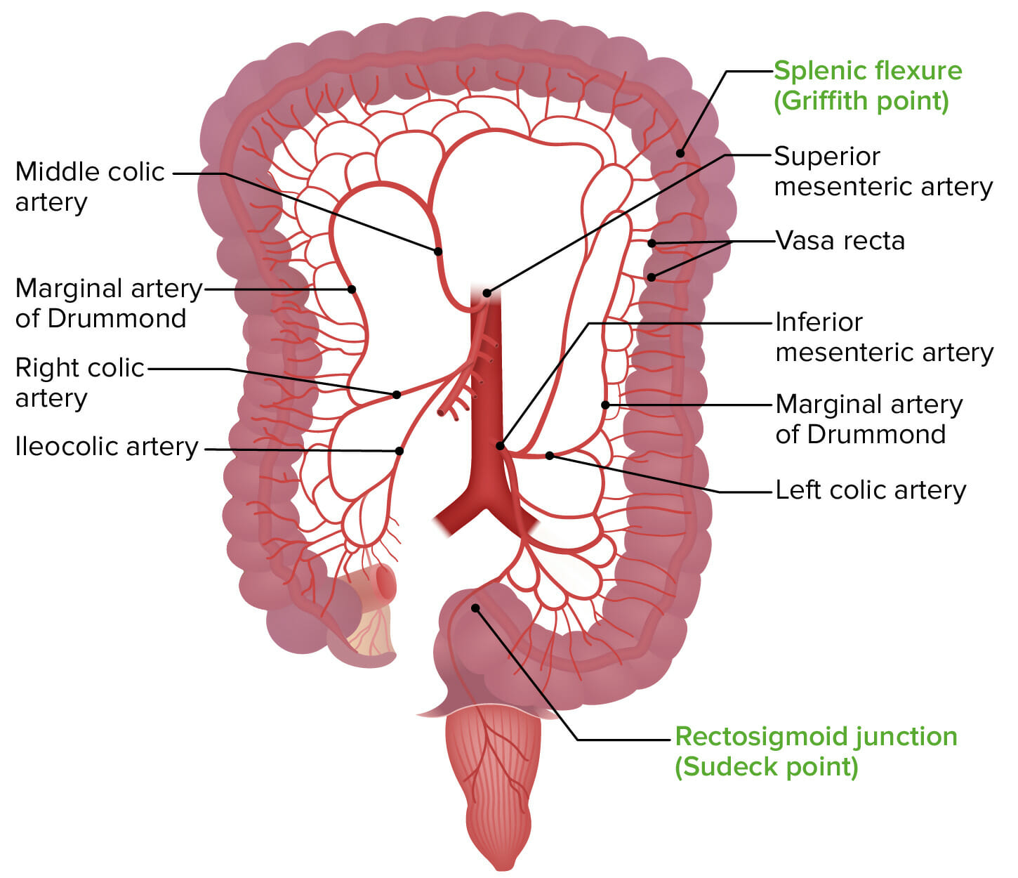
The main vessels affected in mesenteric ischemia Ischemia A hypoperfusion of the blood through an organ or tissue caused by a pathologic constriction or obstruction of its blood vessels, or an absence of blood circulation. Ischemic Cell Damage include:
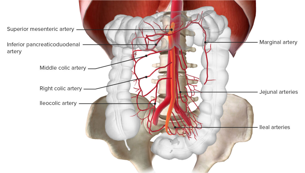
  • Superior mesenteric artery Superior mesenteric artery A large vessel supplying the whole length of the small intestine except the superior part of the duodenum. It also supplies the cecum and the ascending part of the colon and about half the transverse part of the colon. It arises from the anterior surface of the aorta below the celiac artery at the level of the first lumbar vertebra. Small Intestine: Anatomy (SMA):
  • Inferior mesenteric artery Inferior mesenteric artery The artery supplying nearly all the left half of the transverse colon, the whole of the descending colon, the sigmoid colon, and the greater part of the rectum. It is smaller than the superior mesenteric artery and arises from the aorta above its bifurcation into the common iliac arteries. Small Intestine: Anatomy: supplies the hindgut Hindgut Development of the Abdominal Organs (distal ⅓ of transverse colon Transverse colon The segment of large intestine between ascending colon and descending colon. It passes from the right colic flexure across the abdomen, then turns sharply at the left colonic flexure into the descending colon. Colon, Cecum, and Appendix: Anatomy to anal canal above pectinate line Pectinate line Rectum and Anal Canal: Anatomy)

Pathophysiology

  • The intestinal tract has a ↑ metabolic rate → requires ↑ blood flow Blood flow Blood flow refers to the movement of a certain volume of blood through the vasculature over a given unit of time (e.g., mL per minute). Vascular Resistance, Flow, and Mean Arterial Pressure
  • Disruption in blood flow Blood flow Blood flow refers to the movement of a certain volume of blood through the vasculature over a given unit of time (e.g., mL per minute). Vascular Resistance, Flow, and Mean Arterial Pressure → ischemic changes within 15 minutes:
    • May cause a reactive vasospasm → ↓ collateral blood flow Blood flow Blood flow refers to the movement of a certain volume of blood through the vasculature over a given unit of time (e.g., mL per minute). Vascular Resistance, Flow, and Mean Arterial Pressure
    • Bowel hypoxia Hypoxia Sub-optimal oxygen levels in the ambient air of living organisms. Ischemic Cell Damage → bowel wall spasm → symptoms:
      • Vomiting Vomiting The forcible expulsion of the contents of the stomach through the mouth. Hypokalemia
      • Diarrhea Diarrhea Diarrhea is defined as ≥ 3 watery or loose stools in a 24-hour period. There are a multitude of etiologies, which can be classified based on the underlying mechanism of disease. The duration of symptoms (acute or chronic) and characteristics of the stools (e.g., watery, bloody, steatorrheic, mucoid) can help guide further diagnostic evaluation. Diarrhea
    • May depend on:
      • Adequacy of perfusion
      • Any collateral circulation Circulation The movement of the blood as it is pumped through the cardiovascular system. ABCDE Assessment
      • Number of vessels affected
  • Mucosal damage and sloughing occurs within 3 hours and can lead to:
    • Gastrointestinal bleeding Gastrointestinal bleeding Gastrointestinal bleeding (GIB) is a symptom of multiple diseases within the gastrointestinal (GI) tract. Gastrointestinal bleeding is designated as upper or lower based on the etiology’s location to the ligament of Treitz. Depending on the location of the bleeding, the patient may present with hematemesis (vomiting blood), melena (black, tarry stool), or hematochezia (fresh blood in stools). Gastrointestinal Bleeding and visceral pain Visceral pain Pain originating from internal organs (viscera) associated with autonomic phenomena (pallor; sweating; nausea; and vomiting). It often becomes a referred pain. Pain: Types and Pathways
    • Release of vasoactive mediators, bacteria Bacteria Bacteria are prokaryotic single-celled microorganisms that are metabolically active and divide by binary fission. Some of these organisms play a significant role in the pathogenesis of diseases. Bacteriology, and toxic byproducts; resulting in:
  • Bowel wall becomes cyanotic and edematous → fluids released into peritoneal cavity Peritoneal Cavity The space enclosed by the peritoneum. It is divided into two portions, the greater sac and the lesser sac or omental bursa, which lies behind the stomach. The two sacs are connected by the foramen of winslow, or epiploic foramen. Peritoneum: Anatomy
  • Necrosis Necrosis The death of cells in an organ or tissue due to disease, injury or failure of the blood supply. Ischemic Cell Damage occurs within 6 hours → peritoneal signs

Related videos

Acute Mesenteric Ischemia

Epidemiology

  • Exact prevalence Prevalence The total number of cases of a given disease in a specified population at a designated time. It is differentiated from incidence, which refers to the number of new cases in the population at a given time. Measures of Disease Frequency is unknown.
  • Accounts for 0.1% of all hospital admissions
  • Generally seen in patients Patients Individuals participating in the health care system for the purpose of receiving therapeutic, diagnostic, or preventive procedures. Clinician–Patient Relationship > 60-years-old
  • Mortality Mortality All deaths reported in a given population. Measures of Health Status rate: 60%

Etiology

  • Arterial embolism:
    • Most common type (50%)
    • Median age is 70-years-old.
    • Frequently due to a cardiac embolus:
      • Atrial dysrhythmias, such as atrial fibrillation Atrial fibrillation Atrial fibrillation (AF or Afib) is a supraventricular tachyarrhythmia and the most common kind of arrhythmia. It is caused by rapid, uncontrolled atrial contractions and uncoordinated ventricular responses. Atrial Fibrillation
      • Heart failure Heart Failure A heterogeneous condition in which the heart is unable to pump out sufficient blood to meet the metabolic need of the body. Heart failure can be caused by structural defects, functional abnormalities (ventricular dysfunction), or a sudden overload beyond its capacity. Chronic heart failure is more common than acute heart failure which results from sudden insult to cardiac function, such as myocardial infarction. Total Anomalous Pulmonary Venous Return (TAPVR)
      • Myocardial ischemia Myocardial ischemia A disorder of cardiac function caused by insufficient blood flow to the muscle tissue of the heart. The decreased blood flow may be due to narrowing of the coronary arteries (coronary artery disease), to obstruction by a thrombus (coronary thrombosis), or less commonly, to diffuse narrowing of arterioles and other small vessels within the heart. Coronary Heart Disease or infarction
      • Ventricular aneurysm Ventricular aneurysm A localized bulging or dilatation in the muscle wall of a heart (myocardium), usually in the left ventricle. Blood-filled aneurysms are dangerous because they may burst. Fibrous aneurysms interfere with the heart function through the loss of contractility. True aneurysm is bound by the vessel wall or cardiac wall. False aneurysms are hematoma caused by myocardial rupture. Myocardial Infarction
      • Valvular disease and endocarditis Endocarditis Endocarditis is an inflammatory disease involving the inner lining (endometrium) of the heart, most commonly affecting the cardiac valves. Both infectious and noninfectious etiologies lead to vegetations on the valve leaflets. Patients may present with nonspecific symptoms such as fever and fatigue. Endocarditis
    • Most commonly affected artery is the SMA due to:
  • Arterial thrombosis Arterial Thrombosis Hypercoagulable States:
    • 2nd most common type (25%)
    • Often seen in individuals with a history of chronic mesenteric ischemia Ischemia A hypoperfusion of the blood through an organ or tissue caused by a pathologic constriction or obstruction of its blood vessels, or an absence of blood circulation. Ischemic Cell Damage symptoms
    • Acute occlusion occurs in severely narrowed segments of the vessel:
      • Usually due to underlying atherosclerosis Atherosclerosis Atherosclerosis is a common form of arterial disease in which lipid deposition forms a plaque in the blood vessel walls. Atherosclerosis is an incurable disease, for which there are clearly defined risk factors that often can be reduced through a change in lifestyle and behavior of the patient. Atherosclerosis
      • Most often at the vessel origin
    • Risk factors include:
      • Diabetes Diabetes Diabetes mellitus (DM) is a metabolic disease characterized by hyperglycemia and dysfunction of the regulation of glucose metabolism by insulin. Type 1 DM is diagnosed mostly in children and young adults as the result of autoimmune destruction of β cells in the pancreas and the resulting lack of insulin. Type 2 DM has a significant association with obesity and is characterized by insulin resistance. Diabetes Mellitus
      • Hypertension Hypertension Hypertension, or high blood pressure, is a common disease that manifests as elevated systemic arterial pressures. Hypertension is most often asymptomatic and is found incidentally as part of a routine physical examination or during triage for an unrelated medical encounter. Hypertension
      • Tobacco use
      • Hyperlipidemia
  • Non-occlusive mesenteric ischemia Ischemia A hypoperfusion of the blood through an organ or tissue caused by a pathologic constriction or obstruction of its blood vessels, or an absence of blood circulation. Ischemic Cell Damage:
    • 3rd most common type (5%–15%)
    • Can result in recurrent and repetitive injury to the bowel
    • Causes:
      • Hypoperfusion states (e.g., sepsis Sepsis Systemic inflammatory response syndrome with a proven or suspected infectious etiology. When sepsis is associated with organ dysfunction distant from the site of infection, it is called severe sepsis. When sepsis is accompanied by hypotension despite adequate fluid infusion, it is called septic shock. Sepsis and Septic Shock, hypovolemia Hypovolemia Sepsis in Children, ↓ cardiac output Cardiac output The volume of blood passing through the heart per unit of time. It is usually expressed as liters (volume) per minute so as not to be confused with stroke volume (volume per beat). Cardiac Mechanics, hemodialysis Hemodialysis Procedures which temporarily or permanently remedy insufficient cleansing of body fluids by the kidneys. Crush Syndrome
      • Vasospasm or vasoconstriction Vasoconstriction The physiological narrowing of blood vessels by contraction of the vascular smooth muscle. Vascular Resistance, Flow, and Mean Arterial Pressure (e.g., vasopressors Vasopressors Sepsis in Children, cocaine Cocaine An alkaloid ester extracted from the leaves of plants including coca. It is a local anesthetic and vasoconstrictor and is clinically used for that purpose, particularly in the eye, ear, nose, and throat. It also has powerful central nervous system effects similar to the amphetamines and is a drug of abuse. Cocaine, like amphetamines, acts by multiple mechanisms on brain catecholaminergic neurons; the mechanism of its reinforcing effects is thought to involve inhibition of dopamine uptake. Local Anesthetics)
  • Venous thrombosis Venous thrombosis The formation or presence of a blood clot (thrombus) within a vein. Budd-Chiari Syndrome:
    • Least common type (5%–10%)
    • Frequently involves superior mesenteric vein 
    • Risk factors include:
      • Thrombotic disorders or hypercoagulable Hypercoagulable Hypercoagulable states (also referred to as thrombophilias) are a group of hematologic diseases defined by an increased risk of clot formation (i.e., thrombosis) due to either an increase in procoagulants, a decrease in anticoagulants, or a decrease in fibrinolysis. Hypercoagulable States states (e.g., young women taking oral contraceptives)
      • Malignancy Malignancy Hemothorax causing venous compression Compression Blunt Chest Trauma
      • Inflammation Inflammation Inflammation is a complex set of responses to infection and injury involving leukocytes as the principal cellular mediators in the body’s defense against pathogenic organisms. Inflammation is also seen as a response to tissue injury in the process of wound healing. The 5 cardinal signs of inflammation are pain, heat, redness, swelling, and loss of function. Inflammation (e.g., pancreatitis Pancreatitis Inflammation of the pancreas. Pancreatitis is classified as acute unless there are computed tomographic or endoscopic retrograde cholangiopancreatographic findings of chronic pancreatitis. The two most common forms of acute pancreatitis are alcoholic pancreatitis and gallstone pancreatitis. Acute Pancreatitis, intra-abdominal infection)
      • Venous congestion (e.g., portal hypertension Portal hypertension Portal hypertension is increased pressure in the portal venous system. This increased pressure can lead to splanchnic vasodilation, collateral blood flow through portosystemic anastomoses, and increased hydrostatic pressure. There are a number of etiologies, including cirrhosis, right-sided congestive heart failure, schistosomiasis, portal vein thrombosis, hepatitis, and Budd-Chiari syndrome. Portal Hypertension from cirrhosis Cirrhosis Cirrhosis is a late stage of hepatic parenchymal necrosis and scarring (fibrosis) most commonly due to hepatitis C infection and alcoholic liver disease. Patients may present with jaundice, ascites, and hepatosplenomegaly. Cirrhosis can also cause complications such as hepatic encephalopathy, portal hypertension, portal vein thrombosis, and hepatorenal syndrome. Cirrhosis)
      • Trauma

Clinical presentation

  • Classic triad:
    1. Diffuse abdominal pain Abdominal Pain Acute Abdomen out of proportion to the physical exam:
      • Severe pain Pain An unpleasant sensation induced by noxious stimuli which are detected by nerve endings of nociceptive neurons. Pain: Types and Pathways 
      • Not worsened or reproduced with palpation Palpation Application of fingers with light pressure to the surface of the body to determine consistency of parts beneath in physical diagnosis; includes palpation for determining the outlines of organs. Dermatologic Examination
      • Abdomen remains soft
      • May be unresponsive to pain Pain An unpleasant sensation induced by noxious stimuli which are detected by nerve endings of nociceptive neurons. Pain: Types and Pathways medication
    2. Gut emptying:
      • Vomiting Vomiting The forcible expulsion of the contents of the stomach through the mouth. Hypokalemia
      • Diarrhea Diarrhea Diarrhea is defined as ≥ 3 watery or loose stools in a 24-hour period. There are a multitude of etiologies, which can be classified based on the underlying mechanism of disease. The duration of symptoms (acute or chronic) and characteristics of the stools (e.g., watery, bloody, steatorrheic, mucoid) can help guide further diagnostic evaluation. Diarrhea
    3. History of cardiovascular disease or embolic event
  • Indications of bowel infarction:
    • Peritonitis Peritonitis Inflammation of the peritoneum lining the abdominal cavity as the result of infectious, autoimmune, or chemical processes. Primary peritonitis is due to infection of the peritoneal cavity via hematogenous or lymphatic spread and without intra-abdominal source. Secondary peritonitis arises from the abdominal cavity itself through rupture or abscess of intra-abdominal organs. Penetrating Abdominal Injury:
      • Rigidity Rigidity Continuous involuntary sustained muscle contraction which is often a manifestation of basal ganglia diseases. When an affected muscle is passively stretched, the degree of resistance remains constant regardless of the rate at which the muscle is stretched. This feature helps to distinguish rigidity from muscle spasticity. Megacolon
      • Guarding
      • No bowel sounds
    • Septic shock Septic shock Sepsis associated with hypotension or hypoperfusion despite adequate fluid resuscitation. Perfusion abnormalities may include, but are not limited to lactic acidosis; oliguria; or acute alteration in mental status. Sepsis and Septic Shock:
      • Fever Fever Fever is defined as a measured body temperature of at least 38°C (100.4°F). Fever is caused by circulating endogenous and/or exogenous pyrogens that increase levels of prostaglandin E2 in the hypothalamus. Fever is commonly associated with chills, rigors, sweating, and flushing of the skin. Fever
      • Tachycardia Tachycardia Abnormally rapid heartbeat, usually with a heart rate above 100 beats per minute for adults. Tachycardia accompanied by disturbance in the cardiac depolarization (cardiac arrhythmia) is called tachyarrhythmia. Sepsis in Children
      • Hypotension Hypotension Hypotension is defined as low blood pressure, specifically < 90/60 mm Hg, and is most commonly a physiologic response. Hypotension may be mild, serious, or life threatening, depending on the cause. Hypotension
    • Hematochezia Hematochezia Gastrointestinal Bleeding

Diagnosis

Acute mesenteric ischemia Ischemia A hypoperfusion of the blood through an organ or tissue caused by a pathologic constriction or obstruction of its blood vessels, or an absence of blood circulation. Ischemic Cell Damage requires a high index of suspicion to allow a timely diagnosis.
  • Laboratory studies:
    • Nonspecific
    • Findings that should draw suspicion:
      • ↑ Lactic acid and lactate dehydrogenase Lactate Dehydrogenase Osteosarcoma
      • Metabolic acidosis Acidosis A pathologic condition of acid accumulation or depletion of base in the body. The two main types are respiratory acidosis and metabolic acidosis, due to metabolic acid build up. Respiratory Acidosis (↓ bicarbonate Bicarbonate Inorganic salts that contain the -HCO3 radical. They are an important factor in determining the ph of the blood and the concentration of bicarbonate ions is regulated by the kidney. Levels in the blood are an index of the alkali reserve or buffering capacity. Electrolytes)
      • Leukocytosis Leukocytosis A transient increase in the number of leukocytes in a body fluid. West Nile Virus with a left shift Left Shift Yersinia pestis/Plague
      • amylase Amylase A group of amylolytic enzymes that cleave starch, glycogen, and related alpha-1, 4-glucans. Digestion and Absorption
  • Electrocardiogram Electrocardiogram An electrocardiogram (ECG) is a graphic representation of the electrical activity of the heart plotted against time. Adhesive electrodes are affixed to the skin surface allowing measurement of cardiac impulses from many angles. The ECG provides 3-dimensional information about the conduction system of the heart, the myocardium, and other cardiac structures. Electrocardiogram (ECG) ( ECG ECG An electrocardiogram (ECG) is a graphic representation of the electrical activity of the heart plotted against time. Adhesive electrodes are affixed to the skin surface allowing measurement of cardiac impulses from many angles. The ECG provides 3-dimensional information about the conduction system of the heart, the myocardium, and other cardiac structures. Electrocardiogram (ECG)) should be performed to evaluate for cardiac dysrhythmia.
  • Imaging studies:
    • Radiographs: 
      • Nonspecific
      • Possible findings: ileus Ileus A condition caused by the lack of intestinal peristalsis or intestinal motility without any mechanical obstruction. This interference of the flow of intestinal contents often leads to intestinal obstruction. Ileus may be classified into postoperative, inflammatory, metabolic, neurogenic, and drug-induced. Small Bowel Obstruction and pneumatosis intestinalis Pneumatosis intestinalis A condition characterized by the presence of multiple gas-filled cysts in the intestinal wall, the submucosa and/or subserosa of the intestine. The majority of the cysts are found in the jejunum and the ileum. Necrotizing Enterocolitis (gas within the wall of the intestine)
      • Rule-out free air ( perforation Perforation A pathological hole in an organ, blood vessel or other soft part of the body, occurring in the absence of external force. Esophagitis)
    • Duplex ultrasound of mesenteric vessels: assesses the SMA and celiac artery for blood flow Blood flow Blood flow refers to the movement of a certain volume of blood through the vasculature over a given unit of time (e.g., mL per minute). Vascular Resistance, Flow, and Mean Arterial Pressure
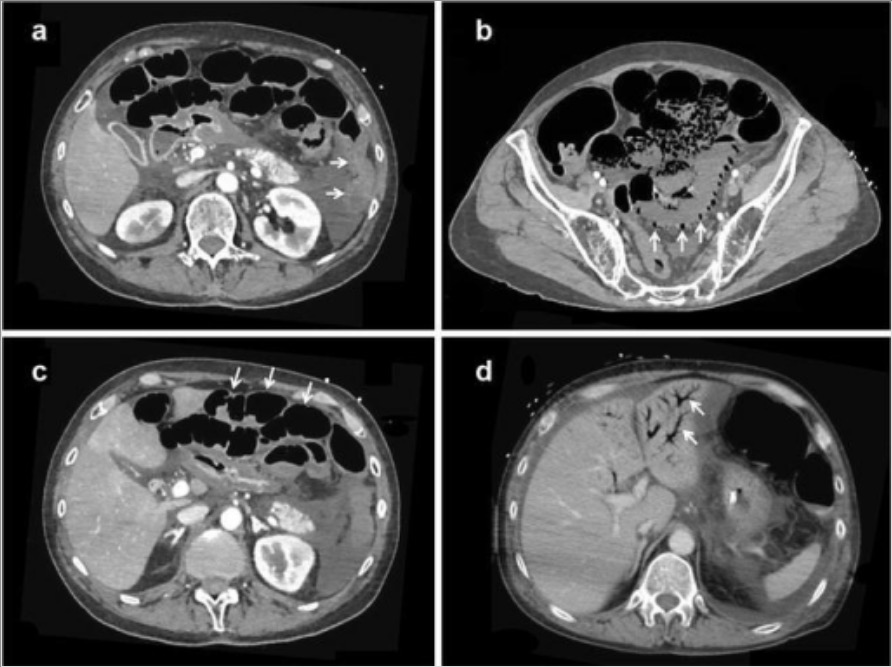
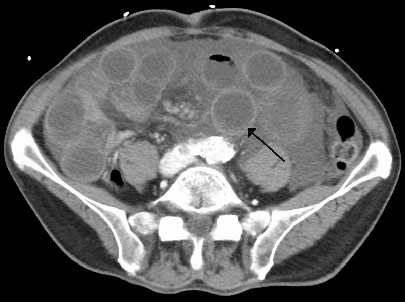
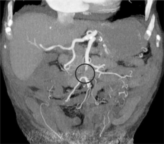
    • Computed tomography (CT) with angiography Angiography Radiography of blood vessels after injection of a contrast medium. Cardiac Surgery:
      • Preferred method for diagnosis
      • Requires intravenous (IV) contrast (no oral contrast)
      • Identifies occluded mesenteric vessels
      • Collateral vessels may be present.
      • Evaluates bowel for signs of ischemia Ischemia A hypoperfusion of the blood through an organ or tissue caused by a pathologic constriction or obstruction of its blood vessels, or an absence of blood circulation. Ischemic Cell Damage (e.g., wall thickening, pneumatosis intestinalis Pneumatosis intestinalis A condition characterized by the presence of multiple gas-filled cysts in the intestinal wall, the submucosa and/or subserosa of the intestine. The majority of the cysts are found in the jejunum and the ileum. Necrotizing Enterocolitis, portal venous gas Portal Venous Gas Imaging of the Intestines)
      • Bowel dilation Bowel dilation Large Bowel Obstruction and lack of bowel wall enhancement may be seen.
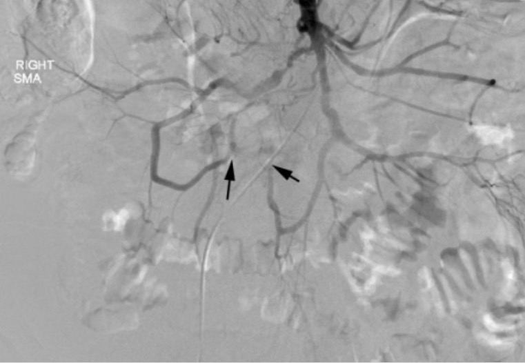
    • Mesenteric angiogram:
      • Identifies occluded mesenteric vessels in real-time under fluoroscopy Fluoroscopy Production of an image when x-rays strike a fluorescent screen. X-rays
      • Used for endovascular interventions, such as angioplasty Angioplasty Reconstruction or repair of a blood vessel, which includes the widening of a pathological narrowing of an artery or vein by the removal of atheromatous plaque material and/or the endothelial lining as well, or by dilatation (balloon angioplasty) to compress an atheroma. Except for endarterectomy, usually these procedures are performed via catheterization as minimally invasive endovascular procedures. Cardiac Surgery and/or stenting
      • May be used for surgical planning
      • Limited availability, requires an endovascular specialist
      • Requires IV contrast
  • Exploratory laparotomy Exploratory Laparotomy Laparotomy and Laparoscopy:
    • Imaging may be skipped in patients Patients Individuals participating in the health care system for the purpose of receiving therapeutic, diagnostic, or preventive procedures. Clinician–Patient Relationship with clear evidence of advanced ischemia Ischemia A hypoperfusion of the blood through an organ or tissue caused by a pathologic constriction or obstruction of its blood vessels, or an absence of blood circulation. Ischemic Cell Damage (e.g, sepsis Sepsis Systemic inflammatory response syndrome with a proven or suspected infectious etiology. When sepsis is associated with organ dysfunction distant from the site of infection, it is called severe sepsis. When sepsis is accompanied by hypotension despite adequate fluid infusion, it is called septic shock. Sepsis and Septic Shock, peritoneal signs); it may be safer and more expedient.
    • Goal is to identify and prevent impending perforation Perforation A pathological hole in an organ, blood vessel or other soft part of the body, occurring in the absence of external force. Esophagitis.

Management

  • Initial management:
    • Early fluid resuscitation Resuscitation The restoration to life or consciousness of one apparently dead. . Neonatal Respiratory Distress Syndrome with crystalloid Crystalloid Isotonic solutions of mineral salts, such as ringer’s lactate and sodium chloride (saline solution), used in fluid therapy to rehydrate blood volume. Intravenous Fluids to maintain hemodynamic stability
    • Broad-spectrum Broad-Spectrum Fluoroquinolones antibiotics to cover anaerobic and gram-negative organisms
    • Nasogastric (NG) tube for decompression of distended stomach Stomach The stomach is a muscular sac in the upper left portion of the abdomen that plays a critical role in digestion. The stomach develops from the foregut and connects the esophagus with the duodenum. Structurally, the stomach is C-shaped and forms a greater and lesser curvature and is divided grossly into regions: the cardia, fundus, body, and pylorus. Stomach: Anatomy and small bowel Small bowel The small intestine is the longest part of the GI tract, extending from the pyloric orifice of the stomach to the ileocecal junction. The small intestine is the major organ responsible for chemical digestion and absorption of nutrients. It is divided into 3 segments: the duodenum, the jejunum, and the ileum. Small Intestine: Anatomy
    • Correction of any electrolyte abnormalities
    • Pain Pain An unpleasant sensation induced by noxious stimuli which are detected by nerve endings of nociceptive neurons. Pain: Types and Pathways control
    • Avoid vasopressors Vasopressors Sepsis in Children, if possible. Options if vasopressors Vasopressors Sepsis in Children must be used include dobutamine Dobutamine A catecholamine derivative with specificity for beta-1 adrenergic receptors. Sympathomimetic Drugs, low-dose dopamine Dopamine One of the catecholamine neurotransmitters in the brain. It is derived from tyrosine and is the precursor to norepinephrine and epinephrine. Dopamine is a major transmitter in the extrapyramidal system of the brain, and important in regulating movement. Receptors and Neurotransmitters of the CNS, or milrinone Milrinone A positive inotropic cardiotonic agent with vasodilator properties. It inhibits camp phosphodiesterase type 3 activity in myocardium and vascular smooth muscle. Milrinone is a derivative of amrinone and has 20-30 times the inotropic potency of amrinone. Phosphodiesterase Inhibitors.
  • Systemic anticoagulation Anticoagulation Pulmonary Hypertension Drugs:
    • Heparin
    • Used to prevent thrombus formation or propagation Propagation Propagation refers to how the electrical signal spreads to every myocyte in the heart. Cardiac Physiology
  • Endovascular angioplasty Angioplasty Reconstruction or repair of a blood vessel, which includes the widening of a pathological narrowing of an artery or vein by the removal of atheromatous plaque material and/or the endothelial lining as well, or by dilatation (balloon angioplasty) to compress an atheroma. Except for endarterectomy, usually these procedures are performed via catheterization as minimally invasive endovascular procedures. Cardiac Surgery
    • With stenting, embolectomy, or thrombolytics Thrombolytics Thrombolytics, also known as fibrinolytics, include recombinant tissue plasminogen activator (TPa) (i.e., alteplase, reteplase, and tenecteplase), urokinase, and streptokinase. The agents promote the breakdown of a blood clot by converting plasminogen to plasmin, which then degrades fibrin. Thrombolytics
    • Indications:
      • Severe comorbidities Comorbidities The presence of co-existing or additional diseases with reference to an initial diagnosis or with reference to the index condition that is the subject of study. Comorbidity may affect the ability of affected individuals to function and also their survival; it may be used as a prognostic indicator for length of hospital stay, cost factors, and outcome or survival. St. Louis Encephalitis Virus
      • No signs of advanced bowel ischemia Ischemia A hypoperfusion of the blood through an organ or tissue caused by a pathologic constriction or obstruction of its blood vessels, or an absence of blood circulation. Ischemic Cell Damage or peritonitis Peritonitis Inflammation of the peritoneum lining the abdominal cavity as the result of infectious, autoimmune, or chemical processes. Primary peritonitis is due to infection of the peritoneal cavity via hematogenous or lymphatic spread and without intra-abdominal source. Secondary peritonitis arises from the abdominal cavity itself through rupture or abscess of intra-abdominal organs. Penetrating Abdominal Injury
      • Short duration of symptoms
  • Surgery:
    • Indications:
      • If endovascular approach fails
      • If any concern for bowel ischemia Ischemia A hypoperfusion of the blood through an organ or tissue caused by a pathologic constriction or obstruction of its blood vessels, or an absence of blood circulation. Ischemic Cell Damage on exam
    • Options:
  • Special considerations:
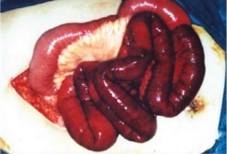
Mesenteric ischemia surgery

Surgical findings in mesenteric ischemia:
Picture of eviscerated small bowel during an exploratory laparotomy with healthy pink bowel to the top left and dark, dusky, ischemic bowel to the lower right

Image: “Segmental gangrene” by Department of Obstetrics and Gynecology, Kaohsiung Chang Gung Memorial Hospital and Chang Gung University College of Medicine, Kaohsiung City 833, Taiwan. License: CC BY 4.0

Complications

  • Reperfusion injury Reperfusion injury Adverse functional, metabolic, or structural changes in ischemic tissues resulting from the restoration of blood flow to the tissue (reperfusion), including swelling; hemorrhage; necrosis; and damage from free radicals. The most common instance is myocardial reperfusion injury. Ischemic Cell Damage 
  • Infection and sepsis Sepsis Systemic inflammatory response syndrome with a proven or suspected infectious etiology. When sepsis is associated with organ dysfunction distant from the site of infection, it is called severe sepsis. When sepsis is accompanied by hypotension despite adequate fluid infusion, it is called septic shock. Sepsis and Septic Shock
  • Short bowel syndrome Short bowel syndrome Short bowel syndrome is a malabsorptive condition most commonly associated with extensive intestinal resection for etiologies such as Crohn’s disease, bowel obstruction, trauma, radiation therapy, and vascular insufficiency. The short length of bowel results in insufficient surface area for fluid and electrolyte absorption. Short Bowel Syndrome

Chronic Mesenteric Ischemia

Epidemiology

  • Rare
  • 60% of cases are inpatients > 60-years-old.
  • More common in women

Etiology

Chronic mesenteric ischemia Ischemia A hypoperfusion of the blood through an organ or tissue caused by a pathologic constriction or obstruction of its blood vessels, or an absence of blood circulation. Ischemic Cell Damage is caused by a progressive stenosis Stenosis Hypoplastic Left Heart Syndrome (HLHS) of ≥ 2 arteries Arteries Arteries are tubular collections of cells that transport oxygenated blood and nutrients from the heart to the tissues of the body. The blood passes through the arteries in order of decreasing luminal diameter, starting in the largest artery (the aorta) and ending in the small arterioles. Arteries are classified into 3 types: large elastic arteries, medium muscular arteries, and small arteries and arterioles. Arteries: Histology, resulting in episodes of blood flow Blood flow Blood flow refers to the movement of a certain volume of blood through the vasculature over a given unit of time (e.g., mL per minute). Vascular Resistance, Flow, and Mean Arterial Pressure supply and demand mismatch (usually after eating).

  • The majority of cases are due to atherosclerotic disease narrowing the origin of the mesenteric vessels.
  • Rare causes:
    • Vasculitis Vasculitis Inflammation of any one of the blood vessels, including the arteries; veins; and rest of the vasculature system in the body. Systemic Lupus Erythematosus
    • Fibromuscular dysplasia Fibromuscular dysplasia Fibromuscular dysplasia (FMD) is a nonatherosclerotic, noninflammatory, medium-sized angiopathy due to fibroplasia of the vessel wall. The condition leads to complications related to arterial stenosis, aneurysm, or dissection. Fibromuscular Dysplasia
    • Artery dissection
    • Retroperitoneal Retroperitoneal Peritoneum: Anatomy fibrosis Fibrosis Any pathological condition where fibrous connective tissue invades any organ, usually as a consequence of inflammation or other injury. Bronchiolitis Obliterans
    • Median arcuate ligament syndrome: celiac artery compression Compression Blunt Chest Trauma by the arcuate ligament of the diaphragm Diaphragm The diaphragm is a large, dome-shaped muscle that separates the thoracic cavity from the abdominal cavity. The diaphragm consists of muscle fibers and a large central tendon, which is divided into right and left parts. As the primary muscle of inspiration, the diaphragm contributes 75% of the total inspiratory muscle force. Diaphragm: Anatomy
    • Endovascular repair of the aorta Aorta The main trunk of the systemic arteries. Mediastinum and Great Vessels: Anatomy

Clinical presentation

  • Episodic abdominal pain Abdominal Pain Acute Abdomen:
    • Referred to as “intestinal angina” 
    • Dull and crampy
    • Epigastric
    • Varies in intensity
    • Postprandial:
      • Occurs within 1 hour after eating
      • Resolves over the following 2 hours
  • Food fear:
  • Abdominal bruit Abdominal Bruit Abdominal Examination: present in 50% of patients Patients Individuals participating in the health care system for the purpose of receiving therapeutic, diagnostic, or preventive procedures. Clinician–Patient Relationship
  • Less common signs and symptoms:
    • Nausea Nausea An unpleasant sensation in the stomach usually accompanied by the urge to vomit. Common causes are early pregnancy, sea and motion sickness, emotional stress, intense pain, food poisoning, and various enteroviruses. Antiemetics and vomiting Vomiting The forcible expulsion of the contents of the stomach through the mouth. Hypokalemia
    • Early satiety Early Satiety Bariatric Surgery
    • Diarrhea Diarrhea Diarrhea is defined as ≥ 3 watery or loose stools in a 24-hour period. There are a multitude of etiologies, which can be classified based on the underlying mechanism of disease. The duration of symptoms (acute or chronic) and characteristics of the stools (e.g., watery, bloody, steatorrheic, mucoid) can help guide further diagnostic evaluation. Diarrhea
    • Gastrointestinal bleeding Gastrointestinal bleeding Gastrointestinal bleeding (GIB) is a symptom of multiple diseases within the gastrointestinal (GI) tract. Gastrointestinal bleeding is designated as upper or lower based on the etiology’s location to the ligament of Treitz. Depending on the location of the bleeding, the patient may present with hematemesis (vomiting blood), melena (black, tarry stool), or hematochezia (fresh blood in stools). Gastrointestinal Bleeding
  • Beware of acute-on-chronic mesenteric ischemia Ischemia A hypoperfusion of the blood through an organ or tissue caused by a pathologic constriction or obstruction of its blood vessels, or an absence of blood circulation. Ischemic Cell Damage:
    • May occur due to thrombus formation in a patient with chronically stenotic vessels.
    • Associated with a ↑ morbidity Morbidity The proportion of patients with a particular disease during a given year per given unit of population. Measures of Health Status and mortality Mortality All deaths reported in a given population. Measures of Health Status
    • Symptoms similar to acute mesenteric ischemia Ischemia A hypoperfusion of the blood through an organ or tissue caused by a pathologic constriction or obstruction of its blood vessels, or an absence of blood circulation. Ischemic Cell Damage.

Diagnosis

Vascular imaging studies are used to make the diagnosis.

  • CT angiography Angiography Radiography of blood vessels after injection of a contrast medium. Cardiac Surgery:
    • Preferred diagnostic modality
    • Should demonstrate high-grade stenosis Stenosis Hypoplastic Left Heart Syndrome (HLHS) of ≥ 2 major mesenteric arteries Arteries Arteries are tubular collections of cells that transport oxygenated blood and nutrients from the heart to the tissues of the body. The blood passes through the arteries in order of decreasing luminal diameter, starting in the largest artery (the aorta) and ending in the small arterioles. Arteries are classified into 3 types: large elastic arteries, medium muscular arteries, and small arteries and arterioles. Arteries: Histology
  • Duplex ultrasound of mesenteric vessels: can be used as a screening Screening Preoperative Care study

Management

  • Conservative treatment:
    • Preferred for:
      • Incidental diagnosis
      • No overt clinical manifestations
    • Focuses on limiting the progression of atherosclerosis Atherosclerosis Atherosclerosis is a common form of arterial disease in which lipid deposition forms a plaque in the blood vessel walls. Atherosclerosis is an incurable disease, for which there are clearly defined risk factors that often can be reduced through a change in lifestyle and behavior of the patient. Atherosclerosis:
      • Smoking Smoking Willful or deliberate act of inhaling and exhaling smoke from burning substances or agents held by hand. Interstitial Lung Diseases cessation
      • Glycemic control
      • Antiplatelet therapy
  • Nutritional support:
    • Necessary in patients Patients Individuals participating in the health care system for the purpose of receiving therapeutic, diagnostic, or preventive procedures. Clinician–Patient Relationship with significant weight loss Weight loss Decrease in existing body weight. Bariatric Surgery
    • Parenteral nutrition Parenteral nutrition The administering of nutrients for assimilation and utilization by a patient who cannot maintain adequate nutrition by enteral feeding alone. Nutrients are administered by a route other than the alimentary canal (e.g., intravenously, subcutaneously). Central Venous Catheter may be considered for severe cases.
  • Revascularization Revascularization Thromboangiitis Obliterans (Buerger’s Disease):
    • Indications:
      • Symptoms and documented stenosis Stenosis Hypoplastic Left Heart Syndrome (HLHS) on imaging
      • Peritonitis Peritonitis Inflammation of the peritoneum lining the abdominal cavity as the result of infectious, autoimmune, or chemical processes. Primary peritonitis is due to infection of the peritoneal cavity via hematogenous or lymphatic spread and without intra-abdominal source. Secondary peritonitis arises from the abdominal cavity itself through rupture or abscess of intra-abdominal organs. Penetrating Abdominal Injury
      • Gastrointestinal hemorrhage Gastrointestinal hemorrhage Bleeding in any segment of the gastrointestinal tract from esophagus to rectum. Peptic Ulcer Disease
    • Goal: Prevent future bowel infarction.
    • Options:
      • Endovascular approach with stenting and/or angioplasty Angioplasty Reconstruction or repair of a blood vessel, which includes the widening of a pathological narrowing of an artery or vein by the removal of atheromatous plaque material and/or the endothelial lining as well, or by dilatation (balloon angioplasty) to compress an atheroma. Except for endarterectomy, usually these procedures are performed via catheterization as minimally invasive endovascular procedures. Cardiac Surgery
      • Open surgical approach for endarterectomy Endarterectomy Surgical excision, performed under general anesthesia, of the atheromatous tunica intima of an artery. When reconstruction of an artery is performed as an endovascular procedure through a catheter, it is called atherectomy. Intestinal Ischemia or bypass

Differential Diagnosis

  • Small bowel obstruction Bowel obstruction Any impairment, arrest, or reversal of the normal flow of intestinal contents toward the anal canal. Ascaris/Ascariasis: an interruption of intraluminal contents through the small bowel Small bowel The small intestine is the longest part of the GI tract, extending from the pyloric orifice of the stomach to the ileocecal junction. The small intestine is the major organ responsible for chemical digestion and absorption of nutrients. It is divided into 3 segments: the duodenum, the jejunum, and the ileum. Small Intestine: Anatomy due to a mechanical or functional problem. Patients Patients Individuals participating in the health care system for the purpose of receiving therapeutic, diagnostic, or preventive procedures. Clinician–Patient Relationship present with abdominal pain Abdominal Pain Acute Abdomen, distention, nausea Nausea An unpleasant sensation in the stomach usually accompanied by the urge to vomit. Common causes are early pregnancy, sea and motion sickness, emotional stress, intense pain, food poisoning, and various enteroviruses. Antiemetics, vomiting Vomiting The forcible expulsion of the contents of the stomach through the mouth. Hypokalemia, and obstipation Obstipation Large Bowel Obstruction. Diagnosis is confirmed with abdominal imaging. Conservative management is usually the 1st step; nil per os (NPO), IV fluids IV fluids Intravenous fluids are one of the most common interventions administered in medicine to approximate physiologic bodily fluids. Intravenous fluids are divided into 2 categories: crystalloid and colloid solutions. Intravenous fluids have a wide variety of indications, including intravascular volume expansion, electrolyte manipulation, and maintenance fluids. Intravenous Fluids, and NG tube decompression. Some patients Patients Individuals participating in the health care system for the purpose of receiving therapeutic, diagnostic, or preventive procedures. Clinician–Patient Relationship fail conservative management and require surgery to relieve the obstruction.
  • Spontaneous bacterial peritonitis Spontaneous Bacterial Peritonitis Ascites ( SBP SBP Ascites): an acute bacterial infection of the peritoneal fluid (a well-known complication in patients Patients Individuals participating in the health care system for the purpose of receiving therapeutic, diagnostic, or preventive procedures. Clinician–Patient Relationship with cirrhosis Cirrhosis Cirrhosis is a late stage of hepatic parenchymal necrosis and scarring (fibrosis) most commonly due to hepatitis C infection and alcoholic liver disease. Patients may present with jaundice, ascites, and hepatosplenomegaly. Cirrhosis can also cause complications such as hepatic encephalopathy, portal hypertension, portal vein thrombosis, and hepatorenal syndrome. Cirrhosis). Individuals present with abdominal pain Abdominal Pain Acute Abdomen, distension, fever Fever Fever is defined as a measured body temperature of at least 38°C (100.4°F). Fever is caused by circulating endogenous and/or exogenous pyrogens that increase levels of prostaglandin E2 in the hypothalamus. Fever is commonly associated with chills, rigors, sweating, and flushing of the skin. Fever, and chills Chills The sudden sensation of being cold. It may be accompanied by shivering. Fever. Physical exam demonstrates a significantly tender abdomen. If SBP SBP Ascites is suspected, patients Patients Individuals participating in the health care system for the purpose of receiving therapeutic, diagnostic, or preventive procedures. Clinician–Patient Relationship require paracentesis Paracentesis A procedure in which fluid is withdrawn from a body cavity or organ via a trocar and cannula, needle, or other hollow instrument. Portal Hypertension with ascitic fluid Ascitic fluid The serous fluid of ascites, the accumulation of fluids in the peritoneal cavity. Ascites analysis and culture. The treatment of choice is antibiotics.
  • Acute cholecystitis Acute cholecystitis Acute inflammation of the gallbladder wall. It is characterized by the presence of abdominal pain; fever; and leukocytosis. Gallstone obstruction of the cystic duct is present in approximately 90% of the cases. Cholecystitis: inflammation Inflammation Inflammation is a complex set of responses to infection and injury involving leukocytes as the principal cellular mediators in the body’s defense against pathogenic organisms. Inflammation is also seen as a response to tissue injury in the process of wound healing. The 5 cardinal signs of inflammation are pain, heat, redness, swelling, and loss of function. Inflammation of the gallbladder Gallbladder The gallbladder is a pear-shaped sac, located directly beneath the liver, that sits on top of the superior part of the duodenum. The primary functions of the gallbladder include concentrating and storing up to 50 mL of bile. Gallbladder and Biliary Tract: Anatomy resulting from sustained gallstone impaction of the cystic Cystic Fibrocystic Change duct. Patients Patients Individuals participating in the health care system for the purpose of receiving therapeutic, diagnostic, or preventive procedures. Clinician–Patient Relationship present with colicky, upper abdominal pain Abdominal Pain Acute Abdomen, nausea Nausea An unpleasant sensation in the stomach usually accompanied by the urge to vomit. Common causes are early pregnancy, sea and motion sickness, emotional stress, intense pain, food poisoning, and various enteroviruses. Antiemetics, and vomiting Vomiting The forcible expulsion of the contents of the stomach through the mouth. Hypokalemia. Inflammation Inflammation Inflammation is a complex set of responses to infection and injury involving leukocytes as the principal cellular mediators in the body’s defense against pathogenic organisms. Inflammation is also seen as a response to tissue injury in the process of wound healing. The 5 cardinal signs of inflammation are pain, heat, redness, swelling, and loss of function. Inflammation of the gallbladder Gallbladder The gallbladder is a pear-shaped sac, located directly beneath the liver, that sits on top of the superior part of the duodenum. The primary functions of the gallbladder include concentrating and storing up to 50 mL of bile. Gallbladder and Biliary Tract: Anatomy and gallstones Gallstones Cholelithiasis (gallstones) is the presence of stones in the gallbladder. Most gallstones are cholesterol stones, while the rest are composed of bilirubin (pigment stones) and other mixed components. Patients are commonly asymptomatic but may present with biliary colic (intermittent pain in the right upper quadrant). Cholelithiasis are seen on ultrasound or CT. Management includes IV fluids IV fluids Intravenous fluids are one of the most common interventions administered in medicine to approximate physiologic bodily fluids. Intravenous fluids are divided into 2 categories: crystalloid and colloid solutions. Intravenous fluids have a wide variety of indications, including intravascular volume expansion, electrolyte manipulation, and maintenance fluids. Intravenous Fluids, antibiotics, and cholecystectomy Cholecystectomy Cholecystectomy is a surgical procedure performed with the goal of resecting and extracting the gallbladder. It is one of the most common abdominal surgeries performed in the Western world. Cholecystectomy is performed for symptomatic cholelithiasis, cholecystitis, gallbladder polyps > 0.5 cm, porcelain gallbladder, choledocholithiasis and gallstone pancreatitis, and rarely, for gallbladder cancer. Cholecystectomy.
  • Peptic ulcer Peptic ulcer Peptic ulcer disease (PUD) refers to the full-thickness ulcerations of duodenal or gastric mucosa. The ulcerations form when exposure to acid and digestive enzymes overcomes mucosal defense mechanisms. The most common etiologies include Helicobacter pylori (H. pylori) infection and prolonged use of non-steroidal anti-inflammatory drugs (NSAIDs). Peptic Ulcer Disease disease: ulcerations located in the stomach Stomach The stomach is a muscular sac in the upper left portion of the abdomen that plays a critical role in digestion. The stomach develops from the foregut and connects the esophagus with the duodenum. Structurally, the stomach is C-shaped and forms a greater and lesser curvature and is divided grossly into regions: the cardia, fundus, body, and pylorus. Stomach: Anatomy and/or duodenum Duodenum The shortest and widest portion of the small intestine adjacent to the pylorus of the stomach. It is named for having the length equal to about the width of 12 fingers. Small Intestine: Anatomy. Peptic ulcer Peptic ulcer Peptic ulcer disease (PUD) refers to the full-thickness ulcerations of duodenal or gastric mucosa. The ulcerations form when exposure to acid and digestive enzymes overcomes mucosal defense mechanisms. The most common etiologies include Helicobacter pylori (H. pylori) infection and prolonged use of non-steroidal anti-inflammatory drugs (NSAIDs). Peptic Ulcer Disease disease may be caused by Helicobacter pylori Helicobacter pylori A spiral bacterium active as a human gastric pathogen. It is a gram-negative, urease-positive, curved or slightly spiral organism initially isolated in 1982 from patients with lesions of gastritis or peptic ulcers in Western Australia. Helicobacter pylori was originally classified in the genus campylobacter, but RNA sequencing, cellular fatty acid profiles, growth patterns, and other taxonomic characteristics indicate that the micro-organism should be included in the genus Helicobacter. It has been officially transferred to Helicobacter gen. Helicobacter ( H. pylori H. pylori A spiral bacterium active as a human gastric pathogen. It is a gram-negative, urease-positive, curved or slightly spiral organism initially isolated in 1982 from patients with lesions of gastritis or peptic ulcers in Western Australia. Helicobacter pylori was originally classified in the genus campylobacter, but RNA sequencing, cellular fatty acid profiles, growth patterns, and other taxonomic characteristics indicate that the micro-organism should be included in the genus Helicobacter. It has been officially transferred to Helicobacter gen. Helicobacter) infection, medications (e.g., nonsteroidal antiinflammatory drugs Nonsteroidal Antiinflammatory Drugs Nonsteroidal antiinflammatory drugs (NSAIDs) are a class of medications consisting of aspirin, reversible NSAIDs, and selective NSAIDs. NSAIDs are used as antiplatelet, analgesic, antipyretic, and antiinflammatory agents. Nonsteroidal Antiinflammatory Drugs (NSAIDs)), lifestyle factors, or hypersecretory conditions. Patients Patients Individuals participating in the health care system for the purpose of receiving therapeutic, diagnostic, or preventive procedures. Clinician–Patient Relationship may have dyspepsia Dyspepsia Impaired digestion, especially after eating. Lactose Intolerance, postprandial pain Pain An unpleasant sensation induced by noxious stimuli which are detected by nerve endings of nociceptive neurons. Pain: Types and Pathways, early satiety Early Satiety Bariatric Surgery, nausea Nausea An unpleasant sensation in the stomach usually accompanied by the urge to vomit. Common causes are early pregnancy, sea and motion sickness, emotional stress, intense pain, food poisoning, and various enteroviruses. Antiemetics, or evidence of bleeding. The diagnostic test of choice is an upper endoscopy Endoscopy Procedures of applying endoscopes for disease diagnosis and treatment. Endoscopy involves passing an optical instrument through a small incision in the skin i.e., percutaneous; or through a natural orifice and along natural body pathways such as the digestive tract; and/or through an incision in the wall of a tubular structure or organ, i.e. Transluminal, to examine or perform surgery on the interior parts of the body. Gastroesophageal Reflux Disease (GERD). Management includes lifestyle changes, H. pylori H. pylori A spiral bacterium active as a human gastric pathogen. It is a gram-negative, urease-positive, curved or slightly spiral organism initially isolated in 1982 from patients with lesions of gastritis or peptic ulcers in Western Australia. Helicobacter pylori was originally classified in the genus campylobacter, but RNA sequencing, cellular fatty acid profiles, growth patterns, and other taxonomic characteristics indicate that the micro-organism should be included in the genus Helicobacter. It has been officially transferred to Helicobacter gen. Helicobacter treatment, and proton pump Pump ACES and RUSH: Resuscitation Ultrasound Protocols inhibitors.
  • Diverticulitis Diverticulitis Inflammation of a diverticulum or diverticula. Diverticular Disease: inflammation Inflammation Inflammation is a complex set of responses to infection and injury involving leukocytes as the principal cellular mediators in the body’s defense against pathogenic organisms. Inflammation is also seen as a response to tissue injury in the process of wound healing. The 5 cardinal signs of inflammation are pain, heat, redness, swelling, and loss of function. Inflammation or infection of a colonic diverticulum Diverticulum A pouch or sac opening from the colon. Diverticular Disease; potentially complicated by perforation Perforation A pathological hole in an organ, blood vessel or other soft part of the body, occurring in the absence of external force. Esophagitis, abscess Abscess Accumulation of purulent material in tissues, organs, or circumscribed spaces, usually associated with signs of infection. Chronic Granulomatous Disease, or fistula Fistula Abnormal communication most commonly seen between two internal organs, or between an internal organ and the surface of the body. Anal Fistula formation. Patients Patients Individuals participating in the health care system for the purpose of receiving therapeutic, diagnostic, or preventive procedures. Clinician–Patient Relationship present with diffuse or left lower quadrant Left Lower quadrant Anterior Abdominal Wall: Anatomy abdominal pain Abdominal Pain Acute Abdomen, nausea Nausea An unpleasant sensation in the stomach usually accompanied by the urge to vomit. Common causes are early pregnancy, sea and motion sickness, emotional stress, intense pain, food poisoning, and various enteroviruses. Antiemetics, vomiting Vomiting The forcible expulsion of the contents of the stomach through the mouth. Hypokalemia, and fever Fever Fever is defined as a measured body temperature of at least 38°C (100.4°F). Fever is caused by circulating endogenous and/or exogenous pyrogens that increase levels of prostaglandin E2 in the hypothalamus. Fever is commonly associated with chills, rigors, sweating, and flushing of the skin. Fever. Diagnosis is made with a CT scan. Management includes antibiotics and, in cases of perforation Perforation A pathological hole in an organ, blood vessel or other soft part of the body, occurring in the absence of external force. Esophagitis, surgical bowel resection.

References

  1. Pearl, G. and Gilani, R. (2020). Acute mesenteric arterial occlusion. Collins, K.A (Ed.), Uptodate. Retrieved January 24, 2021, from https://www.uptodate.com/contents/acute-mesenteric-arterial-occlusion
  2. Tendler, D.A. and Lamont, J.T. (2021). Chronic mesenteric ischemia. Collins, K.A. (Ed.), Uptodate. Retrieved January 24, 2021 from
  3. https://www.uptodate.com/contents/chronic-mesenteric-ischemia
  4. Tendler, D.A. and Lamont, J.T. (2020). Overview of intestinal ischemia in adults. Collins, K.A. (Ed.), Uptodate. Retrieved January 24, 2021 from https://www.uptodate.com/contents/overview-of-intestinal-ischemia-in-adults
  5. Dang, C.V. (2020). Acute Mesenteric Ischemia. Geibel, J. (Ed.), Medscape. Retrieved January 25, 2021, from https://emedicine.medscape.com/article/189146-overview
  6. Alrays, A., and Piper, M.H. (2019). Chronic mesenteric ischemia. In Cagir, B. (Ed.), Medscape. Retrieved January 29, 2021, from https://emedicine.medscape.com/article/183683-overview
  7. Ansari, P. (2020). Acute mesenteric ischemia. [online] MSD Manual Professional Version. Retrieved January 29, 2021, from https://www.merckmanuals.com/professional/gastrointestinal-disorders/acute-abdomen-and-surgical-gastroenterology/acute-mesenteric-ischemia
  8. Monita, M.M., and Gonzalez, L. (2020). Acute mesenteric ischemia. [online] StatPearls. Retrieved January 29, 2021, from https://www.ncbi.nlm.nih.gov/books/NBK431068/
  9. Patel, R., Waheed, A., and Costanza, M. (2020). Chronic mesenteric ischemia. [online] StatPearls. Retrieved January 29, 2021, from https://www.ncbi.nlm.nih.gov/books/NBK430748/
  10. Tilsed, J.V.T., Casamassima, A., et al. (2016). ESTES guidelines: acute mesenteric ischaemia. European Journal of Trauma and Emergency Surgery, 42:253-70. https://www.ncbi.nlm.nih.gov/pmc/articles/PMC4830881/

Create your free account or log in to continue reading!

Sign up now and get free access to Lecturio with concept pages, medical videos, and questions for your medical education.

User Reviews

Unwrap New Skills This Holiday 🎄 Save 30% on all plans now!

Details