Advertisement

Advertisement

Advertisement

Advertisement

Meniscus Tear

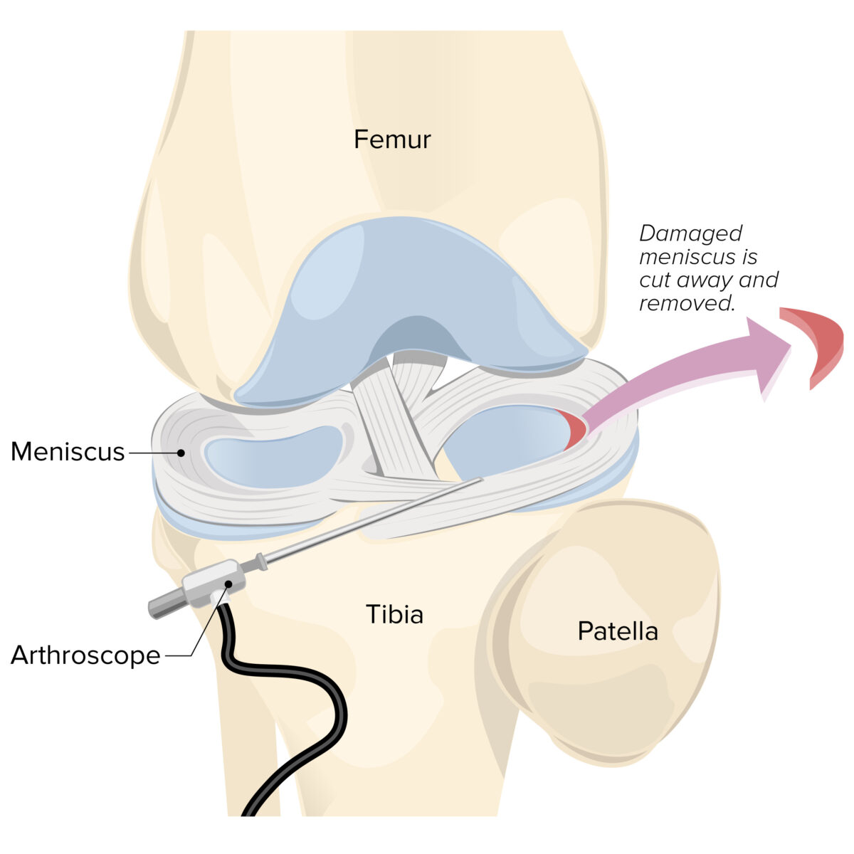
The menisci are fibrocartilaginous wedge-shaped structures between the distal femur and proximal tibia Tibia The second longest bone of the skeleton. It is located on the medial side of the lower leg, articulating with the fibula laterally, the talus distally, and the femur proximally. Knee Joint: Anatomy that stabilize and dissipate weight-bearing forces at the knee joint Knee joint The knee joint is made up of the articulations between the femur, tibia, and patella bones, and is one of the largest and most complex joints of the human body. The knee is classified as a synovial hinge joint, which primarily allows for flexion and extension with a more limited degree of translation and rotation. Knee Joint: Anatomy. A meniscus tear is an injury to the meniscus caused by rotational or shearing forces Shearing forces Vascular Resistance, Flow, and Mean Arterial Pressure across the tibiofemoral joint Tibiofemoral joint Knee Joint: Anatomy. Clinical presentation in a young patient with a traumatic tear includes a history of a twisting or rotational injury followed by pain Pain An unpleasant sensation induced by noxious stimuli which are detected by nerve endings of nociceptive neurons. Pain: Types and Pathways at the joint line with a small effusion. Clinical presentation in an older patient includes weight-bearing pain Pain An unpleasant sensation induced by noxious stimuli which are detected by nerve endings of nociceptive neurons. Pain: Types and Pathways at the joint line that mimics and accompanies progressive pain Pain An unpleasant sensation induced by noxious stimuli which are detected by nerve endings of nociceptive neurons. Pain: Types and Pathways due to the degenerative changes Degenerative Changes Spinal Stenosis of osteoarthritis Osteoarthritis Osteoarthritis (OA) is the most common form of arthritis, and is due to cartilage destruction and changes of the subchondral bone. The risk of developing this disorder increases with age, obesity, and repetitive joint use or trauma. Patients develop gradual joint pain, stiffness lasting < 30 minutes, and decreased range of motion. Osteoarthritis. Mechanical complaints (e.g., joint locking, catching, or clicking) are also common. Diagnosis can be challenging clinically and is often confirmed by diagnostic imaging or by direct visualization ( arthroscopy Arthroscopy Endoscopic examination, therapy and surgery of the joint. Knee Ligament Injuries). Management can be conservative or surgical, depending on the individual situation.

Last updated: Mar 29, 2023
Editorial responsibility: Stanley Oiseth, Lindsay Jones, Evelin Maza

Advertisement

Advertisement

Advertisement

Advertisement

Advertisement

Advertisement

Overview

Definition

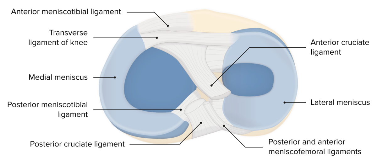
  • Menisci are wedge-shaped fibrocartilaginous structures found in the knee joint Knee joint The knee joint is made up of the articulations between the femur, tibia, and patella bones, and is one of the largest and most complex joints of the human body. The knee is classified as a synovial hinge joint, which primarily allows for flexion and extension with a more limited degree of translation and rotation. Knee Joint: Anatomy.
  • They cover the articular surface of the tibial plateau Plateau Cardiac Physiology.
  • They are essential for the normal function of the knee:
    • Absorb shock Shock Shock is a life-threatening condition associated with impaired circulation that results in tissue hypoxia. The different types of shock are based on the underlying cause: distributive (↑ cardiac output (CO), ↓ systemic vascular resistance (SVR)), cardiogenic (↓ CO, ↑ SVR), hypovolemic (↓ CO, ↑ SVR), obstructive (↓ CO), and mixed. Types of Shock
    • Distribute load
    • Increase stability
    • Provide lubrication
  • In younger patients Patients Individuals participating in the health care system for the purpose of receiving therapeutic, diagnostic, or preventive procedures. Clinician–Patient Relationship, tears are generally acute traumatic injuries (e.g., sports injuries, occupational injuries).
  • In older patient, tears are more commonly degenerative (accompanied by degenerative osteoarthritis Osteoarthritis Osteoarthritis (OA) is the most common form of arthritis, and is due to cartilage destruction and changes of the subchondral bone. The risk of developing this disorder increases with age, obesity, and repetitive joint use or trauma. Patients develop gradual joint pain, stiffness lasting < 30 minutes, and decreased range of motion. Osteoarthritis).
Menisci of the knee

Menisci of the knee

Image by Lecturio. License: CC BY-NC-SA 4.0

Epidemiology

  • Exact incidence Incidence The number of new cases of a given disease during a given period in a specified population. It also is used for the rate at which new events occur in a defined population. It is differentiated from prevalence, which refers to all cases in the population at a given time. Measures of Disease Frequency is unknown, but meniscus tear is a fairly common injury, especially with sports activities.
  • Tears of the medial meniscus are more common (because of attachment to the medial collateral ligament Medial collateral ligament Knee Joint: Anatomy).
  • Anterior cruciate ligament Anterior Cruciate Ligament A strong ligament of the knee that originates from the posteromedial portion of the lateral condyle of the femur, passes anteriorly and inferiorly between the condyles, and attaches to the depression in front of the intercondylar eminence of the tibia. Knee Joint: Anatomy ( ACL ACL A strong ligament of the knee that originates from the posteromedial portion of the lateral condyle of the femur, passes anteriorly and inferiorly between the condyles, and attaches to the depression in front of the intercondylar eminence of the tibia. Knee Joint: Anatomy) tears often have an associated medial meniscus tear.
  • Men are at increased risk compared to women.
  • Bimodal distribution Bimodal distribution Measures of Central Tendency and Dispersion:
    • Common in younger men: associated with athletic injuries 
    • Common in older adults: associated with degenerative changes Degenerative Changes Spinal Stenosis of the meniscus 

Etiology

Types of meniscal tears

There are several types of tear patterns in meniscal tears:
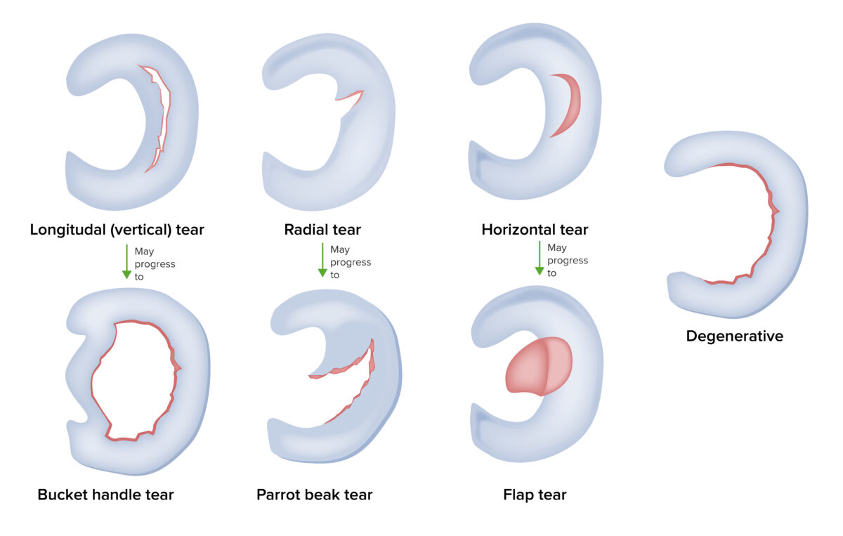
Types of meniscus tears

Types of meniscus tears

Image by Lecturio.

Pathophysiology

Clinical anatomy:

  • C-shaped wedges of fibrocartilage Fibrocartilage A type of cartilage whose matrix contains large bundles of collagen type I. Fibrocartilage is typically found in the intervertebral disk; pubic symphysis; tibial menisci; and articular discs in synovial joints. Cartilage: Histology between the distal femur and proximal tibia Tibia The second longest bone of the skeleton. It is located on the medial side of the lower leg, articulating with the fibula laterally, the talus distally, and the femur proximally. Knee Joint: Anatomy:
    • The medial meniscus is semilunar in shape.
    • The lateral meniscus is more circular in shape.
  • 70% type I collagen Type I Collagen Ehlers-Danlos Syndrome
  • Several ligaments and the joint capsule Capsule An envelope of loose gel surrounding a bacterial cell which is associated with the virulence of pathogenic bacteria. Some capsules have a well-defined border, whereas others form a slime layer that trails off into the medium. Most capsules consist of relatively simple polysaccharides but there are some bacteria whose capsules are made of polypeptides. Bacteroides attach to the meniscus in order to improve stability.
  • Blood supply is peripheral:

Traumatic mechanisms for tears:

Clinical Presentation

History

  • Often a challenging diagnosis with variable Variable Variables represent information about something that can change. The design of the measurement scales, or of the methods for obtaining information, will determine the data gathered and the characteristics of that data. As a result, a variable can be qualitative or quantitative, and may be further classified into subgroups. Types of Variables symptoms
  • Patients Patients Individuals participating in the health care system for the purpose of receiving therapeutic, diagnostic, or preventive procedures. Clinician–Patient Relationship are often able to continue to participate in the initial activity.
  • Assess mechanism of injury:
  • Patients Patients Individuals participating in the health care system for the purpose of receiving therapeutic, diagnostic, or preventive procedures. Clinician–Patient Relationship often report hearing a pop at the time of injury followed by localized pain Pain An unpleasant sensation induced by noxious stimuli which are detected by nerve endings of nociceptive neurons. Pain: Types and Pathways.
  • Mechanical symptoms are common:
    • Catching
    • Locking
    • Buckling
    • Sensation of giving way
  • Severity of symptoms often related to type of meniscal tear:
    • A bucket-handle tear may cause significant locking and associated pain Pain An unpleasant sensation induced by noxious stimuli which are detected by nerve endings of nociceptive neurons. Pain: Types and Pathways.
    • A small vertical tear may cause only intermittent vague symptoms.
  • Joint effusion Joint Effusion Septic Arthritis may develop over a few hours (more quickly with other associated injuries).

Physical examination

  • Inspection Inspection Dermatologic Examination:
  • Palpation Palpation Application of fingers with light pressure to the surface of the body to determine consistency of parts beneath in physical diagnosis; includes palpation for determining the outlines of organs. Dermatologic Examination:
    • Joint line tenderness
    • Evaluate for effusion: fluid shift Fluid Shift Translocation of body fluids from one compartment to another, such as from the vascular to the interstitial compartments. Fluid shifts are associated with profound changes in vascular permeability and water-electrolyte imbalance. The shift can also be from the lower body to the upper body as in conditions of weightlessness. Volume Depletion and Dehydration test
  • Range of motion Range of motion The distance and direction to which a bone joint can be extended. Range of motion is a function of the condition of the joints, muscles, and connective tissues involved. Joint flexibility can be improved through appropriate muscle strength exercises. Examination of the Upper Limbs (ROM): Does the patient have full flexion Flexion Examination of the Upper Limbs and extension Extension Examination of the Upper Limbs?
  • Ligamentous stability:
  • Meniscal tests: Multiple tests have been described, with variable Variable Variables represent information about something that can change. The design of the measurement scales, or of the methods for obtaining information, will determine the data gathered and the characteristics of that data. As a result, a variable can be qualitative or quantitative, and may be further classified into subgroups. Types of Variables sensitivity and specificity Sensitivity and Specificity Binary classification measures to assess test results. Sensitivity or recall rate is the proportion of true positives. Specificity is the probability of correctly determining the absence of a condition. Epidemiological Values of Diagnostic Tests:
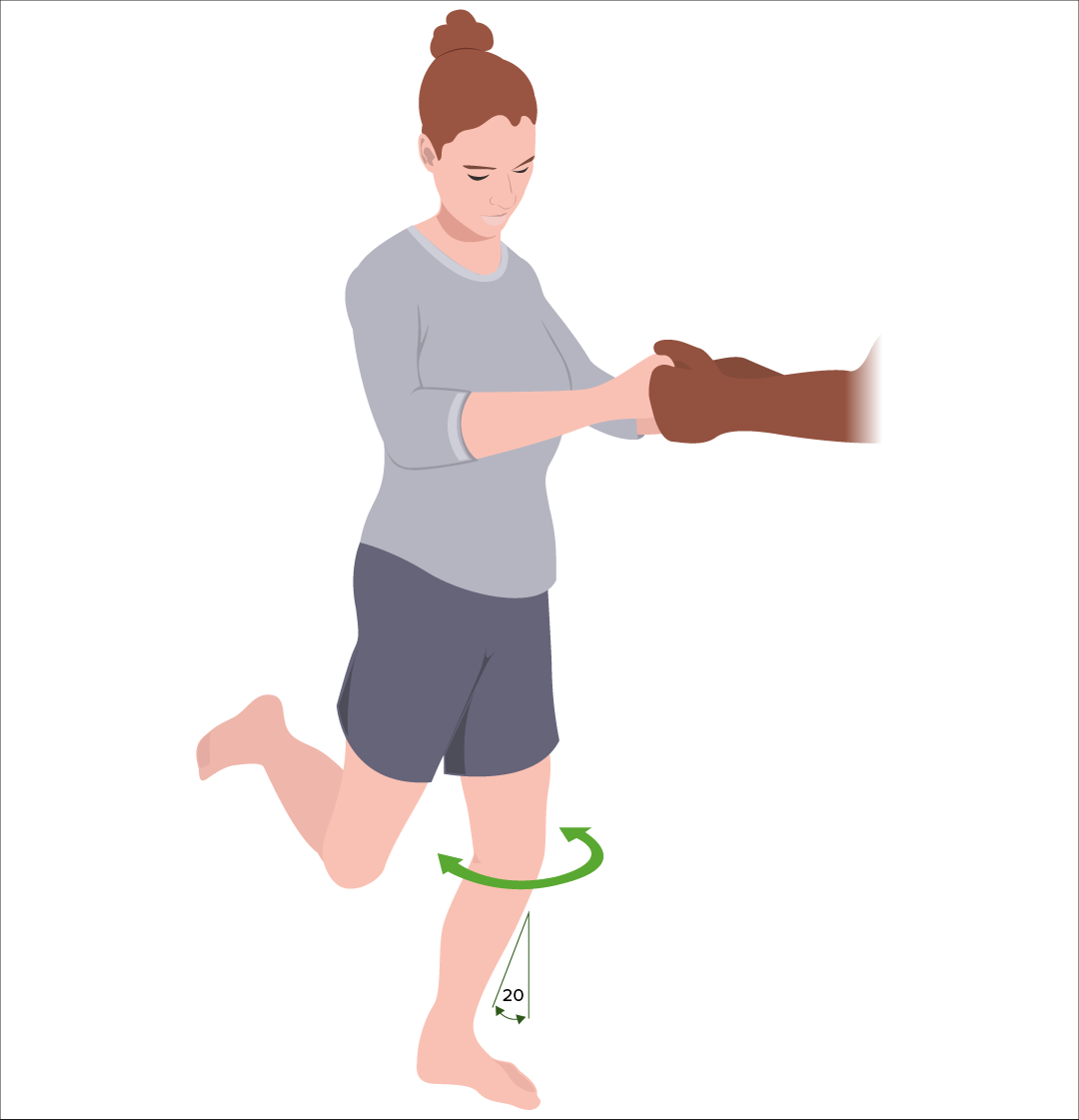
    • Thessaly test:
      • The patient is weight-bearing (flat-footed) on 1 leg Leg The lower leg, or just “leg” in anatomical terms, is the part of the lower limb between the knee and the ankle joint. The bony structure is composed of the tibia and fibula bones, and the muscles of the leg are grouped into the anterior, lateral, and posterior compartments by extensions of fascia. Leg: Anatomy flexed at 20 degrees and asked to initiate internal and external rotation External Rotation Examination of the Upper Limbs.
      • A positive test is joint line discomfort or complaints of locking or catching.
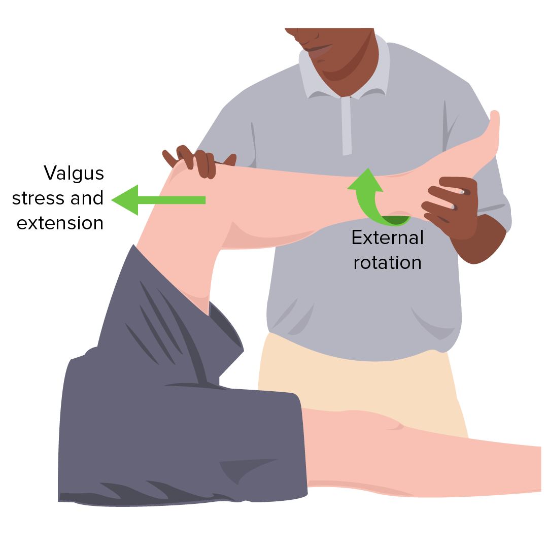
    • McMurray test:
      • The patient is supine, and the knee is taken through its ROM with an associated varus or valgus force with internal and external rotation External Rotation Examination of the Upper Limbs of the foot Foot The foot is the terminal portion of the lower limb, whose primary function is to bear weight and facilitate locomotion. The foot comprises 26 bones, including the tarsal bones, metatarsal bones, and phalanges. The bones of the foot form longitudinal and transverse arches and are supported by various muscles, ligaments, and tendons. Foot: Anatomy.
      • A positive test includes popping or clicking of the knee with associated pain Pain An unpleasant sensation induced by noxious stimuli which are detected by nerve endings of nociceptive neurons. Pain: Types and Pathways.
    • Bounce home test:
      • The patient is supine, and their heel is supported while the examiner passively flexes the knee. The knee is then passively allowed to extend under the influence of gravity.
      • The knee should normally extend fully, or “bounce home,” with a sharp end feel.
      • Positive if incomplete extension Extension Examination of the Upper Limbs of the knee or has a rubbery end feel 
    • Childress test:
    • Apley compression Compression Blunt Chest Trauma and distraction test:
      • The patient is prone, with the knee flexed to 90 degrees.
      • The examiner internally and externally rotates the tibia Tibia The second longest bone of the skeleton. It is located on the medial side of the lower leg, articulating with the fibula laterally, the talus distally, and the femur proximally. Knee Joint: Anatomy while applying compression Compression Blunt Chest Trauma then distraction.
      • Positive for ligament injury if rotation Rotation Motion of an object in which either one or more points on a line are fixed. It is also the motion of a particle about a fixed point. X-rays plus distraction is more painful or increased rotation Rotation Motion of an object in which either one or more points on a line are fixed. It is also the motion of a particle about a fixed point. X-rays compared to normal side
      • Positive for meniscal injury if rotation Rotation Motion of an object in which either one or more points on a line are fixed. It is also the motion of a particle about a fixed point. X-rays plus compression Compression Blunt Chest Trauma is more painful or decreased rotation Rotation Motion of an object in which either one or more points on a line are fixed. It is also the motion of a particle about a fixed point. X-rays compared to normal side

Diagnosis

Diagnosis is usually suspected with a thorough history and physical examination, and often confirmed with diagnostic imaging (e.g., MRI) or by direct visualization (e.g., arthroscopy Arthroscopy Endoscopic examination, therapy and surgery of the joint. Knee Ligament Injuries).

Laboratory studies

  • Only indicated if evaluating for other possible diagnoses:
    • Infection
    • Rheumatological disease
  • Arthrocentesis Arthrocentesis Puncture and aspiration of fluid (e.g., synovial fluid) from a joint cavity. It is used sometimes to irrigate or administer drugs into a joint cavity. Septic Arthritis generally not indicated:
    • May be indicated in the evaluation for other diagnoses
    • May provide symptomatic relief when there is a large effusion

Imaging

X-ray X-ray Penetrating electromagnetic radiation emitted when the inner orbital electrons of an atom are excited and release radiant energy. X-ray wavelengths range from 1 pm to 10 nm. Hard x-rays are the higher energy, shorter wavelength x-rays. Soft x-rays or grenz rays are less energetic and longer in wavelength. The short wavelength end of the x-ray spectrum overlaps the gamma rays wavelength range. The distinction between gamma rays and x-rays is based on their radiation source. Pulmonary Function Tests:
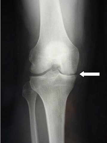
  • Indicated to evaluate for associated acute bony injury or underlying OA OA Osteoarthritis (OA) is the most common form of arthritis, and is due to cartilage destruction and changes of the subchondral bone. The risk of developing this disorder increases with age, obesity, and repetitive joint use or trauma. Patients develop gradual joint pain, stiffness lasting < 30 minutes, and decreased range of motion. Osteoarthritis changes
  • Anteroposterior (AP), lateral, oblique, sunrise, and weight-bearing projections of the knee are ideal.
X-ray demonstrating medial joint space narrowing

X-ray demonstrating medial joint space narrowing

Image: “Postoperative radiography at 3 month follow-up” by Steinmetz S, Bonnomet F, Rahme M, Adam P, Ehlinger M. License: CC BY 4.0

Ultrasound:

  • Safe and inexpensive
  • Able to visualize peripheral meniscus tears
  • Limited ability to visualize deep structures of the knee

MRI:

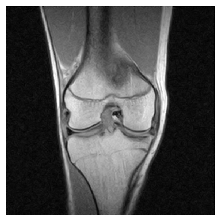
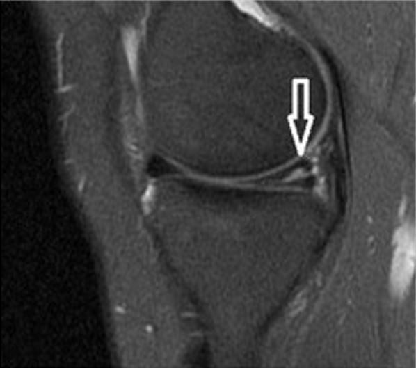
  • Imaging modality of choice for diagnosis and characterization of meniscal tears
  • Accuracy greater than 90%
  • Not always indicated prior to arthroscopy Arthroscopy Endoscopic examination, therapy and surgery of the joint. Knee Ligament Injuries: may give additional information regarding injuries to associated ligaments and cartilage Cartilage Cartilage is a type of connective tissue derived from embryonic mesenchyme that is responsible for structural support, resilience, and the smoothness of physical actions. Perichondrium (connective tissue membrane surrounding cartilage) compensates for the absence of vasculature in cartilage by providing nutrition and support. Cartilage: Histology

Arthroscopy Arthroscopy Endoscopic examination, therapy and surgery of the joint. Knee Ligament Injuries:
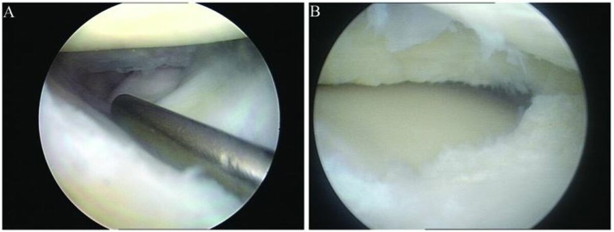
  • Gold standard for diagnosis of a meniscal tear
  • Provides direct visualization of the meniscus and other associated tissues
  • A diagnostic arthroscopy Arthroscopy Endoscopic examination, therapy and surgery of the joint. Knee Ligament Injuries can be converted to therapeutic arthroscopy Arthroscopy Endoscopic examination, therapy and surgery of the joint. Knee Ligament Injuries if pathologic findings are present.
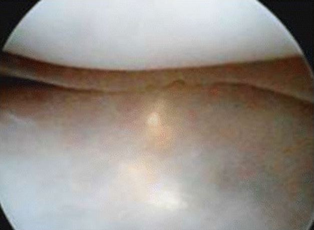
Arthroscopy with meniscus repair

Arthroscopy with arthroscopic meniscus repair:
A: discoid lateral meniscus with longitudinal tear
B: discoid lateral meniscus following partial resection

Image: “Discoid lateral meniscus with longitudinal tear” by Cao H, Zhang Y, Qian W, Cheng XH, Ke Y, Guo XP. License: CC BY 3.0

Management

Management of meniscal tears is dependent on the type of tear, age of the patient, occupation of the patient, associated mechanical symptoms, and other associated injuries (e.g., ACL tear ACL tear Knee Pain).

  • Younger patients Patients Individuals participating in the health care system for the purpose of receiving therapeutic, diagnostic, or preventive procedures. Clinician–Patient Relationship with unstable, painful meniscal tears or associated mechanical symptoms will benefit from arthroscopic surgical management.
  • Older patients Patients Individuals participating in the health care system for the purpose of receiving therapeutic, diagnostic, or preventive procedures. Clinician–Patient Relationship with degenerative meniscal tears may benefit from an initial trial of physical therapy Physical Therapy Becker Muscular Dystrophy (PT).
  • Factors indicating arthroscopy Arthroscopy Endoscopic examination, therapy and surgery of the joint. Knee Ligament Injuries intervention:
    • Loss of motion or a “locked” knee
    • Associated ligamentous injury (e.g., ACL tear ACL tear Knee Pain)
    • Lack of improvement with conservative care

Conservative management

Conservative management indicated as initial treatment in degenerative tears in older patients Patients Individuals participating in the health care system for the purpose of receiving therapeutic, diagnostic, or preventive procedures. Clinician–Patient Relationship or prior to arthroscopy Arthroscopy Endoscopic examination, therapy and surgery of the joint. Knee Ligament Injuries in younger patients Patients Individuals participating in the health care system for the purpose of receiving therapeutic, diagnostic, or preventive procedures. Clinician–Patient Relationship:
  • Rest, ice, compression, and elevation (RICE)
  • Oral analgesics as needed
  • Bracing, knee sleeves, and crutches may be used briefly for symptomatic relief.
  • PT:
    • Begin with ROM, quadriceps strengthening
    • Advance based on symptoms
    • Cross-training for specific exercise if appropriate (e.g., swimming, biking)

Surgical management

  • Arthroscopy Arthroscopy Endoscopic examination, therapy and surgery of the joint. Knee Ligament Injuries indications:
    • Mechanical symptoms
    • Persistent pain Pain An unpleasant sensation induced by noxious stimuli which are detected by nerve endings of nociceptive neurons. Pain: Types and Pathways and effusion
    • Associated ligamentous injuries 
    • Lack of response to initial conservative management
  • Arthroscopy Arthroscopy Endoscopic examination, therapy and surgery of the joint. Knee Ligament Injuries is used diagnostically and therapeutically. 
  • Meniscal repair preferred over partial meniscectomy:
    • For large tears 
    • Meniscal tears in younger patients Patients Individuals participating in the health care system for the purpose of receiving therapeutic, diagnostic, or preventive procedures. Clinician–Patient Relationship
  • Total meniscectomy has been shown to accelerate osteoarthritis Osteoarthritis Osteoarthritis (OA) is the most common form of arthritis, and is due to cartilage destruction and changes of the subchondral bone. The risk of developing this disorder increases with age, obesity, and repetitive joint use or trauma. Patients develop gradual joint pain, stiffness lasting < 30 minutes, and decreased range of motion. Osteoarthritis and is avoided.
  • Meniscal transplantation with a cadaveric allograft is occasionally used in younger patients Patients Individuals participating in the health care system for the purpose of receiving therapeutic, diagnostic, or preventive procedures. Clinician–Patient Relationship with significant meniscal trauma.

Rehabilitation

  • Rehabilitation depends on:
    • Age of patient 
    • Occupational/sports involvement 
    • Type of meniscal injury
    • Type of management of the meniscal tear (conservative vs. surgical)
  • Goals of rehabilitation:
    • Progressive ROM
    • Aerobic conditioning and strength training 
    • Return to normal activity levels
  • Rehabilitation after meniscal repair or meniscus transplant is similar, except for restrictions on ROM and weight-bearing status.

Complications

Prognosis Prognosis A prediction of the probable outcome of a disease based on a individual’s condition and the usual course of the disease as seen in similar situations. Non-Hodgkin Lymphomas

Long-term outcomes depend on:

  • Type of tear
  • Type of repair 
  • Amount of meniscus removed during surgical procedures 
  • Age of patient
  • Underlying condition of the knee

Clinical Relevance

Differential diagnosis

  • Iliotibial band Iliotibial band Thigh: Anatomy syndrome: cause of lateral knee pain Pain An unpleasant sensation induced by noxious stimuli which are detected by nerve endings of nociceptive neurons. Pain: Types and Pathways, attributed to the repetitive friction between the iliotibial band Iliotibial band Thigh: Anatomy and the lateral femoral condyle ( overuse injury Overuse Injury Osgood-Schlatter Disease). Iliotibial band Iliotibial band Thigh: Anatomy syndrome is frequently seen in athletes and military personnel. Diagnosis is clinical. Conservative treatment includes RICE and PT. Corticosteroid injections may be utilized in refractory cases. 
  • Knee osteochondritis Osteochondritis Inflammation of a bone and its overlaying cartilage. Osteochondritis Dissecans dissecans ( OCD OCD Obsessive-compulsive disorder (OCD) is a condition characterized by obsessions (recurring and intrusive thoughts, urges, or images) and/or compulsions (repetitive actions the person is compelled to perform) that are time-consuming and associated with functional impairment. Obsessive-compulsive Disorder (OCD)): an orthopedic disorder characterized by the detachment of a segment of subchondral bone Subchondral Bone Osteochondritis Dissecans and cartilage Cartilage Cartilage is a type of connective tissue derived from embryonic mesenchyme that is responsible for structural support, resilience, and the smoothness of physical actions. Perichondrium (connective tissue membrane surrounding cartilage) compensates for the absence of vasculature in cartilage by providing nutrition and support. Cartilage: Histology due to focal aseptic necrosis Focal Aseptic Necrosis Osteochondritis Dissecans. The disorder may occur at any age, but it is most commonly seen in adolescents who participate in competitive sports. Diagnosis is by X-ray X-ray Penetrating electromagnetic radiation emitted when the inner orbital electrons of an atom are excited and release radiant energy. X-ray wavelengths range from 1 pm to 10 nm. Hard x-rays are the higher energy, shorter wavelength x-rays. Soft x-rays or grenz rays are less energetic and longer in wavelength. The short wavelength end of the x-ray spectrum overlaps the gamma rays wavelength range. The distinction between gamma rays and x-rays is based on their radiation source. Pulmonary Function Tests. Conservative treatment includes RICE and PT. Arthroscopic intervention may be required in refractory cases. 
  • Lumbosacral radiculopathy Radiculopathy Disease involving a spinal nerve root which may result from compression related to intervertebral disk displacement; spinal cord injuries; spinal diseases; and other conditions. Clinical manifestations include radicular pain, weakness, and sensory loss referable to structures innervated by the involved nerve root. Rheumatoid Arthritis: pain Pain An unpleasant sensation induced by noxious stimuli which are detected by nerve endings of nociceptive neurons. Pain: Types and Pathways caused by lumbar nerve root irritation or compression Compression Blunt Chest Trauma. Lumbosacral radiculopathy Radiculopathy Disease involving a spinal nerve root which may result from compression related to intervertebral disk displacement; spinal cord injuries; spinal diseases; and other conditions. Clinical manifestations include radicular pain, weakness, and sensory loss referable to structures innervated by the involved nerve root. Rheumatoid Arthritis is commonly attributed to disk herniation Herniation Omphalocele, vertebral degeneration, and neuroforaminal narrowing. Presentation includes low back pain Pain An unpleasant sensation induced by noxious stimuli which are detected by nerve endings of nociceptive neurons. Pain: Types and Pathways with dermatomal Dermatomal Dermatologic Examination radiation Radiation Emission or propagation of acoustic waves (sound), electromagnetic energy waves (such as light; radio waves; gamma rays; or x-rays), or a stream of subatomic particles (such as electrons; neutrons; protons; or alpha particles). Osteosarcoma to lower limbs, accompanied by paresthesias Paresthesias Subjective cutaneous sensations (e.g., cold, warmth, tingling, pressure, etc.) that are experienced spontaneously in the absence of stimulation. Posterior Cord Syndrome, myotomal muscle weakness, and hyporeflexia Hyporeflexia Duchenne Muscular Dystrophy. Diagnosis is clinical and confirmed by MRI. Conservative treatment includes RICE and PT. Refractory cases may require epidural steroid injection or surgical decompression. 

Associated conditions/injuries

  • Anterior and posterior cruciate ligament Posterior Cruciate Ligament A strong ligament of the knee that originates from the anterolateral surface of the medial condyle of the femur, passes posteriorly and inferiorly between the condyles, and attaches to the posterior intercondylar area of the tibia. Knee Joint: Anatomy injury: anterior cruciate ligament Anterior Cruciate Ligament A strong ligament of the knee that originates from the posteromedial portion of the lateral condyle of the femur, passes anteriorly and inferiorly between the condyles, and attaches to the depression in front of the intercondylar eminence of the tibia. Knee Joint: Anatomy ( ACL ACL A strong ligament of the knee that originates from the posteromedial portion of the lateral condyle of the femur, passes anteriorly and inferiorly between the condyles, and attaches to the depression in front of the intercondylar eminence of the tibia. Knee Joint: Anatomy) injuries occur most frequently secondary to non-contact, deceleration Deceleration A decrease in the rate of speed. Blunt Chest Trauma injuries, and they are also seen in contact sports secondary to trauma. Posterior cruciate injuries are seen in contact sports and high-energy trauma such as motor vehicle accidents Motor Vehicle Accidents Spinal Cord Injuries. Both injuries may have associated meniscal trauma. 
  • Patellofemoral pain syndrome Patellofemoral pain syndrome A syndrome characterized by retropatellar or peripatellar pain resulting from physical and biochemical changes in the patellofemoral joint. The pain is most prominent when ascending or descending stairs, squatting, or sitting with flexed knees. There is a lack of consensus on the etiology and treatment. The syndrome is often confused with (or accompanied by) chondromalacia patellae, the latter describing a pathological condition of the cartilage and not a syndrome. Examination of the Lower Limbs: the presence of vague anterior knee pain Pain An unpleasant sensation induced by noxious stimuli which are detected by nerve endings of nociceptive neurons. Pain: Types and Pathways, generally without any structural changes in the articular cartilage Cartilage Cartilage is a type of connective tissue derived from embryonic mesenchyme that is responsible for structural support, resilience, and the smoothness of physical actions. Perichondrium (connective tissue membrane surrounding cartilage) compensates for the absence of vasculature in cartilage by providing nutrition and support. Cartilage: Histology that would be considered pathological. Patellofemoral pain syndrome Patellofemoral pain syndrome A syndrome characterized by retropatellar or peripatellar pain resulting from physical and biochemical changes in the patellofemoral joint. The pain is most prominent when ascending or descending stairs, squatting, or sitting with flexed knees. There is a lack of consensus on the etiology and treatment. The syndrome is often confused with (or accompanied by) chondromalacia patellae, the latter describing a pathological condition of the cartilage and not a syndrome. Examination of the Lower Limbs may be considered in a patient with meniscal symptoms.
  • Tibia Tibia The second longest bone of the skeleton. It is located on the medial side of the lower leg, articulating with the fibula laterally, the talus distally, and the femur proximally. Knee Joint: Anatomy and fibula Fibula The bone of the lower leg lateral to and smaller than the tibia. In proportion to its length, it is the most slender of the long bones. Leg: Anatomy fractures: intra-articular fractures of the distal femur or proximal tibia Tibia The second longest bone of the skeleton. It is located on the medial side of the lower leg, articulating with the fibula laterally, the talus distally, and the femur proximally. Knee Joint: Anatomy may have associated meniscal injuries.
  • Osteoarthritis Osteoarthritis Osteoarthritis (OA) is the most common form of arthritis, and is due to cartilage destruction and changes of the subchondral bone. The risk of developing this disorder increases with age, obesity, and repetitive joint use or trauma. Patients develop gradual joint pain, stiffness lasting < 30 minutes, and decreased range of motion. Osteoarthritis: the most common form of arthritis Arthritis Acute or chronic inflammation of joints. Osteoarthritis. Osteoarthritis Osteoarthritis Osteoarthritis (OA) is the most common form of arthritis, and is due to cartilage destruction and changes of the subchondral bone. The risk of developing this disorder increases with age, obesity, and repetitive joint use or trauma. Patients develop gradual joint pain, stiffness lasting < 30 minutes, and decreased range of motion. Osteoarthritis is due to cartilage Cartilage Cartilage is a type of connective tissue derived from embryonic mesenchyme that is responsible for structural support, resilience, and the smoothness of physical actions. Perichondrium (connective tissue membrane surrounding cartilage) compensates for the absence of vasculature in cartilage by providing nutrition and support. Cartilage: Histology destruction and changes of the subchondral bone Subchondral Bone Osteochondritis Dissecans in weight-bearing joints. The risk of developing this disorder increases with age, obesity Obesity Obesity is a condition associated with excess body weight, specifically with the deposition of excessive adipose tissue. Obesity is considered a global epidemic. Major influences come from the western diet and sedentary lifestyles, but the exact mechanisms likely include a mixture of genetic and environmental factors. Obesity, and trauma. Degenerative changes Degenerative Changes Spinal Stenosis are also commonly present, and meniscal injuries predispose the patient for the development of premature osteoarthritis Premature Osteoarthritis Overview of Bone Fractures

References

  1. Lafferty, PM, & Cole, PA. (2020). In Browner, BD, et al. (Eds.), Tibial Plateau Fractures. pp. 2181–2276. https://pubmed.ncbi.nlm.nih.gov/29261932/
  2. Hansen, JT. (2019). Lower limb. In Netter’s Clinical Anatomy. pp. 291–366. https://www.worldcat.org/title/netters-clinical-anatomy/oclc/1017817783
  3. Raj, MA, & Bubnis, MA. (2021). Knee meniscal tears. StatPearls. Treasure Island (FL): StatPearls Publishing. http://www.ncbi.nlm.nih.gov/books/NBK431067/ 
  4. Dennis, A, et al. (2021). Meniscal injury of the knee. UpToDate. Retrieved July 16, 2021, from https://www.uptodate.com/contents/meniscal-injury-of-the-knee
  5. Lento, P, Marshall, B, & Akuthota, V. (2020). Chapter 72: Meniscal injuries. In Frontera, W.R., et al. (Eds.). pp. 403–409. Elsevier.

Create your free account or log in to continue reading!

Sign up now and get free access to Lecturio with concept pages, medical videos, and questions for your medical education.

User Reviews

Unwrap New Skills This Holiday 🎄 Save 30% on all plans now!

Details