Advertisement

Advertisement

Advertisement

Advertisement

Mediastinitis

Mediastinitis refers to an infection or inflammation Inflammation Inflammation is a complex set of responses to infection and injury involving leukocytes as the principal cellular mediators in the body's defense against pathogenic organisms. Inflammation is also seen as a response to tissue injury in the process of wound healing. The 5 cardinal signs of inflammation are pain, heat, redness, swelling, and loss of function. Inflammation involving the mediastinum Mediastinum The mediastinum is the thoracic area between the 2 pleural cavities. The mediastinum contains vital structures of the circulatory, respiratory, digestive, and nervous systems including the heart and esophagus, and major thoracic vessels. Mediastinum and Great Vessels: Anatomy (a region in the thoracic cavity containing the heart, thymus Thymus A single, unpaired primary lymphoid organ situated in the mediastinum, extending superiorly into the neck to the lower edge of the thyroid gland and inferiorly to the fourth costal cartilage. It is necessary for normal development of immunologic function early in life. By puberty, it begins to involute and much of the tissue is replaced by fat. Lymphatic Drainage System: Anatomy gland, portions of the esophagus Esophagus The esophagus is a muscular tube-shaped organ of around 25 centimeters in length that connects the pharynx to the stomach. The organ extends from approximately the 6th cervical vertebra to the 11th thoracic vertebra and can be divided grossly into 3 parts: the cervical part, the thoracic part, and the abdominal part. Esophagus: Anatomy, and trachea Trachea The trachea is a tubular structure that forms part of the lower respiratory tract. The trachea is continuous superiorly with the larynx and inferiorly becomes the bronchial tree within the lungs. The trachea consists of a support frame of semicircular, or C-shaped, rings made out of hyaline cartilage and reinforced by collagenous connective tissue. Trachea: Anatomy). Acute mediastinitis can be caused by bacterial infection due to direct contamination, hematogenous Hematogenous Hepatocellular Carcinoma (HCC) and Liver Metastases or lymphatic spread, or extension Extension Examination of the Upper Limbs of infection from nearby structures. Chronic mediastinitis, also known as fibrosing mediastinitis, is commonly related to chronic inflammatory conditions that cause the proliferation of connective tissue Connective tissue Connective tissues originate from embryonic mesenchyme and are present throughout the body except inside the brain and spinal cord. The main function of connective tissues is to provide structural support to organs. Connective tissues consist of cells and an extracellular matrix. Connective Tissue: Histology. Mediastinitis is treated with broad-spectrum Broad-Spectrum Fluoroquinolones antibiotics and surgery, as well as supportive care, depending on the etiology. Mortality Mortality All deaths reported in a given population. Measures of Health Status from this condition is high.

Last updated: Jan 18, 2024

Editorial responsibility: Stanley Oiseth, Lindsay Jones, Evelin Maza

Advertisement

Advertisement

Advertisement

Advertisement

Advertisement

Advertisement

Advertisement

Advertisement

Anatomy and Etiology

Anatomy

Mediastinum Mediastinum The mediastinum is the thoracic area between the 2 pleural cavities. The mediastinum contains vital structures of the circulatory, respiratory, digestive, and nervous systems including the heart and esophagus, and major thoracic vessels. Mediastinum and Great Vessels: Anatomy:
  • Central compartment of thoracic cavity bordered by:
    • Lung pleural sacs laterally
    • Thoracic outlet Thoracic Outlet Thoracic Outlet Syndrome above
    • Diaphragm Diaphragm The diaphragm is a large, dome-shaped muscle that separates the thoracic cavity from the abdominal cavity. The diaphragm consists of muscle fibers and a large central tendon, which is divided into right and left parts. As the primary muscle of inspiration, the diaphragm contributes 75% of the total inspiratory muscle force. Diaphragm: Anatomy below
  • Structures inside mediastinum Mediastinum The mediastinum is the thoracic area between the 2 pleural cavities. The mediastinum contains vital structures of the circulatory, respiratory, digestive, and nervous systems including the heart and esophagus, and major thoracic vessels. Mediastinum and Great Vessels: Anatomy:
    • Heart
    • Thymus Thymus A single, unpaired primary lymphoid organ situated in the mediastinum, extending superiorly into the neck to the lower edge of the thyroid gland and inferiorly to the fourth costal cartilage. It is necessary for normal development of immunologic function early in life. By puberty, it begins to involute and much of the tissue is replaced by fat. Lymphatic Drainage System: Anatomy
    • Great vessels (vena cava, aorta Aorta The main trunk of the systemic arteries. Mediastinum and Great Vessels: Anatomy, pulmonary arteries Arteries Arteries are tubular collections of cells that transport oxygenated blood and nutrients from the heart to the tissues of the body. The blood passes through the arteries in order of decreasing luminal diameter, starting in the largest artery (the aorta) and ending in the small arterioles. Arteries are classified into 3 types: large elastic arteries, medium muscular arteries, and small arteries and arterioles. Arteries: Histology)
    • Esophagus Esophagus The esophagus is a muscular tube-shaped organ of around 25 centimeters in length that connects the pharynx to the stomach. The organ extends from approximately the 6th cervical vertebra to the 11th thoracic vertebra and can be divided grossly into 3 parts: the cervical part, the thoracic part, and the abdominal part. Esophagus: Anatomy
    • Distal portion of trachea Trachea The trachea is a tubular structure that forms part of the lower respiratory tract. The trachea is continuous superiorly with the larynx and inferiorly becomes the bronchial tree within the lungs. The trachea consists of a support frame of semicircular, or C-shaped, rings made out of hyaline cartilage and reinforced by collagenous connective tissue. Trachea: Anatomy
    • Mainstem bronchi Bronchi The larger air passages of the lungs arising from the terminal bifurcation of the trachea. They include the largest two primary bronchi which branch out into secondary bronchi, and tertiary bronchi which extend into bronchioles and pulmonary alveoli. Bronchial Tree: Anatomy
    • Phrenic nerve Phrenic nerve The motor nerve of the diaphragm. The phrenic nerve fibers originate in the cervical spinal column (mostly C4) and travel through the cervical plexus to the diaphragm. Diaphragm: Anatomy
    • Vagus nerve Vagus nerve The 10th cranial nerve. The vagus is a mixed nerve which contains somatic afferents (from skin in back of the ear and the external auditory meatus), visceral afferents (from the pharynx, larynx, thorax, and abdomen), parasympathetic efferents (to the thorax and abdomen), and efferents to striated muscle (of the larynx and pharynx). Pharynx: Anatomy (cranial nerve (CN) 10)
Mediastinum

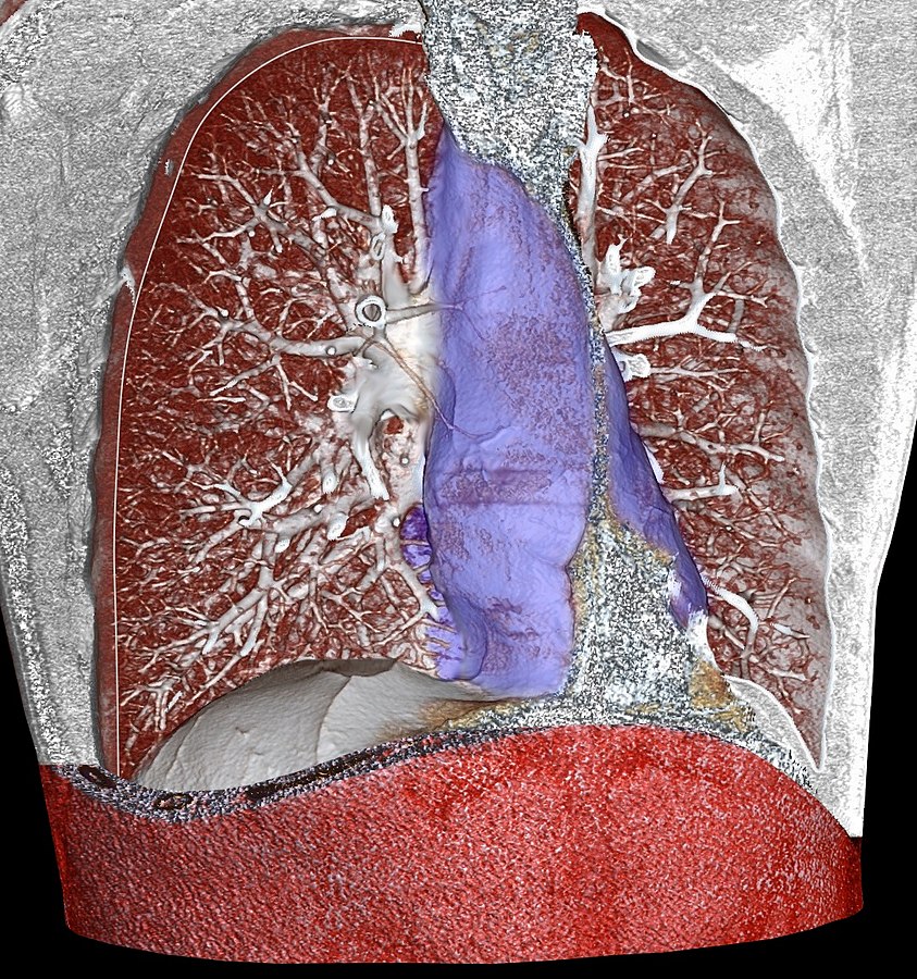
Three-dimensional rendering of high-resolution CT identifying the mediastinum:
The area highlighted in blue is the mediastinum. This area is bordered by the lung pleural sacs laterally, the thoracic outlet above, and the diaphragm below. The structures contained in the mediastinum include the heart, thymus, great vessels (vena cava, aorta, pulmonary arteries), esophagus, distal portion of trachea, mainstem bronchi, and phrenic and vagus nerves.

Image: “Mediastinum” by Mikael Häggström. License: CC0

Etiology

Acute mediastinitis:

  • Esophageal perforation Perforation A pathological hole in an organ, blood vessel or other soft part of the body, occurring in the absence of external force. Esophagitis:
  • Postoperative mediastinitis:
    • One of the most common causes
    • 1%–2% of cardiac surgeries
    • Fever Fever Fever is defined as a measured body temperature of at least 38°C (100.4°F). Fever is caused by circulating endogenous and/or exogenous pyrogens that increase levels of prostaglandin E2 in the hypothalamus. Fever is commonly associated with chills, rigors, sweating, and flushing of the skin. Fever, chest pain Pain An unpleasant sensation induced by noxious stimuli which are detected by nerve endings of nociceptive neurons. Pain: Types and Pathways, purulent or erythematous chest wound
    • Most patients Patients Individuals participating in the health care system for the purpose of receiving therapeutic, diagnostic, or preventive procedures. Clinician–Patient Relationship present within 1 month of surgery.
  • Descending necrotizing mediastinitis:
    • Usually direct extension Extension Examination of the Upper Limbs (retropharyngeal abscess Abscess Accumulation of purulent material in tissues, organs, or circumscribed spaces, usually associated with signs of infection. Chronic Granulomatous Disease, tonsillitis Tonsillitis Tonsillitis is inflammation of the pharynx or pharyngeal tonsils, and therefore is also called pharyngitis. An infectious etiology in the setting of tonsillitis is referred to as infectious pharyngitis, which is caused by viruses (most common), bacteria, or fungi. Tonsillitis, pharyngitis Pharyngitis Pharyngitis is an inflammation of the back of the throat (pharynx). Pharyngitis is usually caused by an upper respiratory tract infection, which is viral in most cases. It typically results in a sore throat and fever. Other symptoms may include a runny nose, cough, headache, and hoarseness. Pharyngitis, epiglottitis Epiglottitis Epiglottitis (or “supraglottitis”) is an inflammation of the epiglottis and adjacent supraglottic structures. The majority of cases are caused by bacterial infection. Symptoms are rapid in onset and severe. Epiglottitis)
    • Worsening sore throat Sore throat Pharyngitis is an inflammation of the back of the throat (pharynx). Pharyngitis is usually caused by an upper respiratory tract infection, which is viral in most cases. It typically results in a sore throat and fever. Other symptoms may include a runny nose, cough, headache, and hoarseness. Pharyngitis and/or neck Neck The part of a human or animal body connecting the head to the rest of the body. Peritonsillar Abscess swelling Swelling Inflammation
    • Fever Fever Fever is defined as a measured body temperature of at least 38°C (100.4°F). Fever is caused by circulating endogenous and/or exogenous pyrogens that increase levels of prostaglandin E2 in the hypothalamus. Fever is commonly associated with chills, rigors, sweating, and flushing of the skin. Fever, chest pain Pain An unpleasant sensation induced by noxious stimuli which are detected by nerve endings of nociceptive neurons. Pain: Types and Pathways, shortness of breath Shortness of breath Dyspnea is the subjective sensation of breathing discomfort. Dyspnea is a normal manifestation of heavy physical or psychological exertion, but also may be caused by underlying conditions (both pulmonary and extrapulmonary). Dyspnea
    • Has become less frequent due to antibiotic usage

Chronic mediastinitis:

Fibrosing mediastinitis:

  • Related to long-standing inflammatory conditions
  • Marked by growth of acellular collagen Collagen A polypeptide substance comprising about one third of the total protein in mammalian organisms. It is the main constituent of skin; connective tissue; and the organic substance of bones (bone and bones) and teeth (tooth). Connective Tissue: Histology and fibrous Fibrous Fibrocystic Change tissue within chest
  • Granulomatous mediastinitis ( histoplasmosis Histoplasmosis Histoplasmosis is an infection caused by Histoplasma capsulatum, a dimorphic fungus. Transmission is through inhalation, and exposure to soils containing bird or bat droppings increases the risk of infection. Most infections are asymptomatic; however, immunocompromised individuals generally develop acute pulmonary infection, chronic infection, or even disseminated disease. Histoplasma/Histoplasmosis, tuberculosis Tuberculosis Tuberculosis (TB) is an infectious disease caused by Mycobacterium tuberculosis complex bacteria. The bacteria usually attack the lungs but can also damage other parts of the body. Approximately 30% of people around the world are infected with this pathogen, with the majority harboring a latent infection. Tuberculosis spreads through the air when a person with active pulmonary infection coughs or sneezes. Tuberculosis infections Infections Invasion of the host organism by microorganisms or their toxins or by parasites that can cause pathological conditions or diseases. Chronic Granulomatous Disease)
  • Non-granulomatous fibrosing mediastinitis (reaction to drugs or radiation Radiation Emission or propagation of acoustic waves (sound), electromagnetic energy waves (such as light; radio waves; gamma rays; or x-rays), or a stream of subatomic particles (such as electrons; neutrons; protons; or alpha particles). Osteosarcoma therapy)
  • Autoimmune diseases Autoimmune diseases Disorders that are characterized by the production of antibodies that react with host tissues or immune effector cells that are autoreactive to endogenous peptides. Selective IgA Deficiency (Behcet’s disease)

Related videos

Diagnosis

History

  • Symptoms:
    • Cough
    • Fevers
    • Sore throat Sore throat Pharyngitis is an inflammation of the back of the throat (pharynx). Pharyngitis is usually caused by an upper respiratory tract infection, which is viral in most cases. It typically results in a sore throat and fever. Other symptoms may include a runny nose, cough, headache, and hoarseness. Pharyngitis
    • Chest pain Pain An unpleasant sensation induced by noxious stimuli which are detected by nerve endings of nociceptive neurons. Pain: Types and Pathways 
    • Painful swallowing Swallowing The act of taking solids and liquids into the gastrointestinal tract through the mouth and throat. Gastrointestinal Motility
    • Severe vomiting Vomiting The forcible expulsion of the contents of the stomach through the mouth. Hypokalemia
  • History of:
    • Recent surgery
    • Autoimmune disease

Physical exam

Acute mediastinitis:

  • “Ill appearing”
  • Fever Fever Fever is defined as a measured body temperature of at least 38°C (100.4°F). Fever is caused by circulating endogenous and/or exogenous pyrogens that increase levels of prostaglandin E2 in the hypothalamus. Fever is commonly associated with chills, rigors, sweating, and flushing of the skin. Fever
  • Chills Chills The sudden sensation of being cold. It may be accompanied by shivering. Fever
  • Tachycardia Tachycardia Abnormally rapid heartbeat, usually with a heart rate above 100 beats per minute for adults. Tachycardia accompanied by disturbance in the cardiac depolarization (cardiac arrhythmia) is called tachyarrhythmia. Sepsis in Children
  • Chest pain Pain An unpleasant sensation induced by noxious stimuli which are detected by nerve endings of nociceptive neurons. Pain: Types and Pathways
  • Odynophagia Odynophagia Epiglottitis
  • CN deficits (IX, X, XI, and XII)
  • Leukocytosis Leukocytosis A transient increase in the number of leukocytes in a body fluid. West Nile Virus
  • Wound drainage or purulent discharge Purulent Discharge Dacryocystitis

Chronic mediastinitis:

  • Insidious progression
  • Constricted airway Airway ABCDE Assessment causes cough and shortness of breath Shortness of breath Dyspnea is the subjective sensation of breathing discomfort. Dyspnea is a normal manifestation of heavy physical or psychological exertion, but also may be caused by underlying conditions (both pulmonary and extrapulmonary). Dyspnea.
  • Constricted esophagus Esophagus The esophagus is a muscular tube-shaped organ of around 25 centimeters in length that connects the pharynx to the stomach. The organ extends from approximately the 6th cervical vertebra to the 11th thoracic vertebra and can be divided grossly into 3 parts: the cervical part, the thoracic part, and the abdominal part. Esophagus: Anatomy causes difficulty swallowing Swallowing The act of taking solids and liquids into the gastrointestinal tract through the mouth and throat. Gastrointestinal Motility.
  • Constricted blood vessels cause hemoptysis Hemoptysis Hemoptysis is defined as the expectoration of blood originating in the lower respiratory tract. Hemoptysis is a consequence of another disease process and can be classified as either life threatening or non-life threatening. Hemoptysis can result in significant morbidity and mortality due to both drowning (reduced gas exchange as the lungs fill with blood) and hemorrhagic shock. Hemoptysis and pain Pain An unpleasant sensation induced by noxious stimuli which are detected by nerve endings of nociceptive neurons. Pain: Types and Pathways in chest.

Laboratory tests

  • CBC: elevated leukocytes Leukocytes White blood cells. These include granular leukocytes (basophils; eosinophils; and neutrophils) as well as non-granular leukocytes (lymphocytes and monocytes). White Myeloid Cells: Histology (nonspecific for acute infection)
  • Inflammatory markers:
  • Mediastinal aspiration:
    • Invasive
    • Allows for culturing and targeting of infectious agent 
  • Blood cultures:
    • May reveal bacterial pathogen
    • Especially postoperative cases

Imaging

  • Chest X-ray X-ray Penetrating electromagnetic radiation emitted when the inner orbital electrons of an atom are excited and release radiant energy. X-ray wavelengths range from 1 pm to 10 nm. Hard x-rays are the higher energy, shorter wavelength x-rays. Soft x-rays or grenz rays are less energetic and longer in wavelength. The short wavelength end of the x-ray spectrum overlaps the gamma rays wavelength range. The distinction between gamma rays and x-rays is based on their radiation source. Pulmonary Function Tests:
  • CT scan:
    • Mediastinal abscess Abscess Accumulation of purulent material in tissues, organs, or circumscribed spaces, usually associated with signs of infection. Chronic Granulomatous Disease/hilar mass Mass Three-dimensional lesion that occupies a space within the breast Imaging of the Breast
    • Swelling Swelling Inflammation
    • If pericardial involvement:
      • Pericardial thickening
      • Pericardial effusion Pericardial effusion Fluid accumulation within the pericardium. Serous effusions are associated with pericardial diseases. Hemopericardium is associated with trauma. Lipid-containing effusion (chylopericardium) results from leakage of thoracic duct. Severe cases can lead to cardiac tamponade. Pericardial Effusion and Cardiac Tamponade
      • Pneumopericardium
    • Fibrosing mediastinitis
      • Calcifications
      • Infiltrating mass Mass Three-dimensional lesion that occupies a space within the breast Imaging of the Breast

Management

Acute mediastinitis:

Chronic fibrosing mediastinitis:

  • Lack of consensus
  • Treat underlying condition when known.
  • Medication:
  • Surgical decompression of affected structure

Clinical Relevance

  • Retropharyngeal abscess Abscess Accumulation of purulent material in tissues, organs, or circumscribed spaces, usually associated with signs of infection. Chronic Granulomatous Disease: located behind posterior pharyngeal wall in retropharyngeal space Retropharyngeal space Retropharyngeal Abscess. Usually presents with stiff neck Neck The part of a human or animal body connecting the head to the rest of the body. Peritonsillar Abscess and difficulty swallowing Swallowing The act of taking solids and liquids into the gastrointestinal tract through the mouth and throat. Gastrointestinal Motility. Can pass from parapharyngeal to retropharyngeal space Retropharyngeal space Retropharyngeal Abscess and then behind esophagus Esophagus The esophagus is a muscular tube-shaped organ of around 25 centimeters in length that connects the pharynx to the stomach. The organ extends from approximately the 6th cervical vertebra to the 11th thoracic vertebra and can be divided grossly into 3 parts: the cervical part, the thoracic part, and the abdominal part. Esophagus: Anatomy into mediastinum Mediastinum The mediastinum is the thoracic area between the 2 pleural cavities. The mediastinum contains vital structures of the circulatory, respiratory, digestive, and nervous systems including the heart and esophagus, and major thoracic vessels. Mediastinum and Great Vessels: Anatomy. Treatment involves drainage and antibiotic administration.
  • Epiglottitis Epiglottitis Epiglottitis (or “supraglottitis”) is an inflammation of the epiglottis and adjacent supraglottic structures. The majority of cases are caused by bacterial infection. Symptoms are rapid in onset and severe. Epiglottitis: life-threatening inflammation Inflammation Inflammation is a complex set of responses to infection and injury involving leukocytes as the principal cellular mediators in the body’s defense against pathogenic organisms. Inflammation is also seen as a response to tissue injury in the process of wound healing. The 5 cardinal signs of inflammation are pain, heat, redness, swelling, and loss of function. Inflammation of epiglottis Epiglottis A thin leaf-shaped cartilage that is covered with laryngeal mucosa and situated posterior to the root of the tongue and hyoid bone. During swallowing, the epiglottis folds back over the larynx inlet thus prevents foods from entering the airway. Larynx: Anatomy and surrounding structures usually secondary to bacterial infection. Symptoms are rapid in onset and severe. Can cause airway Airway ABCDE Assessment obstruction leading to difficulty in breathing, stridor Stridor Laryngomalacia and Tracheomalacia, and bluish discoloration of skin Skin The skin, also referred to as the integumentary system, is the largest organ of the body. The skin is primarily composed of the epidermis (outer layer) and dermis (deep layer). The epidermis is primarily composed of keratinocytes that undergo rapid turnover, while the dermis contains dense layers of connective tissue. Skin: Structure and Functions, ultimately leading to death. Diagnosis is made on direct visualization of the epiglottis Epiglottis A thin leaf-shaped cartilage that is covered with laryngeal mucosa and situated posterior to the root of the tongue and hyoid bone. During swallowing, the epiglottis folds back over the larynx inlet thus prevents foods from entering the airway. Larynx: Anatomy by direct fiberoptic laryngoscopy in the operating room. 
  • Histoplasmosis Histoplasmosis Histoplasmosis is an infection caused by Histoplasma capsulatum, a dimorphic fungus. Transmission is through inhalation, and exposure to soils containing bird or bat droppings increases the risk of infection. Most infections are asymptomatic; however, immunocompromised individuals generally develop acute pulmonary infection, chronic infection, or even disseminated disease. Histoplasma/Histoplasmosis: rare fungal infection of the lungs Lungs Lungs are the main organs of the respiratory system. Lungs are paired viscera located in the thoracic cavity and are composed of spongy tissue. The primary function of the lungs is to oxygenate blood and eliminate CO2. Lungs: Anatomy caused by inhaling Histoplasma capsulatum Histoplasma capsulatum Histoplasma/Histoplasmosis spores Spores The reproductive elements of lower organisms, such as bacteria; fungi; and cryptogamic plants. Anthrax, often found in bat and bird droppings, which may lead to granulomatous mediastinitis. Presents with cough, shortness of breath Shortness of breath Dyspnea is the subjective sensation of breathing discomfort. Dyspnea is a normal manifestation of heavy physical or psychological exertion, but also may be caused by underlying conditions (both pulmonary and extrapulmonary). Dyspnea, and difficulty breathing, accompanied by mediastinal or hilar lymph nodes Lymph Nodes They are oval or bean shaped bodies (1 – 30 mm in diameter) located along the lymphatic system. Lymphatic Drainage System: Anatomy (or masses), or by arthralgias and erythema Erythema Redness of the skin produced by congestion of the capillaries. This condition may result from a variety of disease processes. Chalazion nodosum. Treatment involves antifungal Antifungal Azoles medication of varied durations and strengths depending on severity of infection.
  • Tuberculosis Tuberculosis Tuberculosis (TB) is an infectious disease caused by Mycobacterium tuberculosis complex bacteria. The bacteria usually attack the lungs but can also damage other parts of the body. Approximately 30% of people around the world are infected with this pathogen, with the majority harboring a latent infection. Tuberculosis spreads through the air when a person with active pulmonary infection coughs or sneezes. Tuberculosis ( TB TB Tuberculosis (TB) is an infectious disease caused by Mycobacterium tuberculosis complex bacteria. The bacteria usually attack the lungs but can also damage other parts of the body. Approximately 30% of people around the world are infected with this pathogen, with the majority harboring a latent infection. Tuberculosis spreads through the air when a person with active pulmonary infection coughs or sneezes. Tuberculosis): disease caused by Mycobacterium tuberculosis Mycobacterium tuberculosis Tuberculosis (TB) is an infectious disease caused by Mycobacterium tuberculosis complex bacteria. The bacteria usually attack the lungs but can also damage other parts of the body. Approximately 30% of people around the world are infected with this pathogen, with the majority harboring a latent infection. Tuberculosis spreads through the air when a person with active pulmonary infection coughs or sneezes. Tuberculosis. Bacteria Bacteria Bacteria are prokaryotic single-celled microorganisms that are metabolically active and divide by binary fission. Some of these organisms play a significant role in the pathogenesis of diseases. Bacteriology usually attack the lungs Lungs Lungs are the main organs of the respiratory system. Lungs are paired viscera located in the thoracic cavity and are composed of spongy tissue. The primary function of the lungs is to oxygenate blood and eliminate CO2. Lungs: Anatomy, but can also affect other parts of the body. Spreads through the air when a person infected with TB TB Tuberculosis (TB) is an infectious disease caused by Mycobacterium tuberculosis complex bacteria. The bacteria usually attack the lungs but can also damage other parts of the body. Approximately 30% of people around the world are infected with this pathogen, with the majority harboring a latent infection. Tuberculosis spreads through the air when a person with active pulmonary infection coughs or sneezes. Tuberculosis of the lungs Lungs Lungs are the main organs of the respiratory system. Lungs are paired viscera located in the thoracic cavity and are composed of spongy tissue. The primary function of the lungs is to oxygenate blood and eliminate CO2. Lungs: Anatomy or throat Throat The pharynx is a component of the digestive system that lies posterior to the nasal cavity, oral cavity, and larynx. The pharynx can be divided into the oropharynx, nasopharynx, and laryngopharynx. Pharyngeal muscles play an integral role in vital processes such as breathing, swallowing, and speaking. Pharynx: Anatomy talks, sneezes, or coughs. Chronic TB TB Tuberculosis (TB) is an infectious disease caused by Mycobacterium tuberculosis complex bacteria. The bacteria usually attack the lungs but can also damage other parts of the body. Approximately 30% of people around the world are infected with this pathogen, with the majority harboring a latent infection. Tuberculosis spreads through the air when a person with active pulmonary infection coughs or sneezes. Tuberculosis infection may cause granulomatous mediastinitis.
  • Caustic ingestion Caustic Ingestion Caustic agents are acidic or alkaline substances that damage tissues severely if ingested. Alkali ingestion typically damages the esophagus via liquefactive necrosis, whereas acids cause more severe gastric injury leading to coagulative necrosis. Caustic Ingestion (Cleaning Products): Caustic agents are acidic or alkaline substances Alkaline Substances Caustic Ingestion (Cleaning Products) that can severely damage organic tissues. Alkali ingestion typically damages the esophagus Esophagus The esophagus is a muscular tube-shaped organ of around 25 centimeters in length that connects the pharynx to the stomach. The organ extends from approximately the 6th cervical vertebra to the 11th thoracic vertebra and can be divided grossly into 3 parts: the cervical part, the thoracic part, and the abdominal part. Esophagus: Anatomy, whereas acids Acids Chemical compounds which yield hydrogen ions or protons when dissolved in water, whose hydrogen can be replaced by metals or basic radicals, or which react with bases to form salts and water (neutralization). An extension of the term includes substances dissolved in media other than water. Acid-Base Balance cause more severe gastric injury. Both kinds can induce laryngeal and tracheobronchial injury Tracheobronchial Injury Thoracic Trauma in Children. Management involves securing airways, checking breathing and circulation Circulation The movement of the blood as it is pumped through the cardiovascular system. ABCDE Assessment, decontamination (remove clothes, skin Skin The skin, also referred to as the integumentary system, is the largest organ of the body. The skin is primarily composed of the epidermis (outer layer) and dermis (deep layer). The epidermis is primarily composed of keratinocytes that undergo rapid turnover, while the dermis contains dense layers of connective tissue. Skin: Structure and Functions irrigation), and endoscopy Endoscopy Procedures of applying endoscopes for disease diagnosis and treatment. Endoscopy involves passing an optical instrument through a small incision in the skin i.e., percutaneous; or through a natural orifice and along natural body pathways such as the digestive tract; and/or through an incision in the wall of a tubular structure or organ, i.e. Transluminal, to examine or perform surgery on the interior parts of the body. Gastroesophageal Reflux Disease (GERD) within 24 hours.
    Esophageal perforation Perforation A pathological hole in an organ, blood vessel or other soft part of the body, occurring in the absence of external force. Esophagitis:
    life-threatening condition resulting from spontaneous rupture (Boerhaave’s syndrome), iatrogenic Iatrogenic Any adverse condition in a patient occurring as the result of treatment by a physician, surgeon, or other health professional, especially infections acquired by a patient during the course of treatment. Anterior Cord Syndrome causes ( endoscopy Endoscopy Procedures of applying endoscopes for disease diagnosis and treatment. Endoscopy involves passing an optical instrument through a small incision in the skin i.e., percutaneous; or through a natural orifice and along natural body pathways such as the digestive tract; and/or through an incision in the wall of a tubular structure or organ, i.e. Transluminal, to examine or perform surgery on the interior parts of the body. Gastroesophageal Reflux Disease (GERD)), infectious causes, or esophageal ulcers. Present with chest pain Pain An unpleasant sensation induced by noxious stimuli which are detected by nerve endings of nociceptive neurons. Pain: Types and Pathways, tachypnea Tachypnea Increased respiratory rate. Pulmonary Examination, and pleural effusion Pleural Effusion Pleural effusion refers to the accumulation of fluid between the layers of the parietal and visceral pleura. Common causes of this condition include infection, malignancy, autoimmune disorders, or volume overload. Clinical manifestations include chest pain, cough, and dyspnea. Pleural Effusion. Management involves antibiotics and surgical repair. Due to its location adjacent to the mediastinum Mediastinum The mediastinum is the thoracic area between the 2 pleural cavities. The mediastinum contains vital structures of the circulatory, respiratory, digestive, and nervous systems including the heart and esophagus, and major thoracic vessels. Mediastinum and Great Vessels: Anatomy, esophageal perforation Perforation A pathological hole in an organ, blood vessel or other soft part of the body, occurring in the absence of external force. Esophagitis may be complicated by acute mediastinitis.

References

  1. Kappus S, King O. Mediastinitis. (2020). StatPearls. Treasure Island (FL): StatPearls Publishing. https://www.ncbi.nlm.nih.gov/books/NBK559266/
  2. Krüger M, Decker S, Schneider JP, Haverich A, Schega O. Therapie der akuten Mediastinitis [Surgical treatment of acute mediastinitis]. Chirurg. 2016 Jun;87(6):478-85. German. doi: 10.1007/s00104-016-0171-8.
  3. Van Wingerden JJ, de Mol BA, van der Horst CM. Defining post-sternotomy mediastinitis for clinical evidence-based studies. (2019). Asian Cardiovasc Thorac Ann 355-63. doi: 10.1177/0218492316639405.
  4. Goh SSC. Post-sternotomy mediastinitis in the modern era. (2017). J Card Surg 556-566. doi: 10.1111/jocs.13189.
  5. Patel M, Lu F, Hannaway M, Hochman K. Fibrosing mediastinitis: a rare complication of histoplasmosis. (2015). BMJ Case Rep. doi: 10.1136/bcr-2015-212774

Create your free account or log in to continue reading!

Sign up now and get free access to Lecturio with concept pages, medical videos, and questions for your medical education.

User Reviews

Unwrap New Skills This Holiday 🎄 Save 30% on all plans now!

Details