Advertisement

Advertisement

Advertisement

Advertisement

Le Fort Fractures

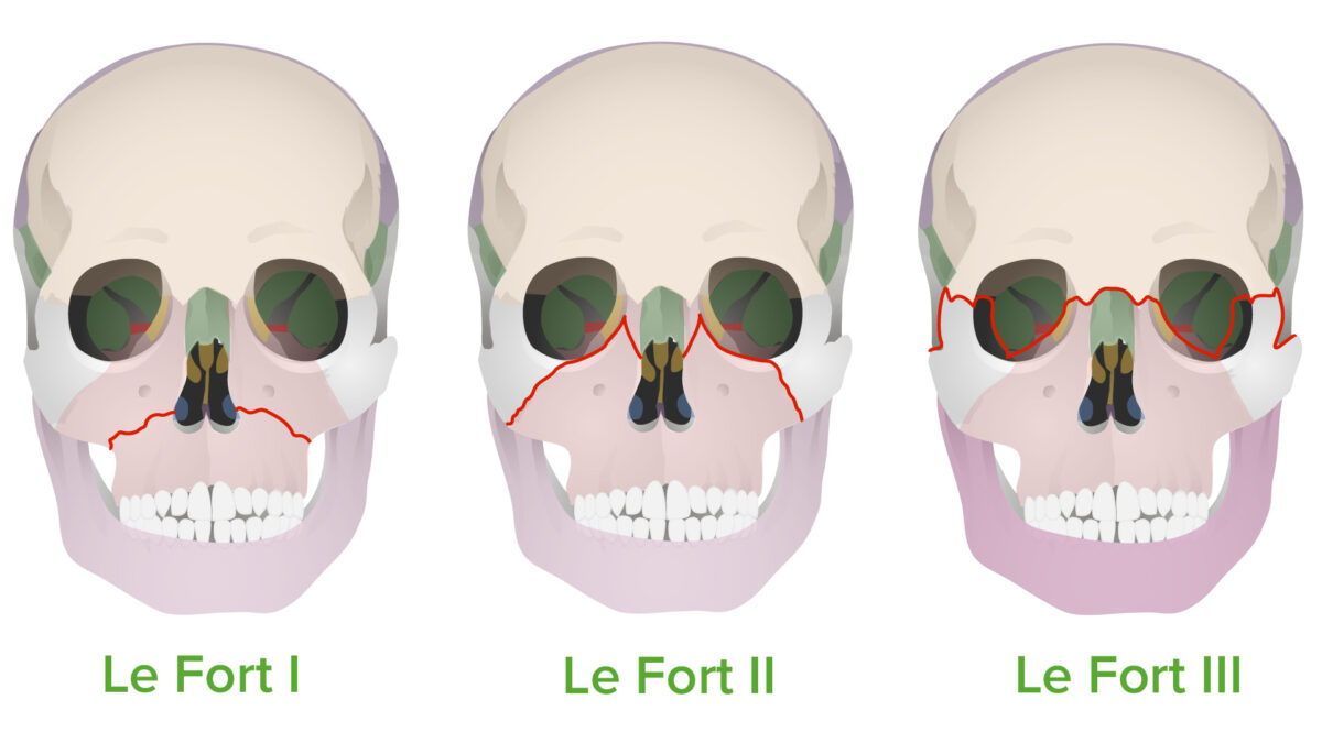
Le Fort fractures are a group midface fracture Fracture A fracture is a disruption of the cortex of any bone and periosteum and is commonly due to mechanical stress after an injury or accident. Open fractures due to trauma can be a medical emergency. Fractures are frequently associated with automobile accidents, workplace injuries, and trauma. Overview of Bone Fractures patterns classified into 3 types: Le Fort I, II, and III. Le Fort fractures represent 10%–20% of all facial fractures and can be caused by any significant blunt trauma to the face, most commonly from motor vehicle accidents Motor Vehicle Accidents Spinal Cord Injuries. Clinical presentation includes severe facial bleeding and edema Edema Edema is a condition in which excess serous fluid accumulates in the body cavity or interstitial space of connective tissues. Edema is a symptom observed in several medical conditions. It can be categorized into 2 types, namely, peripheral (in the extremities) and internal (in an organ or body cavity). Edema and mobility of different bone Bone Bone is a compact type of hardened connective tissue composed of bone cells, membranes, an extracellular mineralized matrix, and central bone marrow. The 2 primary types of bone are compact and spongy. Bones: Structure and Types segments, depending on the type in question. Diagnosis is clinical, supported by imaging techniques. Initial management centers around stabilization of the individual and control of bleeding. Definitive management is surgical. Long-term management concerns preservation and rehabilitation of facial function.

Last updated: Jan 10, 2023

Editorial responsibility: Stanley Oiseth, Lindsay Jones, Evelin Maza

Advertisement

Advertisement

Advertisement

Advertisement

Advertisement

Advertisement

Overview

Definition

  • A group of midface fractures typically brought on by trauma
  • Described by Dr. Rene Le Fort in 1901

Classification

  • Le Fort I (most inferior):
    • Trans-maxillary fracture Fracture A fracture is a disruption of the cortex of any bone and periosteum and is commonly due to mechanical stress after an injury or accident. Open fractures due to trauma can be a medical emergency. Fractures are frequently associated with automobile accidents, workplace injuries, and trauma. Overview of Bone Fractures (also known as Guerin fracture Fracture A fracture is a disruption of the cortex of any bone and periosteum and is commonly due to mechanical stress after an injury or accident. Open fractures due to trauma can be a medical emergency. Fractures are frequently associated with automobile accidents, workplace injuries, and trauma. Overview of Bone Fractures)
    • Bony disruption involves:
      • Superior to the maxillary alveolar process
      • Spans the body of the nasal septum Nasal septum The partition separating the two nasal cavities in the midplane. It is formed by the septal nasal cartilage, parts of skull bones, and membranous parts. Nose Anatomy (External & Internal)
      • May involve pterygoid plates of the sphenoid bone Sphenoid bone An irregular unpaired bone situated at the skull base and wedged between the frontal, temporal, and occipital bones (frontal bone; temporal bone; occipital bone). Sphenoid bone consists of a median body and three pairs of processes resembling a bat with spread wings. The body is hollowed out in its inferior to form two large cavities (sphenoid sinus). Orbit and Extraocular Muscles: Anatomy
  • Le Fort II:
    • Pyramidal fracture Fracture A fracture is a disruption of the cortex of any bone and periosteum and is commonly due to mechanical stress after an injury or accident. Open fractures due to trauma can be a medical emergency. Fractures are frequently associated with automobile accidents, workplace injuries, and trauma. Overview of Bone Fractures
    • Bony disruption involves:
      • Posterolateral portions of the maxillary sinuses
      • Typically passes through the infraorbital foramina
      • Lacrimal and/or ethmoid bones may be involved.
  • Le Fort III (most superior):
    • Craniofacial disjunction: Face is separated from the skull Skull The skull (cranium) is the skeletal structure of the head supporting the face and forming a protective cavity for the brain. The skull consists of 22 bones divided into the viscerocranium (facial skeleton) and the neurocranium. Skull: Anatomy.
    • Also known as horizontal fracture Fracture A fracture is a disruption of the cortex of any bone and periosteum and is commonly due to mechanical stress after an injury or accident. Open fractures due to trauma can be a medical emergency. Fractures are frequently associated with automobile accidents, workplace injuries, and trauma. Overview of Bone Fractures 
    • Bony disruption involves:
      • Superior orbital fissures
      • Lacrimal bones
      • Ethmoid bones
      • Nasal bones
      • Greater wings of the sphenoid bone Sphenoid bone An irregular unpaired bone situated at the skull base and wedged between the frontal, temporal, and occipital bones (frontal bone; temporal bone; occipital bone). Sphenoid bone consists of a median body and three pairs of processes resembling a bat with spread wings. The body is hollowed out in its inferior to form two large cavities (sphenoid sinus). Orbit and Extraocular Muscles: Anatomy
      • Frontozygomatic sutures
    • Eyes are held in place by the optic nerves.
  • Le Fort IV:
    • Characteristics of type III + involvement of the frontal Frontal The bone that forms the frontal aspect of the skull. Its flat part forms the forehead, articulating inferiorly with the nasal bone and the cheek bone on each side of the face. Skull: Anatomy bone Bone Bone is a compact type of hardened connective tissue composed of bone cells, membranes, an extracellular mineralized matrix, and central bone marrow. The 2 primary types of bone are compact and spongy. Bones: Structure and Types

Epidemiology

  • 10%–20% of all facial fractures
  • Usually seen in the context of polytrauma Polytrauma Multitrauma occurs when 2 or more traumatic injuries occur in at least 2 areas of the body. A systematic management approach is necessary for individuals who have undergone trauma to maximize outcomes and reduce the risk of undiscovered injuries. Multitrauma
  • Associated with substance use
  • Decreased incidence Incidence The number of new cases of a given disease during a given period in a specified population. It also is used for the rate at which new events occur in a defined population. It is differentiated from prevalence, which refers to all cases in the population at a given time. Measures of Disease Frequency related to motor Motor Neurons which send impulses peripherally to activate muscles or secretory cells. Nervous System: Histology vehicle crashes owing to better restraint systems

Etiology

  • Any trauma to the face that transfers a significant amount of kinetic energy:
    • Motor vehicle accidents Motor Vehicle Accidents Spinal Cord Injuries
    • Sports injuries
    • Physical assault (e.g., child abuse Child abuse Child abuse is an act or failure to act that results in harm to a child’s health or development. The abuse encompasses neglect as well as physical, sexual, and emotional harm. Seen in all subsets of society, child abuse is a cause of significant morbidity and mortality in the pediatric population. Child Abuse, domestic violence, elder abuse)
    • Falls
  • Velocity of trauma is associated with severity of Le Fort fractures.
Le fort fracture types

Le Fort fracture types:
These fractures follow the “lines of weakness” proposed by Rene Le Fort in 1901.
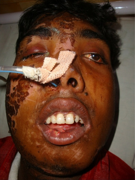
Image by Lecturio.

Pathophysiology

  • Blunt trauma: a significant amount of kinetic energy is directly transferred to the midface.
    • Motor vehicle accidents Motor Vehicle Accidents Spinal Cord Injuries: Unrestrained individual’s face strikes the dashboard.
    • Sports (e.g., football, baseball, and hockey): contact injuries or projectile (e.g., ball or puck) strikes the face 
    • Physical assault: severely battered individuals
    • Falls from significant heights 
    • May also include:
  • Le Fort I: Force is directed downward on the maxillary rim.
  • Le Fort II: Force is directed to the lower or mid maxilla Maxilla One of a pair of irregularly shaped bones that form the upper jaw. A maxillary bone provides tooth sockets for the superior teeth, forms part of the orbit, and contains the maxillary sinus. Skull: Anatomy.
  • Le Fort III: Force impacts the nasal bridge or upper maxilla Maxilla One of a pair of irregularly shaped bones that form the upper jaw. A maxillary bone provides tooth sockets for the superior teeth, forms part of the orbit, and contains the maxillary sinus. Skull: Anatomy.

Clinical Presentation

History

  • Remember: Because of the nature of the fracture Fracture A fracture is a disruption of the cortex of any bone and periosteum and is commonly due to mechanical stress after an injury or accident. Open fractures due to trauma can be a medical emergency. Fractures are frequently associated with automobile accidents, workplace injuries, and trauma. Overview of Bone Fractures, the individual most likely won’t be able to articulate any details about the incident. 
  • First responders or companions will report recent high-energy blunt trauma.
    • Motor Motor Neurons which send impulses peripherally to activate muscles or secretory cells. Nervous System: Histology vehicle accident
    • Fall from a great height
    • Attack with a blunt object
  • The clinician Clinician A physician, nurse practitioner, physician assistant, or another health professional who is directly involved in patient care and has a professional relationship with patients. Clinician–Patient Relationship must determine:
    • The mechanism of trauma
    • Further details if possible

Physical examination

  • Airway Airway ABCDE Assessment:
  • Oral cavity assessment:
    • To determine the presence of hard palate Hard palate The anteriorly located rigid section of the palate. Palate: Anatomy fracture Fracture A fracture is a disruption of the cortex of any bone and periosteum and is commonly due to mechanical stress after an injury or accident. Open fractures due to trauma can be a medical emergency. Fractures are frequently associated with automobile accidents, workplace injuries, and trauma. Overview of Bone Fractures
    • To determine the presence/source of oropharyngeal hemorrhage
  • Nasal cavity Nasal cavity The proximal portion of the respiratory passages on either side of the nasal septum. Nasal cavities, extending from the nares to the nasopharynx, are lined with ciliated nasal mucosa. Nose Anatomy (External & Internal) assessment:
    • To determine the presence of nasal fractures
    • To determine the presence/source of nasopharyngeal hemorrhage
    • Assess for leakage of CSF.
  • Cervical spine Spine The human spine, or vertebral column, is the most important anatomical and functional axis of the human body. It consists of 7 cervical vertebrae, 12 thoracic vertebrae, and 5 lumbar vertebrae and is limited cranially by the skull and caudally by the sacrum. Vertebral Column: Anatomy/ neck Neck The part of a human or animal body connecting the head to the rest of the body. Peritonsillar Abscess assessment:
    • Keep cervical spine Spine The human spine, or vertebral column, is the most important anatomical and functional axis of the human body. It consists of 7 cervical vertebrae, 12 thoracic vertebrae, and 5 lumbar vertebrae and is limited cranially by the skull and caudally by the sacrum. Vertebral Column: Anatomy immobilized during the examination.
    • Cervical spinal fracture Fracture A fracture is a disruption of the cortex of any bone and periosteum and is commonly due to mechanical stress after an injury or accident. Open fractures due to trauma can be a medical emergency. Fractures are frequently associated with automobile accidents, workplace injuries, and trauma. Overview of Bone Fractures should always be suspected until proven otherwise.
  • Facial assessment:
    • Significant facial hemorrhage and edema Edema Edema is a condition in which excess serous fluid accumulates in the body cavity or interstitial space of connective tissues. Edema is a symptom observed in several medical conditions. It can be categorized into 2 types, namely, peripheral (in the extremities) and internal (in an organ or body cavity). Edema are likely to be present.
    • Possible development of early ecchymosis Ecchymosis Extravasation of blood into the skin, resulting in a nonelevated, rounded or irregular, blue or purplish patch, larger than a petechia. Orbital Fractures
    • Dish-face deformity Deformity Examination of the Upper Limbs: flattening of the face (type III)
  • Determine mobile bony segments (with forehead Forehead The part of the face above the eyes. Melasma stabilized):
    • Le Fort I: hard palate Hard palate The anteriorly located rigid section of the palate. Palate: Anatomy
    • Le Fort II: maxilla Maxilla One of a pair of irregularly shaped bones that form the upper jaw. A maxillary bone provides tooth sockets for the superior teeth, forms part of the orbit, and contains the maxillary sinus. Skull: Anatomy and nose Nose The nose is the human body’s primary organ of smell and functions as part of the upper respiratory system. The nose may be best known for inhaling oxygen and exhaling carbon dioxide, but it also contributes to other important functions, such as tasting. The anatomy of the nose can be divided into the external nose and the nasal cavity. Nose Anatomy (External & Internal) 
    • Le Fort III: maxilla Maxilla One of a pair of irregularly shaped bones that form the upper jaw. A maxillary bone provides tooth sockets for the superior teeth, forms part of the orbit, and contains the maxillary sinus. Skull: Anatomy, nose Nose The nose is the human body’s primary organ of smell and functions as part of the upper respiratory system. The nose may be best known for inhaling oxygen and exhaling carbon dioxide, but it also contributes to other important functions, such as tasting. The anatomy of the nose can be divided into the external nose and the nasal cavity. Nose Anatomy (External & Internal), and zygomas
    • Anterior open bite malocclusion:
      • May be associated with fracture Fracture A fracture is a disruption of the cortex of any bone and periosteum and is commonly due to mechanical stress after an injury or accident. Open fractures due to trauma can be a medical emergency. Fractures are frequently associated with automobile accidents, workplace injuries, and trauma. Overview of Bone Fractures and posterior displacement Displacement The process by which an emotional or behavioral response that is appropriate for one situation appears in another situation for which it is inappropriate. Defense Mechanisms of the maxilla Maxilla One of a pair of irregularly shaped bones that form the upper jaw. A maxillary bone provides tooth sockets for the superior teeth, forms part of the orbit, and contains the maxillary sinus. Skull: Anatomy
      • May be associated with accompanying fracture Fracture A fracture is a disruption of the cortex of any bone and periosteum and is commonly due to mechanical stress after an injury or accident. Open fractures due to trauma can be a medical emergency. Fractures are frequently associated with automobile accidents, workplace injuries, and trauma. Overview of Bone Fractures and anterior displacement Displacement The process by which an emotional or behavioral response that is appropriate for one situation appears in another situation for which it is inappropriate. Defense Mechanisms of the maxilla Maxilla One of a pair of irregularly shaped bones that form the upper jaw. A maxillary bone provides tooth sockets for the superior teeth, forms part of the orbit, and contains the maxillary sinus. Skull: Anatomy
  • Orbital/ocular assessment:
    • Bilateral orbital ecchymosis Ecchymosis Extravasation of blood into the skin, resulting in a nonelevated, rounded or irregular, blue or purplish patch, larger than a petechia. Orbital Fractures (also known as raccoon eyes)
    • Widening of the intercanthal space
    • Orbital involvement commonly occurs with types II and III fractures (requires emergent ophthalmologic consultation).
  • Neurologic assessment:
    • Determine mental status and level of consciousness.
    • If level of consciousness is impaired, determine baseline Glasgow Coma Coma Coma is defined as a deep state of unarousable unresponsiveness, characterized by a score of 3 points on the GCS. A comatose state can be caused by a multitude of conditions, making the precise epidemiology and prognosis of coma difficult to determine. Coma Scale Scale Dermatologic Examination score.
    • Assess for cranial nerve function and the presence of focal neurologic injury.
    • Assess for symptoms/signs of increased intracranial pressure Intracranial Pressure Idiopathic Intracranial Hypertension.
    • Assess for symptoms/signs of intracranial hemorrhage Intracranial hemorrhage Subarachnoid hemorrhage (SAH) is a type of cerebrovascular accident (stroke) resulting from intracranial hemorrhage into the subarachnoid space between the arachnoid and the pia mater layers of the meninges surrounding the brain. Most sahs originate from a saccular aneurysm in the circle of willis but may also occur as a result of trauma, uncontrolled hypertension, vasculitis, anticoagulant use, or stimulant use. Subarachnoid Hemorrhage/traumatic brain Brain The part of central nervous system that is contained within the skull (cranium). Arising from the neural tube, the embryonic brain is comprised of three major parts including prosencephalon (the forebrain); mesencephalon (the midbrain); and rhombencephalon (the hindbrain). The developed brain consists of cerebrum; cerebellum; and other structures in the brain stem. Nervous System: Anatomy, Structure, and Classification injury.
Le fort iii fracture

Individual with a Le Fort III fracture:
Note the widening of the intercanthal space and pronounced facial edema. The ED physicians opted for a secure airway through the nose.
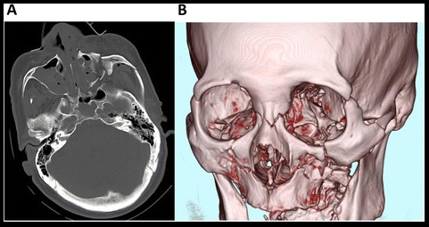
Image: “Preoperative” by Santosh Kumar Yadav et al. License: CC0 1.0

Diagnosis

Clinical diagnosis

  • Prior to assessment for findings specific to Le Fort fractures, the following should be assessed, diagnosed, and appropriately triaged:
    • Airway Airway ABCDE Assessment patency
    • Cervical spine Spine The human spine, or vertebral column, is the most important anatomical and functional axis of the human body. It consists of 7 cervical vertebrae, 12 thoracic vertebrae, and 5 lumbar vertebrae and is limited cranially by the skull and caudally by the sacrum. Vertebral Column: Anatomy stability
    • Life-threatening trauma
    • Life-threatening hemorrhage
    • Hemodynamic status
  • The diagnosis of a Le Fort fracture Fracture A fracture is a disruption of the cortex of any bone and periosteum and is commonly due to mechanical stress after an injury or accident. Open fractures due to trauma can be a medical emergency. Fractures are frequently associated with automobile accidents, workplace injuries, and trauma. Overview of Bone Fractures should be considered and ruled out in any individual with the following:
    • History of significant cranial, facial, or cervical trauma
    • Facial hemorrhage
    • Facial edema Edema Edema is a condition in which excess serous fluid accumulates in the body cavity or interstitial space of connective tissues. Edema is a symptom observed in several medical conditions. It can be categorized into 2 types, namely, peripheral (in the extremities) and internal (in an organ or body cavity). Edema
    • Facial ecchymosis Ecchymosis Extravasation of blood into the skin, resulting in a nonelevated, rounded or irregular, blue or purplish patch, larger than a petechia. Orbital Fractures
  • History and physical examination should be sufficient for initial triage if the individual is stable and clinical suspicion for Le Fort fracture Fracture A fracture is a disruption of the cortex of any bone and periosteum and is commonly due to mechanical stress after an injury or accident. Open fractures due to trauma can be a medical emergency. Fractures are frequently associated with automobile accidents, workplace injuries, and trauma. Overview of Bone Fractures is low.

Imaging

  • Should be obtained only after the individual has been stabilized.
  • Plain films of the face/cervical spine Spine The human spine, or vertebral column, is the most important anatomical and functional axis of the human body. It consists of 7 cervical vertebrae, 12 thoracic vertebrae, and 5 lumbar vertebrae and is limited cranially by the skull and caudally by the sacrum. Vertebral Column: Anatomy:
    • May be useful in the absence of CT to identify superficial fractures
    • Limited utility in identifying deeper fractures and associated soft tissue Soft Tissue Soft Tissue Abscess/vascular injury
    • May be useful in the absence of CT to assess cervical spine Spine The human spine, or vertebral column, is the most important anatomical and functional axis of the human body. It consists of 7 cervical vertebrae, 12 thoracic vertebrae, and 5 lumbar vertebrae and is limited cranially by the skull and caudally by the sacrum. Vertebral Column: Anatomy stability and/or the presence of fracture Fracture A fracture is a disruption of the cortex of any bone and periosteum and is commonly due to mechanical stress after an injury or accident. Open fractures due to trauma can be a medical emergency. Fractures are frequently associated with automobile accidents, workplace injuries, and trauma. Overview of Bone Fractures
  • CT scan:
    • Useful for identification Identification Defense Mechanisms of superficial and deep facial fractures
    • Useful for identification Identification Defense Mechanisms of associated soft tissue Soft Tissue Soft Tissue Abscess/vascular injury
    • Useful in assessment of associated penetrating injury Penetrating Injury Brown-Séquard Syndrome
    • Useful in assessment of associated traumatic brain Brain The part of central nervous system that is contained within the skull (cranium). Arising from the neural tube, the embryonic brain is comprised of three major parts including prosencephalon (the forebrain); mesencephalon (the midbrain); and rhombencephalon (the hindbrain). The developed brain consists of cerebrum; cerebellum; and other structures in the brain stem. Nervous System: Anatomy, Structure, and Classification injury and/or intracranial hemorrhage Intracranial hemorrhage Subarachnoid hemorrhage (SAH) is a type of cerebrovascular accident (stroke) resulting from intracranial hemorrhage into the subarachnoid space between the arachnoid and the pia mater layers of the meninges surrounding the brain. Most sahs originate from a saccular aneurysm in the circle of willis but may also occur as a result of trauma, uncontrolled hypertension, vasculitis, anticoagulant use, or stimulant use. Subarachnoid Hemorrhage 
Ct image of le fort iii fracture

Craniofacial CT (A) and 3-dimensional reconstruction (B) of an individual with a Le Fort III fracture

Image: “Craniofacial computed tomography scan obtained on arrival.” by Momoko Mishima, Tetsuya Yumoto, Hiroaki Hashimoto et al. License: CC BY 4.0

Management

Goals of management

  • Restore nasal and orbital integrity, along with height and projection of the midface
  • Regain proper occlusion of the jaw Jaw The jaw is made up of the mandible, which comprises the lower jaw, and the maxilla, which comprises the upper jaw. The mandible articulates with the temporal bone via the temporomandibular joint (TMJ). The 4 muscles of mastication produce the movements of the TMJ to ensure the efficient chewing of food. Jaw and Temporomandibular Joint: Anatomy
  • Evaluate for and appropriately manage associated injuries:
    • Head injury/traumatic brain Brain The part of central nervous system that is contained within the skull (cranium). Arising from the neural tube, the embryonic brain is comprised of three major parts including prosencephalon (the forebrain); mesencephalon (the midbrain); and rhombencephalon (the hindbrain). The developed brain consists of cerebrum; cerebellum; and other structures in the brain stem. Nervous System: Anatomy, Structure, and Classification injury
    • Intracranial hemorrhage Intracranial hemorrhage Subarachnoid hemorrhage (SAH) is a type of cerebrovascular accident (stroke) resulting from intracranial hemorrhage into the subarachnoid space between the arachnoid and the pia mater layers of the meninges surrounding the brain. Most sahs originate from a saccular aneurysm in the circle of willis but may also occur as a result of trauma, uncontrolled hypertension, vasculitis, anticoagulant use, or stimulant use. Subarachnoid Hemorrhage
    • Increased intracranial pressure Intracranial Pressure Idiopathic Intracranial Hypertension
    • CSF leakage
    • Associated soft tissue Soft Tissue Soft Tissue Abscess/vascular injury
    • Cervical spine Spine The human spine, or vertebral column, is the most important anatomical and functional axis of the human body. It consists of 7 cervical vertebrae, 12 thoracic vertebrae, and 5 lumbar vertebrae and is limited cranially by the skull and caudally by the sacrum. Vertebral Column: Anatomy fracture Fracture A fracture is a disruption of the cortex of any bone and periosteum and is commonly due to mechanical stress after an injury or accident. Open fractures due to trauma can be a medical emergency. Fractures are frequently associated with automobile accidents, workplace injuries, and trauma. Overview of Bone Fractures/ spinal cord Spinal cord The spinal cord is the major conduction pathway connecting the brain to the body; it is part of the CNS. In cross section, the spinal cord is divided into an H-shaped area of gray matter (consisting of synapsing neuronal cell bodies) and a surrounding area of white matter (consisting of ascending and descending tracts of myelinated axons). Spinal Cord: Anatomy injury
    • Ocular injuries/loss of vision Vision Ophthalmic Exam
    • Dental injuries 

Initial triage

Initial management

Surgical management

  • After stabilization and management of life-threatening injuries
  • Maxillofacial surgery:
    • Fracture Fracture A fracture is a disruption of the cortex of any bone and periosteum and is commonly due to mechanical stress after an injury or accident. Open fractures due to trauma can be a medical emergency. Fractures are frequently associated with automobile accidents, workplace injuries, and trauma. Overview of Bone Fractures reduction with plate-and-screws osteosynthesis
    • Proper occlusion ensures adequate repair.
  • Neurosurgery Neurosurgery Neurosurgery is a specialized field focused on the surgical management of pathologies of the brain, spine, spinal cord, and peripheral nerves. General neurosurgery includes cases of trauma and emergencies. There are a number of specialized neurosurgical practices, including oncologic neurosurgery, spinal neurosurgery, and pediatric neurosurgery. Neurosurgery consultation: cases with CSF leaks

Complications

  • Life-threatening exsanguination
  • Intracranial hemorrhage Intracranial hemorrhage Subarachnoid hemorrhage (SAH) is a type of cerebrovascular accident (stroke) resulting from intracranial hemorrhage into the subarachnoid space between the arachnoid and the pia mater layers of the meninges surrounding the brain. Most sahs originate from a saccular aneurysm in the circle of willis but may also occur as a result of trauma, uncontrolled hypertension, vasculitis, anticoagulant use, or stimulant use. Subarachnoid Hemorrhage
  • Traumatic brain Brain The part of central nervous system that is contained within the skull (cranium). Arising from the neural tube, the embryonic brain is comprised of three major parts including prosencephalon (the forebrain); mesencephalon (the midbrain); and rhombencephalon (the hindbrain). The developed brain consists of cerebrum; cerebellum; and other structures in the brain stem. Nervous System: Anatomy, Structure, and Classification injury
  • CSF leak
  • Severe epistaxis Epistaxis Bleeding from the nose. Granulomatosis with Polyangiitis
  • Permanent disfigurement

Prognosis Prognosis A prediction of the probable outcome of a disease based on a individual’s condition and the usual course of the disease as seen in similar situations. Non-Hodgkin Lymphomas

  • Depends on mechanism, severity, and location of injury
  • Potentially life-threatening if not managed promptly 
  • Common morbidities after injury include:
    • Visual problems
    • Difficulty with mastication Mastication The act and process of chewing and grinding food in the mouth. Jaw and Temporomandibular Joint: Anatomy
    • Cranial nerve palsies Cranial Nerve Palsies Cranial nerve palsy is a congenital or acquired dysfunction of 1 or more cranial nerves that will, in turn, lead to focal neurologic abnormalities in movement or autonomic dysfunction of its territory. Head/neck trauma, mass effect, infectious processes, and ischemia/infarction are among the many etiologies for these dysfunctions. Diagnosis is initially clinical and supported by diagnostic aids. Management includes both symptomatic measures and interventions aimed at correcting the underlying cause. Cranial Nerve Palsies
    • Taste/ smell Smell The sense of smell, or olfaction, begins in a small area on the roof of the nasal cavity, which is covered in specialized mucosa. From there, the olfactory nerve transmits the sensory perception of smell via the olfactory pathway. This pathway is composed of the olfactory cells and bulb, the tractus and striae olfactoriae, and the primary olfactory cortex and amygdala. Olfaction: Anatomy abnormalities

Differential Diagnosis

  • Frontal Frontal The bone that forms the frontal aspect of the skull. Its flat part forms the forehead, articulating inferiorly with the nasal bone and the cheek bone on each side of the face. Skull: Anatomy fracture Fracture A fracture is a disruption of the cortex of any bone and periosteum and is commonly due to mechanical stress after an injury or accident. Open fractures due to trauma can be a medical emergency. Fractures are frequently associated with automobile accidents, workplace injuries, and trauma. Overview of Bone Fractures: disruption in the cortex of the frontal Frontal The bone that forms the frontal aspect of the skull. Its flat part forms the forehead, articulating inferiorly with the nasal bone and the cheek bone on each side of the face. Skull: Anatomy bone Bone Bone is a compact type of hardened connective tissue composed of bone cells, membranes, an extracellular mineralized matrix, and central bone marrow. The 2 primary types of bone are compact and spongy. Bones: Structure and Types, usually due to direct blunt or penetrating trauma to the forehead Forehead The part of the face above the eyes. Melasma. Clinical presentation includes active bleeding and edema Edema Edema is a condition in which excess serous fluid accumulates in the body cavity or interstitial space of connective tissues. Edema is a symptom observed in several medical conditions. It can be categorized into 2 types, namely, peripheral (in the extremities) and internal (in an organ or body cavity). Edema, crepitus Crepitus Osteoarthritis in the area of fracture Fracture A fracture is a disruption of the cortex of any bone and periosteum and is commonly due to mechanical stress after an injury or accident. Open fractures due to trauma can be a medical emergency. Fractures are frequently associated with automobile accidents, workplace injuries, and trauma. Overview of Bone Fractures and/or mobility of bone Bone Bone is a compact type of hardened connective tissue composed of bone cells, membranes, an extracellular mineralized matrix, and central bone marrow. The 2 primary types of bone are compact and spongy. Bones: Structure and Types segments, and altered mental status Altered Mental Status Sepsis in Children associated with accompanying traumatic brain Brain The part of central nervous system that is contained within the skull (cranium). Arising from the neural tube, the embryonic brain is comprised of three major parts including prosencephalon (the forebrain); mesencephalon (the midbrain); and rhombencephalon (the hindbrain). The developed brain consists of cerebrum; cerebellum; and other structures in the brain stem. Nervous System: Anatomy, Structure, and Classification injury. Diagnosis is clinical, supported by imaging, and definitive management can be conservative or surgical. 
  • Mandible Mandible The largest and strongest bone of the face constituting the lower jaw. It supports the lower teeth. Jaw and Temporomandibular Joint: Anatomy luxation: describes the unilateral or bilateral uncoupling of the components of the temporomandibular joint. Causes can be traumatic or atraumatic, such as forceful over-opening of the jaw Jaw The jaw is made up of the mandible, which comprises the lower jaw, and the maxilla, which comprises the upper jaw. The mandible articulates with the temporal bone via the temporomandibular joint (TMJ). The 4 muscles of mastication produce the movements of the TMJ to ensure the efficient chewing of food. Jaw and Temporomandibular Joint: Anatomy. Diagnosis is clinical. Management consists of bimanual reduction.
  • Mandible Mandible The largest and strongest bone of the face constituting the lower jaw. It supports the lower teeth. Jaw and Temporomandibular Joint: Anatomy fracture Fracture A fracture is a disruption of the cortex of any bone and periosteum and is commonly due to mechanical stress after an injury or accident. Open fractures due to trauma can be a medical emergency. Fractures are frequently associated with automobile accidents, workplace injuries, and trauma. Overview of Bone Fractures: disruption in the cortex of the mandible Mandible The largest and strongest bone of the face constituting the lower jaw. It supports the lower teeth. Jaw and Temporomandibular Joint: Anatomy, usually due to blunt trauma seen in vehicle accidents or worksite injuries. Clinical presentation includes maxillofacial deformity Deformity Examination of the Upper Limbs and edema Edema Edema is a condition in which excess serous fluid accumulates in the body cavity or interstitial space of connective tissues. Edema is a symptom observed in several medical conditions. It can be categorized into 2 types, namely, peripheral (in the extremities) and internal (in an organ or body cavity). Edema and ecchymosis Ecchymosis Extravasation of blood into the skin, resulting in a nonelevated, rounded or irregular, blue or purplish patch, larger than a petechia. Orbital Fractures in the site of injury. Diagnosis is clinical, supported by imaging, and definitive management is surgical.

References

  1. Phillips, B. J., Turco, L. M. (2017). Le Fort fractures: a collective review. Bulletin of Emergency & Trauma, 5:221.
  2. Rodriguez, E. D., Dorafshar, A. H., Manson, P. N. (2018). Facial injuries. In: Rodriguez, E. D., Losee, J. E., P. C. Neligan, P. C. (Eds.), Plastic Surgery. Volume 3: Craniofacial, Head and Neck Surgery and Pediatric Plastic Surgery, 4th ed. pp. 47–81. Elsevier.
  3. Hedayati, T., Amin, D. P. (2020). Trauma to the face. Chapter 259 of Tintinalli, J. E., Ma, O. J., Yealy, D. M., Meckler, G. D., Stapczynski, J. S., Cline, D. M., Thomas, S. H. (Eds.), Tintinalli’s Emergency Medicine: A Comprehensive Study Guide, 9th ed. McGraw-Hill Education. http://accessmedicine.mhmedical.com/content.aspx?aid=1167028185
  4. Patel, B. C., Wright, T., Waseem, M. (2021). Le fort fractures. StatPearls. Retrieved October 15, 2021, from http://www.ncbi.nlm.nih.gov/books/NBK526060/

Create your free account or log in to continue reading!

Sign up now and get free access to Lecturio with concept pages, medical videos, and questions for your medical education.

User Reviews

Unwrap New Skills This Holiday 🎄 Save 30% on all plans now!

Details